
Article Figures & Data
Figures
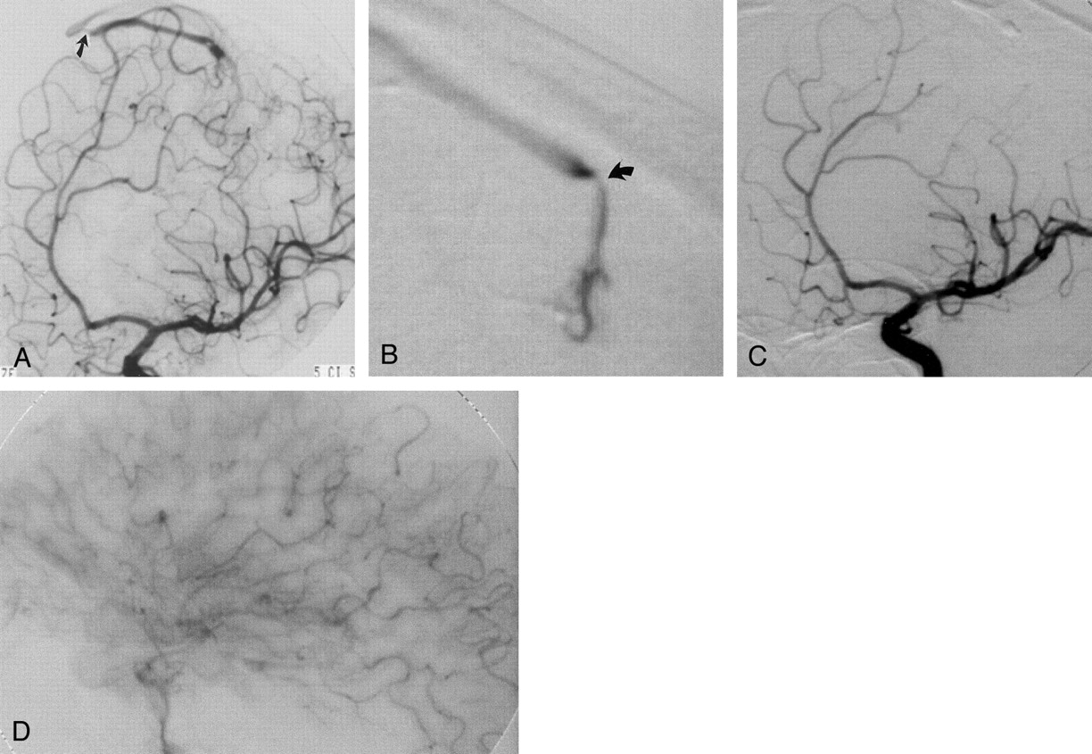
- Fig 1.
Imaging studies in a 31-year-old man (case 2) with a cortical hematoma in the left angular and supramarginal gyri.
A and B, At admission, lateral early (A) and late (B) arterial-phase angiograms of left internal carotid artery do not show evidence of arteriovenous shunting.
C, Oblique left internal carotid arterial-phase angiogram obtained 1 month later, after hemorrhage resorption, shows early venous filling of two cortical veins that drain in the superior sagittal sinus (arrows and arrowheads).
D, Subsequent superselective exploration of the angular branch of the left middle cerebral artery reveals the feeder, the nidus, and the double superficial venous drainage of the micro-AVM. After stable microcatheter positioning, treatment of the lesion was performed by using a single injection (ie, single shot) of a polymerizing agent.
- Fig 2.
Imaging studies in a 43-year-old woman (case 1) with a large hematoma in the left occipitoparietal lobe.
A and B, Frontal (A) and lateral (B) late arterial-phase left vertebral angiograms show a small tangle of arterial blush (curved arrow) and a questionable early venous filling (straight arrow) along the parieto-occipital branches of the left posterior cerebral artery.
C, Superselective exploration of the parieto-occipital branch demonstrates the plexiform structure of the nidus of the micro-AVM, which is characterized by multiple arteriovenous shunts and a single deep draining vein coursing toward the vein of Galen.
D, The lesion could not be embolized because microcatheter tip instability and opacification of functional vessels originated from the distal tract of the feeder. The patient subsequently underwent successful surgery.
- Fig 3.
Imaging studies in a 41-year-old man (case 4) with a large hemorrhage of the left paracentral lobule.
A, Oblique arterial-phase left internal carotid angiogram shows early venous drainage in the absence of a clearly defined nidus. Note stenosis of the draining vein at the junction with the superior sagittal sinus (arrow).
B, Superselective exploration of the paracentral branch of the left callosomarginal artery allows identification of the nidus with demonstration of another stenosis (arrow) in the proximal tract of the draining vein.
C and D, After successful embolization, left internal carotid angiograms show the glue cast of the nidus and the draining vein (C) in the absence of any residual arteriovenous shunting (D). C is an arterial-phase oblique view; D, a capillary-phase lateral view.


Tables
- TABLE 1:
Overview of clinical and diagnostic features and therapy in the 10 patients with cerebral micro-AVMs
Patient No./Age (y)/Sex Neurologic Presentation Hematoma Location and Size Nidus Feeder Vessel Drainage Therapy Clinical Follow-Up Findings 1/43/F Prodrome (1 mo), headache, vomiting, R hemianopsia L occipitoparietal lobe, subarachnoid hemorrhage, 21.98 cm3 Cortical L parieto-occipital branch of PCA Deep Surgery (endovascular therapy failed) R inferior quadrantanopia, 12 mo 2/31/M Prodrome (1 wk), transitory aphasia, seizure L temporoparietal lobe, 6.18 cm3 Cortical L angular branch of MCA Superficial Endovascular, 2 sessions Occasional word-finding difficulties, 24 mo 3/60/F R paresthesias, R arm hypesthesia and paresis L parietal lobe, 4.13 cm3 Cortical R superior parietal branch of CA Superficial ectasia Endovascular Clumsiness of R upper extremity, 24 mo 4/41/M Headache, R hemiparesis L frontal lobe, 8.15 cm3 Cortical L paracentral branch of CA Superficial stenosis Endovascular R limp, 24 mo 5/57/M Coma (GCS 4), L hemiparesis R temporoparietal lobes, 35.15 cm3 Cortical R temporal branch of MCA Deep Surgery L hand plegia, walking with brace, 4 y 6/48/F Coma (GCS 12), hemorrhagic ictus (possibly 13 y previously) R temporal lobe, intraventricular, 3.0 cm3 Deep R posterior temporal branch of PCA Deep Endovascular (surgery failed) Mild cognitive impairment, 24 mo 7/43/M Visual disturbances, seizure R temporal lobe, 9.18 cm3 Cortical R middle temporal branch of MCA Superficial ectasia Surgery Seizures >30 d postoperatively, 24 mo 8/65/F Coma (GCS 8) Inferior cerebellar vermis, 15.18 cm3 Cortical R PICA and 2 aneurysms Superficial Endovascular None, 24 mo 9/38/M Dizziness, headache, vomiting R cerebellar tonsil, 3.18 cm3 Cortical R PICA Superficial Surgery None, 24 mo 10/50/F R hemiplegia, seizure, hemorrhagic ictus (possibly 8 y before) L frontoparietal lobe, 10.5 cm3 Cortical L ascending parietal branch of MCA Superficial Surgery (endovascular therapy failed) R hand weakness and numbness, 2 mo Note.—GCS indicates Glasgow Coma Scale score; CA, callosomarginal artery; MCA, middle cerebral artery; PCA, posterior cerebral artery; PICA, posterior inferior cerebellar artery.
- TABLE 2:
Overview of diagnostic workup results, therapeutic course, angiographic follow-up findings, and outcome
Patient No./Age (y)/Sex Surgical Treatment Endovascular Treatment First DSA Finding Second DSA Finding Superselect DSA Course of Treatment Follow-up Angiography* Barthel Index 1/43/F Yes Yes Negative Questionable 2 First DSA findings negative and control DSA findings (1 mo later) questionable. Microcatheterization was diagnostic. Endovascular treatment failed due to clinically uncomplicated vasospasm of the arterial feeder. Surgical intervention was successful. At 6 mo 100 2/31/M No Yes Negative Questionable 3 First DSA findings negative and control DSA findings (1 mo later) questionable. Microcatheterization was diagnostic. First embolization incomplete. Second (6 mo later) endovascular procedure successful. At 3 mo 100 3/60/F No Yes Positive 1 No data At 6 mo 95 4/41/M No Yes Questionable Positive 1 First DSA findings questionable. Repeat angiographic findings (2 mo later) were diagnostic. Single-shot endovascular treatment (1 wk later) successful. At 6 mo 95 5/57/M Yes No Positive 0 No data None 50 6/48/F Yes Yes Positive 1 Surgery attempted first; failed due to inability to locate AV shunt. Postoperative angiograms showed persistence of AV shunt. Endovascular treatment subsequently performed At 6 mo 100 7/43/M Yes No Questionable Positive First DSA findings questionable. Repeat angiography findings (1 mo later) were diagnostic. Subsequently surgery was successful. At 3 mo 100 8/65/F No Yes Questionable Positive 1 First DSA findings questionable. Repeat angiography findings (2 mo later) were diagnostic. Endovascular treatment (1 mo later) successful. At 3 mo 100 9/38/M Yes No Positive 0 No data At 3 mo 100 10/50/F Yes Yes Positive 1 DSA findings diagnostic. Endovascular treatment failed due to suboptimal microcatheter tip positioning. Patient subsequently underwent surgery. Immediate postoperative angiograms confirmed complete AV shunt resection. None 95 Note.—DSA indicates digital subtraction angiography; AV, arteriovenous.
↵* Performed after the last procedure.
In this issue


Jump to section
Related Articles
Cited By...
- No citing articles found.