Advertisement

Main menu

  • Home
  • Content
    • Current Issue
    • Accepted Manuscripts
    • Article Preview
    • Past Issue Archive
    • AJNR Case Collection
    • Case of the Week Archive
    • Classic Case Archive
    • Case of the Month Archive
  • Special Collections
    • Spinal CSF Leak Articles (Jan 2020-June 2024)
    • 2024 AJNR Journal Awards
    • Most Impactful AJNR Articles
  • Multimedia
    • AJNR Podcast
    • AJNR Scantastics
    • Video Articles
  • For Authors
    • Submit a Manuscript
    • Author Policies
    • Fast publishing of Accepted Manuscripts
    • Graphical Abstract Preparation
    • Manuscript Submission Guidelines
    • Imaging Protocol Submission
    • Submit a Case for the Case Collection
  • About Us
    • About AJNR
    • Editorial Board
  • More
    • Become a Reviewer/Academy of Reviewers
    • Subscribers
    • Permissions
    • Alerts
    • Feedback
    • Advertisers
    • ASNR Home
  • Other Publications
    • ajnr

User menu

  • Alerts
  • Log in

Search

  • Advanced search
American Journal of Neuroradiology

American Journal of Neuroradiology

ASHNR American Society of Functional Neuroradiology ASHNR American Society of Pediatric Neuroradiology ASSR
  • Alerts
  • Log in

Advanced Search

  • Home
  • Content
    • Current Issue
    • Accepted Manuscripts
    • Article Preview
    • Past Issue Archive
    • AJNR Case Collection
    • Case of the Week Archive
    • Classic Case Archive
    • Case of the Month Archive
  • Special Collections
    • Spinal CSF Leak Articles (Jan 2020-June 2024)
    • 2024 AJNR Journal Awards
    • Most Impactful AJNR Articles
  • Multimedia
    • AJNR Podcast
    • AJNR Scantastics
    • Video Articles
  • For Authors
    • Submit a Manuscript
    • Author Policies
    • Fast publishing of Accepted Manuscripts
    • Graphical Abstract Preparation
    • Manuscript Submission Guidelines
    • Imaging Protocol Submission
    • Submit a Case for the Case Collection
  • About Us
    • About AJNR
    • Editorial Board
  • More
    • Become a Reviewer/Academy of Reviewers
    • Subscribers
    • Permissions
    • Alerts
    • Feedback
    • Advertisers
    • ASNR Home
  • Follow AJNR on Twitter
  • Visit AJNR on Facebook
  • Follow AJNR on Instagram
  • Join AJNR on LinkedIn
  • RSS Feeds

Welcome to the new AJNR, Updated Hall of Fame, and more. Read the full announcements.


AJNR is seeking candidates for the position of Associate Section Editor, AJNR Case Collection. Read the full announcement.


OtherINTERVENTIONAL

Delayed Thromboembolic Events 9 Weeks after Endovascular Treatment of an Anterior Communicating Artery Aneurysm: Case Report

Matthew T. Studley, David H. Robinson and John F. Howe
American Journal of Neuroradiology June 2002, 23 (6) 975-977;
Matthew T. Studley
aDepartment of Radiology, Neurosurgery Department, Seattle, WA
  • Find this author on Google Scholar
  • Find this author on PubMed
  • Search for this author on this site
David H. Robinson
aDepartment of Radiology, Neurosurgery Department, Seattle, WA
  • Find this author on Google Scholar
  • Find this author on PubMed
  • Search for this author on this site
John F. Howe
bDepartment of Virginia Mason Medical Center, and Group Health Central, Neurosurgery Department, Seattle, WA
  • Find this author on Google Scholar
  • Find this author on PubMed
  • Search for this author on this site
  • Article
  • Figures & Data
  • Info & Metrics
  • Responses
  • References
  • PDF
Loading

Abstract

Summary: We present a case of delayed thromboembolic events that occurred 9 weeks after endovascular treatment of an anterior communicating artery aneurysm with GDC.

Acute thromboembolic events are a well-known complication of cerebral artery aneurysm treatment with GDC. Periprocedural complication rates have been reported to range between 2.5% and 28% (1–3). These events present as transient ischemic attacks or strokes and usually occur at the time of the procedure or within the first 48 hr (2). However, the occurrence of delayed thromboembolic events (weeks or months after the procedure) in a patient with stable coil packing of the aneurysm sac has not been previously described in the literature.

Case Report

A 54-year-old man presented to the emergency room of an outside hospital with a severe headache of sudden onset. An initial CT study revealed no acute hemorrhage. Because of the patient’s symptoms, MR angiography was performed and revealed an anterior communicating artery aneurysm. The patient was then referred to our medical center, where CT angiography was performed to further define the aneurysm anatomy. CT angiography showed an 11 × 7 mm lobulated anterior communicating artery aneurysm with a 4-mm neck (Fig 1). The geometry of the aneurysm was thought to be suitable for either surgical or endovascular therapy. Both options were offered to the patient who chose to undergo attempted endovascular treatment.

Fig 1.
  • Download figure
  • Open in new tab
  • Download powerpoint
Fig 1.

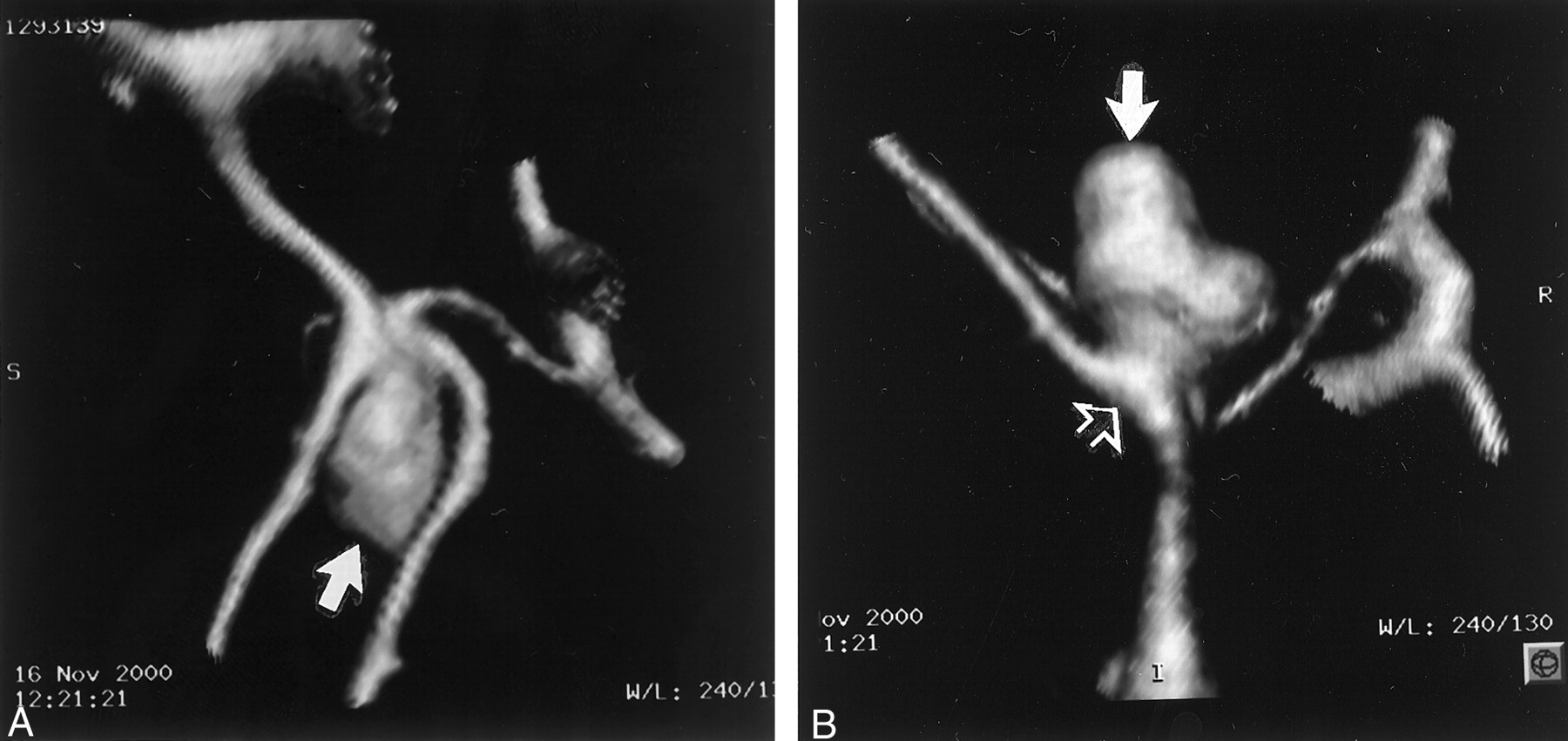
CT angiograms obtained to further define aneurysm anatomy in the case of a 54-year-old man.

A, Water’s projection with 3D reconstruction shows a 11 × 7 mm anterior communicating artery aneurysm (arrow).

B, Lateral view shows a lobulated anterior communicating artery aneurysm (solid arrow) with a 4-mm neck (open arrow).

The aneurysm was treated with 12 Guglielmi detachable coils (GDCs). The postoperative angiograms showed total occlusion of the aneurysm sac with no residual contrast material filling of the aneurysm sac or neck. No coil loops extended beyond the aneurysm sac into the anterior communicating artery, which remained patent (Figs 2 and 3). The patient tolerated the procedure well without any periprocedural complications. CT of the head performed immediately after coil placement showed no intracranial bleed or evidence of acute thromboembolic events. The patient experienced an unremarkable post-procedural course and was discharged several days later with entirely normal results of a neurologic examination. Neither aspirin nor other antiplatelets were administered because of clinical suspicion that this aneurysm may have been an occult rupture at the time of presentation, despite the negative CT findings.

Fig 2.
  • Download figure
  • Open in new tab
  • Download powerpoint
Fig 2.

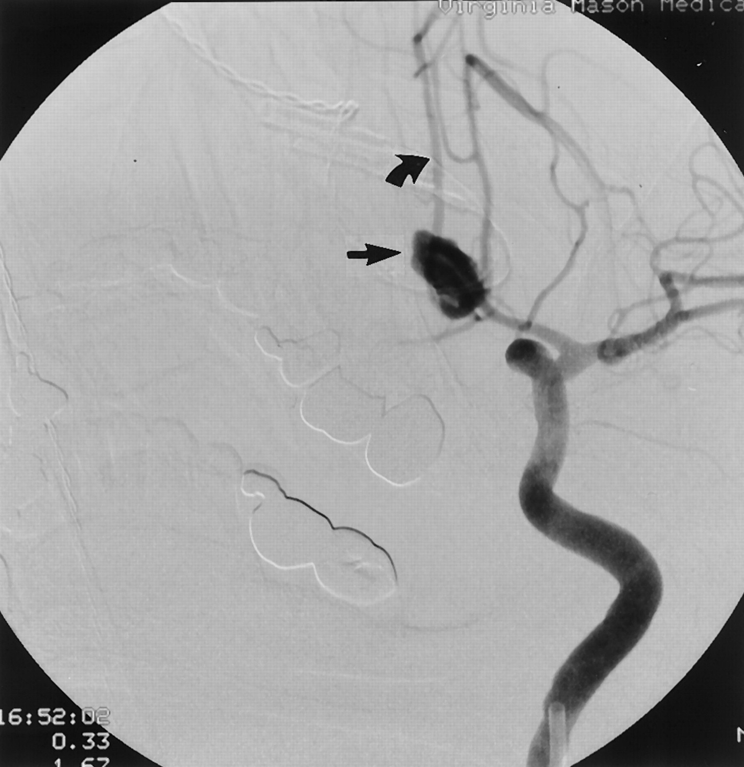
Preoperative injection of the left internal carotid artery shows the anterior communicating artery aneurysm (straight arrow) with cross filling of the right anterior cerebral artery (curved arrow).

Fig 3.
  • Download figure
  • Open in new tab
  • Download powerpoint
Fig 3.

Postoperative angiograms showed total occlusion of the aneurysm sac, with no residual contrast material filling of the aneurysm sac or neck.

A, Magnified postoperative anteroposterior angiogram shows the densely packed aneurysm sac (straight arrow). No coils extend into the well-visualized aneurysm neck (curved arrow).

B, Subtracted postoperative anteroposterior angiogram shows no residual blood flow in the aneurysm sac (solid arrow). Cross filling of the anterior cerebral artery (curved arrow) can be seen.

The patient presented 9 weeks later complaining of right foot drop, which progressed to complete right lower extremity monoplegia over 36 hr. CT of the head performed 36 hr after initial onset of symptoms showed no acute bleed. MR imaging performed 9 hr later showed multiple bilateral diffusion-weighted abnormalities scattered in the distribution of the anterior cerebral arteries, consistent with acute embolic infarcts (Fig 4). No abnormalities were present in the middle or posterior cerebral artery territories, suggesting the anterior communicating artery as the embolic source. Cerebral angiography was performed the next day, and the findings were unchanged from those of the immediate postoperative study. There was no shift in the positioning of the coils or residual filling of the aneurysm sac.

Fig 4.
  • Download figure
  • Open in new tab
  • Download powerpoint
Fig 4.

Axial diffusion-weighted MR images obtained 9 weeks after endovascular treatment.

A and B, Multiple regions of high signal intensity are depicted bilaterally in the distribution of the anterior communicating arteries, consistent with acute embolic infarcts.

The patient was discharged 2 days later after being placed on antiplatelet therapy and showing mild improvement in the right lower extremity weakness. The only remaining neurologic deficit 6 weeks later was mild toe extensor weakness.

Discussion

Thromboembolic events are a well-known complication of endovascular treatment of cerebral aneurysms. They are the most important cause of permanent morbidity associated with the procedure (3). The vast majority of previously reported thromboembolic events associated with coil placement occurred within 48 hr of the procedure (1, 2). Lenthall et al (4) reported a single case of delayed occlusion of the superior cerebellar artery associated with reconfiguration of GDC at the neck of a treated aneurysm. Delayed thromboembolic events occurring de novo with stable coil packing have not previously been described in the literature. In this case, the progressive nature of the patient’s symptoms combined with multiple diffusion abnormalities in the distribution of the anterior cerebral arteries on MR images suggest multiple embolic events originating from the treated anterior communicating artery aneurysm.

Because of the known risk of periprocedural thromboembolic events, heparin is routinely used during GDC placement. However, heparin has limited effectiveness, possibly because of its lack of inhibition of platelet aggregation (3). Although no definitive evidence exists at this time to support the use of antiplatelet medications during or after endovascular aneurysm treatment, the theoretical benefits have led to their use at some institutions.

Postoperative anticoagulation was not used in this case because of a recent institutional experience with a patient who rebled while receiving antiplatelet medications after endovascular treatment of a ruptured intracranial aneurysm. However, no empirical evidence exists to suggest that anticoagulation increases the risk of rebleeding in an endovascularly treated aneurysm.

This case illustrates that thromboembolic events can occur many months after endovascular aneurysm coil placement. Aneurysms with relatively large necks, as in this case, theoretically may be at increased risk of thromboembolic events because of the increased surface area of exposed thrombogenic material. The occurrence of delayed thromboembolic events at 9 weeks suggests that there may be benefits to the use of antiplatelet medications after endovascular coiling extending well beyond the periprocedural time frame. However, the effectiveness of antiplatelet medication in preventing delayed thromboembolic events and the risks of rebleeding of endovascularly treated aneurysms need further investigation.

Conclusion

The occurrence of delayed thromboembolic events 9 weeks after endovascular coil placement suggests that there may be a role for prolonged antiplatelet therapy after GDC aneurysm treatment.

References

  1. ↵
    Viñuela F, Duckwiler D, Mawad M. Guglielmi detachable coil embolization of acute intracranial aneurysm: perioperative anatomical and clinical outcome in 403 patients. J Neurosurg 1997;86:475–482
    CrossRefPubMed
  2. ↵
    Pelz DM, Lownie SP, Fox AJ. Thromboembolic events associated with the treatment of cerebral aneurysms with Guglielmi detachable coils. AJNR Am J Neuroradiol 1998;19:1541–1547
    Abstract
  3. ↵
    Cronqvist ME, Moret J. Detachable coil embolization of intracranial aneurysms. In: Conners JJ, Wojak JC, eds. Interventional Neuroradiology: Strategies and Practical Techniques. Philadelphia: W.B. Saunders Company;1999 :311–131
  4. ↵
    Lenthall RK, McConachie NS, Jaspan T. Delayed reconfiguration of a Guglielmi detachable coil mass associated with late occlusion of an adjacent aneurysm and parent vessel. AJNR Am J Neuroradiol 2000;21:1908–1910
    Abstract/FREE Full Text
  • Received November 19, 2001.
  • Accepted after revision January 14, 2002.
  • Copyright © American Society of Neuroradiology
PreviousNext
Back to top

In this issue

American Journal of Neuroradiology: 23 (6)
American Journal of Neuroradiology
Vol. 23, Issue 6
1 Jun 2002
  • Table of Contents
  • Index by author
Advertisement
Print
Download PDF
Email Article

Thank you for your interest in spreading the word on American Journal of Neuroradiology.

NOTE: We only request your email address so that the person you are recommending the page to knows that you wanted them to see it, and that it is not junk mail. We do not capture any email address.

Enter multiple addresses on separate lines or separate them with commas.
Delayed Thromboembolic Events 9 Weeks after Endovascular Treatment of an Anterior Communicating Artery Aneurysm: Case Report
(Your Name) has sent you a message from American Journal of Neuroradiology
(Your Name) thought you would like to see the American Journal of Neuroradiology web site.
CAPTCHA
This question is for testing whether or not you are a human visitor and to prevent automated spam submissions.
Cite this article
Matthew T. Studley, David H. Robinson, John F. Howe
Delayed Thromboembolic Events 9 Weeks after Endovascular Treatment of an Anterior Communicating Artery Aneurysm: Case Report
American Journal of Neuroradiology Jun 2002, 23 (6) 975-977;

Citation Manager Formats

  • BibTeX
  • Bookends
  • EasyBib
  • EndNote (tagged)
  • EndNote 8 (xml)
  • Medlars
  • Mendeley
  • Papers
  • RefWorks Tagged
  • Ref Manager
  • RIS
  • Zotero
0 Responses
Respond to this article
Share
Bookmark this article
Delayed Thromboembolic Events 9 Weeks after Endovascular Treatment of an Anterior Communicating Artery Aneurysm: Case Report
Matthew T. Studley, David H. Robinson, John F. Howe
American Journal of Neuroradiology Jun 2002, 23 (6) 975-977;
del.icio.us logo Twitter logo Facebook logo Mendeley logo
  • Tweet Widget
  • Facebook Like
  • Google Plus One

Jump to section

  • Article
    • Abstract
    • Case Report
    • Discussion
    • Conclusion
    • References
  • Figures & Data
  • Info & Metrics
  • Responses
  • References
  • PDF

Related Articles

  • No related articles found.
  • PubMed
  • Google Scholar

Cited By...

  • No citing articles found.
  • Crossref
  • Google Scholar

This article has not yet been cited by articles in journals that are participating in Crossref Cited-by Linking.

More in this TOC Section

  • SAVE vs. Solumbra Techniques for Thrombectomy
  • CT Perfusion&Reperfusion in Acute Ischemic Stroke
  • Delayed Reperfusion Post-Thrombectomy&Thrombolysis
Show more Interventional

Similar Articles

Advertisement

Indexed Content

  • Current Issue
  • Accepted Manuscripts
  • Article Preview
  • Past Issues
  • Editorials
  • Editors Choice
  • Fellow Journal Club
  • Letters to the Editor

Cases

  • Case Collection
  • Archive - Case of the Week
  • Archive - Case of the Month
  • Archive - Classic Case

Special Collections

  • Special Collections

Resources

  • News and Updates
  • Turn around Times
  • Submit a Manuscript
  • Author Policies
  • Manuscript Submission Guidelines
  • Evidence-Based Medicine Level Guide
  • Publishing Checklists
  • Graphical Abstract Preparation
  • Imaging Protocol Submission
  • Submit a Case
  • Become a Reviewer/Academy of Reviewers
  • Get Peer Review Credit from Publons

Multimedia

  • AJNR Podcast
  • AJNR SCANtastic
  • Video Articles

About Us

  • About AJNR
  • Editorial Board
  • Not an AJNR Subscriber? Join Now
  • Alerts
  • Feedback
  • Advertise with us
  • Librarian Resources
  • Permissions
  • Terms and Conditions

American Society of Neuroradiology

  • Not an ASNR Member? Join Now

© 2025 by the American Society of Neuroradiology All rights, including for text and data mining, AI training, and similar technologies, are reserved.
Print ISSN: 0195-6108 Online ISSN: 1936-959X

Powered by HighWire