
Abstract
Summary: Closure of a direct carotid cavernous fistula with detachable coils by transpterygoid venous approach to the cavernous sinus is an alternative technique that may be applied in cases in which other techniques offer increased risk or in which other techniques have failed. In this case report, we present the details of the management of a direct carotid cavernous fistula by this method.
The efficacy and safety of detachable balloon occlusion of direct carotid cavernous fistulas are well established (1). However, detachable balloon techniques have required occlusion of the internal carotid artery (ICA) in a substantial percentage of patients (2–4). New approaches to and occlusion methods of carotid cavernous fistulas have been described, with increased focus on preserving ICA flow. These methods include the use of two-balloon techniques (5, 6), GDC (7–11), permanent solidifying agents (12), and even stents. Failure of conventional techniques has even given rise to modifications incorporating other combination therapies (13, 14). Transvenous access to the cavernous sinus is most often achieved via the inferior petrosal sinus or the superior ophthalmic vein (15, 16). Embolization of an indirect carotid cavernous fistula has been described via the contralateral pterygoid plexus (17). We report the successful transvenous embolization of a direct carotid cavernous fistula through the ipsilateral pterygoid plexus.
Case Report
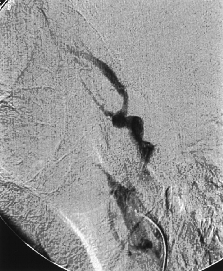
A 71-year-old woman had a 3-week history of left eye chemosis, proptosis, and bruit after an automobile accident. No external trauma to the face or neck was sustained during the accident. The patient did strike her sternum against the steering wheel quite forcibly. Her medical history included pulmonary emphysema, lung carcinoma treated with irradiation and chemotherapy 1 year earlier, and aortofemoral bypass graft to her right common femoral artery. A physical examination revealed light vision only, paralysis of extraocular movement, and decreased sensation in the left V1 distribution. Intraocular pressure was 45 mm Hg. Angiography confirmed a direct Type A left carotid cavernous fistula originating from the inferior aspect of the posterior horizontal C4 segment of the ICA, just beyond a relatively acutely angulated posterior genu (Fig 1). No aneurysm was identified. Venous effluence was through the superior ophthalmic vein, attenuated anteromedially with small connection to the left nasal vein, right nasal and right angular veins, and a single transforaminal channel to the pterygoid plexus. Venous-phase angiography over the face and neck showed the pterygoid plexus, maxillary vein, and both the facial and retromandibular veins draining the fistula (Fig 2).

Lateral-projection angiogram of the left common carotid artery shows a direct carotid cavernous fistula, with the ostium in the inferior C4 portion of the ICA just anterior to the posterior genu. Fistulous flow to the cavernous sinus, with effluence to an attenuated superior ophthalmic vein, a single channel connecting to the pterygoid plexus, is present. No inferior petrosal sinus opacified.

Venous-phase angiogram shows the pterygoid plexus, maxillary veins, facial vein, and retromandibular-external jugular vein channels.
An 80-cm 8F Bright Tip (Cordis Endovascular Systems, Miami Lakes, FL) guide catheter was passed coaxially with a 5F H1 catheter over a 0.035-inch Glidewire (Boston Scientific, Natick, MA) into the left common carotid artery via the left common femoral artery. A medium-detachment-pressure 0.5-cc detachable silicone balloon (Target Therapeutics, Fremont, CA), attached to a heat-shaped Tracker-18 catheter (with 0.010-inch wire coaxially placed through rotating hemostatic valve), was then passed into the left ICA but would not enter the fistula ostium because of the position of the ostium just beyond the posterior ICA genu in the C4 segment, along its inferior wall. Compression of the left common carotid artery deflected the balloon but not sufficiently to allow the balloon to enter the cavernous sinus.
A two-balloon technique and a direct transarterial microcatheter approach were considered but rejected because of the unfavorable position of the ostium, atherosclerotic changes of the ICA origin, and a kink of the midcervical ICA. Because of high intraorbital and intraocular pressures, concern arose for inflating a second balloon beyond the ostium, further pressurizing the venous drainage.
Using a Site Rite (Dymax, Pittsburgh, PA) sonographic guide, a 7F venous sheath was placed in the right femoral vein and a 7F Berenstein catheter was passed, using an angled Glidewire, into the left internal jugular and facial veins. A Rapid Transit catheter was manipulated over a .016-inch Headliner (Target Therapeutics) guidewire into the left maxillary vein, pterygoid plexus, and the single connecting channel into the cavernous sinus (Fig 3). Six Guglielmi detachable coils (one 7 × 30, one 2 × 6 fibered, two 2 × 5 fibered, one 2 × 4 fibered, and one 2 × 3 fibered) were then placed (Fig 4).

Rapid Transit microcatheter has been passed into the cavernous sinus via the pterygoid plexus. Injection of contrast medium shows the venous compartment.

Injection of the left common carotid artery after placement of standard and fibered GDC shows complete occlusion of the fistulous flow.
Angiography showed no retrograde filling of the superior ophthalmic vein. Repeat angiography after all the coils had been placed showed only a small puddle of contrast medium filling near the ostium. Heparin was administered overnight, and a regimen of 325 mg per day acetylsalicylic acid was begun. At 1 month, ophthalmologic signs had improved, but useful vision had not returned.
Discussion
The pterygoid plexus is an extensive network of small vascular channels that overlies the lateral pterygoid muscle and connects to the cavernous sinus via sphenobasilar emissary channels, which travel through the foramina ovale, spinosum, and lacerum. Posteriorly, it drains into the maxillary vein. The maxillary vein communicates with the superficial temporal vein to form the retromandibular vein. It also communicates with the ophthalmic veins through the inferior orbital fissure and with the anterior facial vein via a deep facial branch (18). In our patient, the retromandibular and facial systems opacified from the venous effluence from the pterygoid plexus. Once thought to indicate possible posterior obstruction of the cavernous sinus, angiographic visualization of the pterygoid venous plexus is both common and normal. A review of our available case material from 1980 through 2000 revealed that the pterygoid plexus was visualized in 25 (29%) of 86 direct carotid cavernous fistulas.
Although transarterial detachable balloon occlusion may be a quick, efficient, and safe method for occluding most direct carotid cavernous fistulas and although it remains the preferred treatment of choice to close direct carotid cavernous fistulas, it is not without complexities, limitations, and complications. The transvenous access to the cavernous sinus offers an alternative route and has supplanted transarterial embolization of dural feeders originating from the external carotid artery or the ICA, for indirect (or dural) arteriovenous fistulas of the cavernous sinus (dural carotid cavernous fistulas). Transvenous embolization for direct carotid cavernous fistulas has been primarily used in cases in which transarterial access has been impossible because of unfavorable position or size of the ostium of the fistula or unfavorable slow-flow characteristics of the fistula, both of which imply unfavorable mechanical characteristics of the balloon and catheter system. These factors decrease the likelihood of the balloon’s entering the ostium at the venous side. Additional maneuvers that might facilitate a balloon’s entering the ostium include the following: 1) heat shaping of the catheter tip, with placement of a curved guidewire within the balloon catheter, which may aid in the balloon’s passing along the appropriate wall of the ICA at the ostium; 2) compression of the ipsilateral common carotid artery in the neck or temporary occlusion of the ICA by a second balloon, which leads to reversal of flow in the distal ICA beyond the fistula, which may deflect the balloon appropriately; 3) two-balloon technique, with inflation of a balloon just beyond the fistula, increasing flow through the fistula, which may add sufficient propulsion of the balloon through the ostium.
In lieu of, or after the failure of, the aforementioned methods, transarterial microcatheter placement across the lumen may allow coil placement for fistula closure. The ability to microcatheterize and hold position in the fistula is also limited by the position of the ostium, typically more difficult just beyond the posterior genu along the inferior wall of the C4 segment. This ability may also be facilitated by a nondetachable balloon support. All of these maneuvers were used or considered in this patient before the transvenous approach was attempted. Because of the precarious visual function and marked increase in intraocular pressure, the two-balloon method, requiring occlusion of the ICA beyond the fistula, maximizing flow into the fistula, was not attractive. Severe pain has been encountered with the latter maneuver, and increasing flow through an ostium by occluding an outflow pathway, or even the ICA distally, might cause hemorrhage or further deterioration of vision (19).
Transvenous access is typically most easily achieved through the inferior petrosal sinus or superior ophthalmic vein. In our patient, the inferior petrosal sinus was not visualized but might have served as a potential access. The superior ophthalmic vein was narrowed near the trochlea but drained to the opposite angular vein, precluding a retrograde transfacial approach. A retrograde open catheterization of the superior ophthalmic vein would have been difficult with the degree of orbital induration and would have pressurized the orbit more greatly and threatened total loss of vision immediately. Therefore, the transpterygoid venous approach was chosen, with the expectation that the superior ophthalmic vein and cavernous sinus could be disconnected, with the option that a nondetachable balloon catheter still might be required to prevent coils from prolapsing into the ICA.
Accessing the cavernous sinus through the jugular, facial, and maxillary veins and the pterygoid plexus was expedited by review of the venous phase of the cerebral angiogram that filled these veins through the fistulous flow of the cavernous sinus.
Conclusion
The transpterygoid plexus approach to the cavernous sinus should be recognized as a potential access route for certain direct carotid cavernous fistulas and might be used in cases in which ancillary anatomic or clinical factors obviate the use of extraordinary transarterial or other transvenous methods.
References
- Received January 4, 2002.
- Accepted after revision March 18, 2002.
- Copyright © American Society of Neuroradiology


