Advertisement

Main menu

  • Home
  • Content
    • Current Issue
    • Accepted Manuscripts
    • Article Preview
    • Past Issue Archive
    • AJNR Case Collection
    • Case of the Week Archive
    • Classic Case Archive
    • Case of the Month Archive
  • Special Collections
    • Spinal CSF Leak Articles (Jan 2020-June 2024)
    • 2024 AJNR Journal Awards
    • Most Impactful AJNR Articles
  • Multimedia
    • AJNR Podcast
    • AJNR Scantastics
    • Video Articles
  • For Authors
    • Submit a Manuscript
    • Author Policies
    • Fast publishing of Accepted Manuscripts
    • Graphical Abstract Preparation
    • Manuscript Submission Guidelines
    • Imaging Protocol Submission
    • Submit a Case for the Case Collection
  • About Us
    • About AJNR
    • Editorial Board
  • More
    • Become a Reviewer/Academy of Reviewers
    • Subscribers
    • Permissions
    • Alerts
    • Feedback
    • Advertisers
    • ASNR Home
  • Other Publications
    • ajnr

User menu

  • Alerts
  • Log in

Search

  • Advanced search
American Journal of Neuroradiology

American Journal of Neuroradiology

ASHNR American Society of Functional Neuroradiology ASHNR American Society of Pediatric Neuroradiology ASSR
  • Alerts
  • Log in

Advanced Search

  • Home
  • Content
    • Current Issue
    • Accepted Manuscripts
    • Article Preview
    • Past Issue Archive
    • AJNR Case Collection
    • Case of the Week Archive
    • Classic Case Archive
    • Case of the Month Archive
  • Special Collections
    • Spinal CSF Leak Articles (Jan 2020-June 2024)
    • 2024 AJNR Journal Awards
    • Most Impactful AJNR Articles
  • Multimedia
    • AJNR Podcast
    • AJNR Scantastics
    • Video Articles
  • For Authors
    • Submit a Manuscript
    • Author Policies
    • Fast publishing of Accepted Manuscripts
    • Graphical Abstract Preparation
    • Manuscript Submission Guidelines
    • Imaging Protocol Submission
    • Submit a Case for the Case Collection
  • About Us
    • About AJNR
    • Editorial Board
  • More
    • Become a Reviewer/Academy of Reviewers
    • Subscribers
    • Permissions
    • Alerts
    • Feedback
    • Advertisers
    • ASNR Home
  • Follow AJNR on Twitter
  • Visit AJNR on Facebook
  • Follow AJNR on Instagram
  • Join AJNR on LinkedIn
  • RSS Feeds

Welcome to the new AJNR, Updated Hall of Fame, and more. Read the full announcements.


AJNR is seeking candidates for the position of Associate Section Editor, AJNR Case Collection. Read the full announcement.


Getting new auth cookie, if you see this message a lot, tell someone!
OtherINTERVENTIONAL

Saccular Aneurysm Associated with Posterior Cerebral Artery Fenestration Manifesting as a Subarachnoid Hemorrhage in a Child

Lotfi Hacein-Bey, Cheryl A. Muszynski and Panayiotis N. Varelas
American Journal of Neuroradiology September 2002, 23 (8) 1291-1294;
Lotfi Hacein-Bey
  • Find this author on Google Scholar
  • Find this author on PubMed
  • Search for this author on this site
Cheryl A. Muszynski
  • Find this author on Google Scholar
  • Find this author on PubMed
  • Search for this author on this site
Panayiotis N. Varelas
  • Find this author on Google Scholar
  • Find this author on PubMed
  • Search for this author on this site
  • Article
  • Figures & Data
  • Info & Metrics
  • Responses
  • References
  • PDF
Loading

Abstract

Summary: Fenestrations of the cerebral arteries are uncommon, and aneurysms in association with fenestrations are even more uncommon. We report the case of a saccular aneurysm associated with a posterior cerebral artery fenestration in a child with subarachnoid hemorrhage. The patient was successfully treated with Guglielmi detachable coils. This case is unusual because of the patient’s age and the location of the fenestration.

Fenestrations of the cerebral arteries are uncommon, with a reported angiographic incidence of 0.03–1% (1–3) and a reported postmortem incidence ranging between 1.3%(4) and 5.3%(5). Typical locations for cerebral arterial fenestrations include the anterior communicating artery (6), the vertebrobasilar system, and the anterior cerebral artery (7). Aneurysms in association with fenestrations are even rarer, although those fenestrations are widely believed to predispose patients to aneurysm formation (2, 8).

We report a saccular aneurysm associated with a posterior cerebral artery fenestration in a child with subarachnoid hemorrhage. The patient was successfully treated with endovascular treatment.

Case Report

This 11-year-old boy was in good health until he was found unresponsive in his bed at night. He was taken to a local hospital and gradually became responsive with complaints of headache, severe photophobia, and a stiff neck. Findings from the initial neurologic examination were reportedly normal. Initial laboratory results were normal. A CT scan of the head showed subarachnoid hemorrhage in the posterior fossa. Isotonic fluids were administered, and the patient was transferred to our institution.

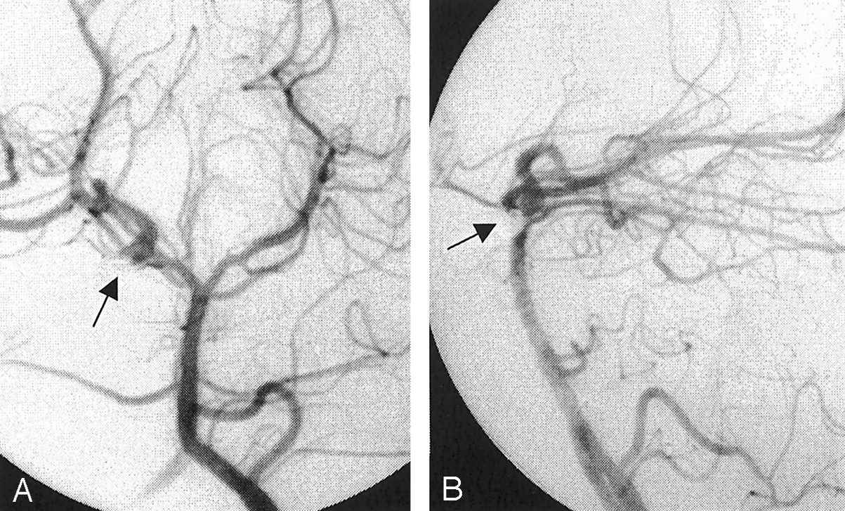
Upon admission, the child complained of occipital and bifrontal headaches and nausea. Mild meningismus was noted. His neurologic examination findings were nonfocal. An MR image obtained the following day showed a round flow void and two tubular structures anterior to the right side of the pons. A cerebral angiogram was obtained (Fig 1); this showed aneurysmal dilatation at the proximal end of a tubular structure overlying the right posterior cerebral artery.

Fig 1.
  • Download figure
  • Open in new tab
  • Download powerpoint
Fig 1.

Cerebral angiograms obtained with a left vertebral artery injection. A saccular aneurysm (arrow) is present at the proximal end of a duplication of the right posterior cerebral artery that bridges the P1-P2 segment and the right P2-P3 segment. The duplicated segment of the right posterior cerebral artery has a stepwise configuration (arrowhead in B).

A, Anteroposterior view.

B, Lateral view.

After a multidisciplinary evaluation involving neurosurgical, neurointerventional, and neurologic inputs, the child and his family were offered endovascular therapy for this aneurysm.

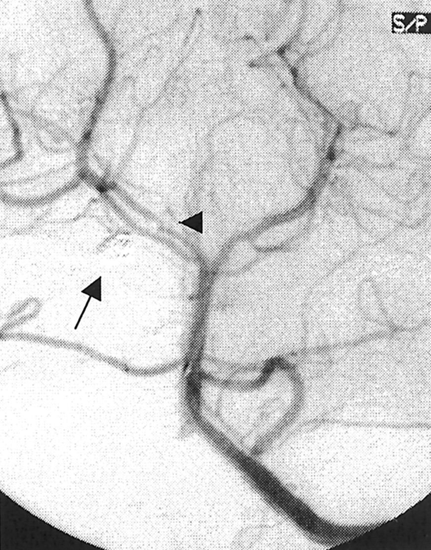
Under general anesthesia, the child underwent transarterial aneurysm obliteration with Guglielmi detachable coils (GDCs). Superselective angiography (Fig 2) confirmed the saccular nature of a broad-based proximal aneurysm in a duplicated segment of the right posterior cerebral artery.

Fig 2.
  • Download figure
  • Open in new tab
  • Download powerpoint
Fig 2.

Superselective angiograms of a duplicated right posterior cerebral artery. A laterally and inferiorly pointing saccular aneurysm (arrow) has a broad connection (arrowhead in B) to the parent artery. The proximal portion of the duplicated segment in the quadrigeminal cistern has a stepwise configuration.

A, Anteroposterior view

B, Lateral view.

Postoperatively, the patient remained in the neurologic intensive care unit for 24 hours and recovered well. He was discharged from the hospital 3 days later after a short course of intravenous heparin (Abbott Laboratories, Abbott Park, IL).

The patient’s headache resolved completely within a week of discharge. As of this writing, he has remained neurologically intact and has resumed all previous activities, including playing soccer without problems. Follow-up angiography performed at 6 months showed persistent aneurysmal obliteration (Fig 3).

Fig 3.
  • Download figure
  • Open in new tab
  • Download powerpoint
Fig 3.

Cerebral angiogram obtained with a left vertebral artery injection. The aneurysm is totally obliterated (arrow). The right posterior cerebral artery duplication is patent.

A, Anteroposterior view.

B, Lateral view.

Discussion

We failed to find in the literature any report of fenestration-associated aneurysm in a child. In one series of 37 patients (3), which was mostly concerned with fenestrations and their possible association with aneurysms, the youngest patient was 18 years old. To our knowledge, this not only is the first report of a ruptured saccular aneurysm associated with a fenestrated posterior cerebral artery but also the first report of such an aneurysm in the pediatric population.

Fenestrations of the cerebral arteries are rare, and a significant discrepancy exists between their reported angiographic incidence of 0.03–1%) (1–3) and their postmortem incidence of 1.3–5.3%(4, 5). This discrepancy has been attributed to both the higher sensitivity of the postmortem examination and the likely under-reporting of fenestrations on angiographic reports due to their uncommon nature (3). A fenestration is a division of the arterial lumen, with resulting separate channels, each with its own endothelial layer and muscularis tunica; the adventitial layer may or may not be shared between channels. At the most, a fenestration results in multiplication, usually duplication, of the vessel. Duplications commonly involve the anterior communicating artery (6–9), where spiderlike connections have been demonstrated in as many as 19% of autopsy specimens (5).

Although uncommon, fenestrations of the vertebrobasilar system with or without associated aneurysms have often been reported (7, 10–14), principally in the Japanese literature. Much less common are aneurysms arising from fenestrations that involve the anterior cerebral artery, (15, 16) the middle cerebral artery, (17–19), or even the posterior inferior cerebellar artery (20).

In a study of anatomic dissections of 100 human brains (21), anomalies of the posterior cerebral artery were noted in 3%of the specimens. These dissections were found to exclusively involve the precommunicating or P1 segment, including one fenestration in this segment.

Another postmortem anatomic study of the posterior cerebral artery segment distal to the posterior communicating artery revealed three possible courses of the distal vessel and revealed a 1.4%incidence of fenestration (22).

The embryologic basis for segmental duplications of cerebral arteries is consistent with an incomplete or absent fusion between primitive neural arteries (23). Defects in the medial layer with a decrease in the smooth muscle and collagen content can be microscopically identified at the proximal and distal edges of the duplicated segment. Defects are more severe at the medial and ventral walls of the proximal juncture and result in saccular aneurysm formation (24–25). In addition to medial defects, flow phenomena at the proximal end of fenestrations, where hemodynamic stress and increased turbulence are present, may contribute to aneurysm formation (10), although aneurysms have also been reported at the distal end of fenestrations (13). However, some have also suggested that the incidence of fenestration-related aneurysms is the same as that of berry aneurysms in the general population (3).

Our patient’s aneurysm was saccular; it arose from the proximal edge of a fenestration and projected ventrally. All of these features are reportedly common for such aneurysms (2–4, 7, 9–10).

The patient’s age and the location of the fenestration both contribute to making this case extremely unusual.

Most fenestration-related aneurysms reported thus far have been treated surgically, although successful transvascular therapy with either detachable balloons (14) or, more recently, GDCs (26–27) is reported.

The anatomic location of this aneurysm was thought to be associated with a relatively high surgical risk. Therefore, our patient was treated with GDCs, which permitted total obliteration of his saccular aneurysm (Fig 3). Interestingly, the placement of a microcatheter across the fenestration resulted in the temporary occlusion of the proximal edge of the fenestration (Fig 4). We elected not to obliterate the dysplastic-appearing duplicated segment because of the possibility of a stroke from occlusion of the perforating arteries, which can arise from either limb of a fenestration. Because medial defects can involve either edge of a fenestration, this young patient will have to be followed up at regular intervals. Soon, such follow-up might be performed in a noninvasive way, at least intermittently, as the accuracy of newer MR imaging techniques (28) approaches that of angiography in routine follow-up.

Fig 4.
  • Download figure
  • Open in new tab
  • Download powerpoint
Fig 4.

Anteroposterior cerebral angiogram obtained with a left vertebral artery injection during aneurysm treatment. The aneurysm is obliterated (arrow). The duplicated segment of the right posterior communicating artery is not seen because the microcatheter is across the fenestration (arrowhead).

Conclusion

To our knowledge, this is the first report of a saccular aneurysm associated with a posterior cerebral artery fenestration in a child. In addition, the aneurysm had ruptured, causing subarachnoid hemorrhage. The patient had an excellent outcome after he was treated with GDCs.

References

  1. ↵
    Takahashi M, Tamakawa Y, Kishikawa T, Kowada M. Fenestration of the basilar artery: report of three cases and review of the literature. Radiology 1973;109:79–82
    PubMed
  2. ↵
    Teal JS, Rumbaugh CL, Bergeron RT, Segall HD. Angiographic demonstration of fenestrations of the intradural intracranial arteries. Radiology 1973;106:123–126
    CrossRefPubMed
  3. ↵
    Sanders WP, Sorek PA, Mehta BA. Fenestration of intracranial arteries with special attention to associated aneurysms and other anomalies. AJNR Am J Neuroradiol 1993;14:675–680
    Abstract/FREE Full Text
  4. ↵
    Hoffman WF, Wilson CB. Fenestrated basilar artery with an associated saccular aneurysm: case report. J Neurosurg 1979;50:262–264
    PubMed
  5. ↵
    Wollschlaeger G, Wollschlaeger PB, Lucas FV, Lopez VF. Experience and result with postmortem cerebral angiography performed as routine procedure of the autopsy. Am J Roentgenol Radium Ther Nucl Med 1967;101:68–87
    PubMed
  6. ↵
    Serizawa T, Saeki N, Yamaura A. Microsurgical anatomy and clinical significance of the anterior communicating artery and its perforating branches. Neurosurgery 1997;40:1211–1216
    CrossRefPubMed
  7. ↵
    Osborn RE, Kirk G. Cerebral arterial fenestration. Comput Radiol 1987;11:141–145
    PubMed
  8. ↵
    Andrews BT, Brant-Zawadzki M, Wilson CB. Variant aneurysms of the fenestrated basilar artery. Neurosurgery 1986;18:204–207
    PubMed
  9. ↵
    San-Galli F, Leman C, Kien P, Khazaal J, Phillips SD, Guerin J. Cerebral arterial fenestrations associated with intracranial saccular aneurysms. Neurosurgery 1992;30:279–283
    PubMed
  10. ↵
    Campos J, Fox AJ, Vinuela F, Lylyk P, Ferguson GG, Drake CG, Peerless SJ. Saccular aneurysms in basilar artery fenestration. AJNR Am J Neuroradiol 1987;8:233–236
    Abstract/FREE Full Text
  11. Hoshimaru M, Hashimoto N, Kikuchi H, Kamijyo Y, Kang Y, Namura S. Aneurysm of the fenestrated basilar artery: report of two cases. Surg Neurol 1992;37:406–409
    PubMed
  12. Imaizumi T, Saito K, Kobayashi T, Sakamoto Y, Komeichi T. Saccular aneurysm associated with fenestration of the distal segment of basilar artery. No Shinkei Geka 1996;24:639–642
    PubMed
  13. ↵
    Fujimura M, Sugawara T, Higuchi H, Oku T, Seki H. A ruptured aneurysm at the distal end of the basilar artery fenestration associated with multiple fenestrations of the vertebrobasilar system: case report. Surg Neurol 1997;47:469–472
    PubMed
  14. ↵
    Picard L, Roy D, Bracard S, Per A, Marchal JC. Aneurysm associated with a fenestrated basilar artery: report of two cases treated by endovascular detachable balloon embolization. AJNR Am J Neuroradiol 1993;14:591–594
    Abstract/FREE Full Text
  15. ↵
    Minakawa T, Kawamata M, Hayano M, Kawakami K. Aneurysms associated with fenestrated anterior cerebral arteries: report of four cases and review of the literature. Surg Neurol 1985;24:284–288
    PubMed
  16. ↵
    Ito J, Washiyama K, Kim CH, Ibuchi Y. Fenestration of the anterior cerebral artery. Neuroradiology 1981;21:277–280
    CrossRefPubMed
  17. ↵
    Serizawa T, Saeki N, Yamaura A. Microsurgical anatomy and clinical significance of the anterior communicating artery and its perforating branches. Neurosurgery 1997;40:1211–1216
  18. Nakamura H, Takada A, Hide T, Ushio Y. Fenestration of the middle cerebral artery associated with an aneurysm: case report. Neurol Med Chir (Tokyo) 1994;34:555–557
  19. ↵
    Ito J, Maeda H, Inoue K, Onishi Y. Fenestration of the middle cerebral artery. Neuroradiology 1977;13:37–39
    PubMed
  20. ↵
    Theodosopoulos PV, Lawton MT. Fenestration of the posteroinferior cerebellar artery: case report. Neurosurgery 2000;47:463–465
    CrossRefPubMed
  21. ↵
    Caruso G, Vincentelli F, Rabehanta P, Giudicelli G, Grisoli F. Anomalies of the P1 segment of the posterior cerebral artery: early bifurcation or duplication, fenestration, common trunk with the superior cerebellar artery. Acta Neurochir (Wien) 1991;109:66–71
  22. ↵
    Milisavljevic M, Marinkovic S, Marinkovic Z, Malobabic S. Anatomic basis for surgical approach to the distal segment of the posterior cerebral artery. Surg Radiol Anat 1988;10:259–266
    PubMed
  23. ↵
    Padget DH. The development of the cranial arteries in the human embryo. Contrib Embryol 1968;32:205–261
  24. ↵
    Black SP, Ansbacher LE. Saccular aneurysm associated with segmental duplication of the basilar artery: a morphological study. J Neurosurg 1984;61:1005–1008
    PubMed
  25. ↵
    Finlay HM, Canham PB. The layered fabric of cerebral artery fenestrations. Stroke 1994;25:1799–1806
    Abstract/FREE Full Text
  26. ↵
    Graves VB, Strother CM, Weir B, Duff TA. Vertebrobasilar junction aneurysms associated with fenestration: treatment with Guglielmi detachable coils. AJNR Am J Neuroradiol 1996;17:35–40
    Abstract
  27. ↵
    Komiyama M, Nakajima H, Nishikawa M, Yasui T, Kan M. Treatment of a saccular aneurysm at the fenestration of the intracranial vertebral artery with Guglielmi detachable coils. Acta Neurochir (Wien) 1999;141:1125–1127
  28. ↵
    Boulin A, Pierot L. Follow-up of intracranial aneurysms treated with detachable coils: comparison of gadolinium-enhanced 3D time-of-flight MR angiography and digital subtraction angiography. Radiology 2001;219:108–113
    PubMed
  • Received September 20, 2001.
  • Accepted after revision April 12, 2002.
  • Copyright © American Society of Neuroradiology
View Abstract
PreviousNext
Back to top

In this issue

American Journal of Neuroradiology: 23 (8)
American Journal of Neuroradiology
Vol. 23, Issue 8
1 Sep 2002
  • Table of Contents
  • Index by author
Advertisement
Print
Download PDF
Email Article

Thank you for your interest in spreading the word on American Journal of Neuroradiology.

NOTE: We only request your email address so that the person you are recommending the page to knows that you wanted them to see it, and that it is not junk mail. We do not capture any email address.

Enter multiple addresses on separate lines or separate them with commas.
Saccular Aneurysm Associated with Posterior Cerebral Artery Fenestration Manifesting as a Subarachnoid Hemorrhage in a Child
(Your Name) has sent you a message from American Journal of Neuroradiology
(Your Name) thought you would like to see the American Journal of Neuroradiology web site.
CAPTCHA
This question is for testing whether or not you are a human visitor and to prevent automated spam submissions.
Cite this article
Lotfi Hacein-Bey, Cheryl A. Muszynski, Panayiotis N. Varelas
Saccular Aneurysm Associated with Posterior Cerebral Artery Fenestration Manifesting as a Subarachnoid Hemorrhage in a Child
American Journal of Neuroradiology Sep 2002, 23 (8) 1291-1294;

Citation Manager Formats

  • BibTeX
  • Bookends
  • EasyBib
  • EndNote (tagged)
  • EndNote 8 (xml)
  • Medlars
  • Mendeley
  • Papers
  • RefWorks Tagged
  • Ref Manager
  • RIS
  • Zotero
0 Responses
Respond to this article
Share
Bookmark this article
Saccular Aneurysm Associated with Posterior Cerebral Artery Fenestration Manifesting as a Subarachnoid Hemorrhage in a Child
Lotfi Hacein-Bey, Cheryl A. Muszynski, Panayiotis N. Varelas
American Journal of Neuroradiology Sep 2002, 23 (8) 1291-1294;
del.icio.us logo Twitter logo Facebook logo Mendeley logo
  • Tweet Widget
  • Facebook Like
  • Google Plus One

Jump to section

  • Article
    • Abstract
    • Case Report
    • Discussion
    • Conclusion
    • References
  • Figures & Data
  • Info & Metrics
  • Responses
  • References
  • PDF

Related Articles

  • No related articles found.
  • PubMed
  • Google Scholar

Cited By...

  • No citing articles found.
  • Crossref
  • Google Scholar

This article has not yet been cited by articles in journals that are participating in Crossref Cited-by Linking.

More in this TOC Section

  • SAVE vs. Solumbra Techniques for Thrombectomy
  • Contrast-Induced Encephalopathy after NeuroIR
  • CT Perfusion&Reperfusion in Acute Ischemic Stroke
Show more Interventional

Similar Articles

Advertisement

Indexed Content

  • Current Issue
  • Accepted Manuscripts
  • Article Preview
  • Past Issues
  • Editorials
  • Editors Choice
  • Fellow Journal Club
  • Letters to the Editor

Cases

  • Case Collection
  • Archive - Case of the Week
  • Archive - Case of the Month
  • Archive - Classic Case

Special Collections

  • Special Collections

Resources

  • News and Updates
  • Turn around Times
  • Submit a Manuscript
  • Author Policies
  • Manuscript Submission Guidelines
  • Evidence-Based Medicine Level Guide
  • Publishing Checklists
  • Graphical Abstract Preparation
  • Imaging Protocol Submission
  • Submit a Case
  • Become a Reviewer/Academy of Reviewers
  • Get Peer Review Credit from Publons

Multimedia

  • AJNR Podcast
  • AJNR SCANtastic
  • Video Articles

About Us

  • About AJNR
  • Editorial Board
  • Not an AJNR Subscriber? Join Now
  • Alerts
  • Feedback
  • Advertise with us
  • Librarian Resources
  • Permissions
  • Terms and Conditions

American Society of Neuroradiology

  • Not an ASNR Member? Join Now

© 2025 by the American Society of Neuroradiology All rights, including for text and data mining, AI training, and similar technologies, are reserved.
Print ISSN: 0195-6108 Online ISSN: 1936-959X

Powered by HighWire