Advertisement

Main menu

  • Home
  • Content
    • Current Issue
    • Accepted Manuscripts
    • Article Preview
    • Past Issue Archive
    • AJNR Case Collection
    • Case of the Week Archive
    • Classic Case Archive
    • Case of the Month Archive
  • Special Collections
    • Spinal CSF Leak Articles (Jan 2020-June 2024)
    • 2024 AJNR Journal Awards
    • Most Impactful AJNR Articles
  • Multimedia
    • AJNR Podcast
    • AJNR Scantastics
    • Video Articles
  • For Authors
    • Submit a Manuscript
    • Author Policies
    • Fast publishing of Accepted Manuscripts
    • Graphical Abstract Preparation
    • Manuscript Submission Guidelines
    • Imaging Protocol Submission
    • Submit a Case for the Case Collection
  • About Us
    • About AJNR
    • Editorial Board
  • More
    • Become a Reviewer/Academy of Reviewers
    • Subscribers
    • Permissions
    • Alerts
    • Feedback
    • Advertisers
    • ASNR Home
  • Other Publications
    • ajnr

User menu

  • Alerts
  • Log in

Search

  • Advanced search
American Journal of Neuroradiology

American Journal of Neuroradiology

ASHNR American Society of Functional Neuroradiology ASHNR American Society of Pediatric Neuroradiology ASSR
  • Alerts
  • Log in

Advanced Search

  • Home
  • Content
    • Current Issue
    • Accepted Manuscripts
    • Article Preview
    • Past Issue Archive
    • AJNR Case Collection
    • Case of the Week Archive
    • Classic Case Archive
    • Case of the Month Archive
  • Special Collections
    • Spinal CSF Leak Articles (Jan 2020-June 2024)
    • 2024 AJNR Journal Awards
    • Most Impactful AJNR Articles
  • Multimedia
    • AJNR Podcast
    • AJNR Scantastics
    • Video Articles
  • For Authors
    • Submit a Manuscript
    • Author Policies
    • Fast publishing of Accepted Manuscripts
    • Graphical Abstract Preparation
    • Manuscript Submission Guidelines
    • Imaging Protocol Submission
    • Submit a Case for the Case Collection
  • About Us
    • About AJNR
    • Editorial Board
  • More
    • Become a Reviewer/Academy of Reviewers
    • Subscribers
    • Permissions
    • Alerts
    • Feedback
    • Advertisers
    • ASNR Home
  • Follow AJNR on Twitter
  • Visit AJNR on Facebook
  • Follow AJNR on Instagram
  • Join AJNR on LinkedIn
  • RSS Feeds

Welcome to the new AJNR, Updated Hall of Fame, and more. Read the full announcements.


AJNR is seeking candidates for the position of Associate Section Editor, AJNR Case Collection. Read the full announcement.


Research ArticleBRAIN

Multisection Proton MR Spectroscopy for Mesial Temporal Lobe Epilepsy

Arístides A. Capizzano, Peter Vermathen, Kenneth D. Laxer, Gerald B. Matson, Andrew A. Maudsley, Brian J. Soher, Norbert W. Schuff and Michael W. Weiner
American Journal of Neuroradiology September 2002, 23 (8) 1359-1368;
Arístides A. Capizzano
  • Find this author on Google Scholar
  • Find this author on PubMed
  • Search for this author on this site
Peter Vermathen
  • Find this author on Google Scholar
  • Find this author on PubMed
  • Search for this author on this site
Kenneth D. Laxer
  • Find this author on Google Scholar
  • Find this author on PubMed
  • Search for this author on this site
Gerald B. Matson
  • Find this author on Google Scholar
  • Find this author on PubMed
  • Search for this author on this site
Andrew A. Maudsley
  • Find this author on Google Scholar
  • Find this author on PubMed
  • Search for this author on this site
Brian J. Soher
  • Find this author on Google Scholar
  • Find this author on PubMed
  • Search for this author on this site
Norbert W. Schuff
  • Find this author on Google Scholar
  • Find this author on PubMed
  • Search for this author on this site
Michael W. Weiner
  • Find this author on Google Scholar
  • Find this author on PubMed
  • Search for this author on this site
  • Article
  • Figures & Data
  • Info & Metrics
  • Responses
  • References
  • PDF
Loading

Article Figures & Data

Figures

  • Tables
  • Fig 1.
    • Download figure
    • Open in new tab
    • Download powerpoint
    Fig 1.

    Parasagittal scout MR images. A, Section positioning of the PRESS sequence. B, Multisection 1H-MRS imaging sequence.

  • Fig 2.
    • Download figure
    • Open in new tab
    • Download powerpoint
    Fig 2.

    Oblique axial proton density–weighted image corresponding to the temporal section of the multisection 1H-MRS sequence of a patient with right mTLE shows regions for voxel selection: 1, insula; 2, temporal operculum; 3, lateral temporal cortex; 4, hippocampal body; 5, cerebellar hemisphere.
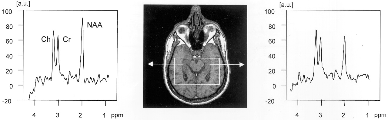
  • Fig 3.
    • Download figure
    • Open in new tab
    • Download powerpoint
    Fig 3.

    Scout axial MR image (center) for PRESS sequence, encompassing both hippocampi. Spectra from right and left hippocampus are shown with NAA reduction on the left side in this case of left mTLE. a.u., absolute units.

  • Fig 4.
    • Download figure
    • Open in new tab
    • Download powerpoint
    Fig 4.

    NAA metabolite images obtained in a control subject. A, Ventricular section (note the low signal intensity from the lateral ventricles). B, Supraventricular section as defined on sagittal scout images (see Fig 1B).
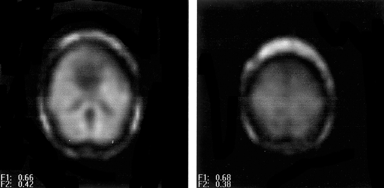
  • Fig 5.
    • Download figure
    • Open in new tab
    • Download powerpoint
    Fig 5.

    NAA metabolite image of a left mTLE shows reduced NAA signal intensity on the anterior left temporal lobe.

Tables

  • Figures
    • View popup
    TABLE 1:

    |NAA| in control subjects and patients in the hippocampus versus other brain regions

    Brain RegionControlsTemporal Lobe Epilepsy
    Ipsilateral*Contralateral†
    Hippocampus (PRESS)3.63 ± 0.793.05 ± 0.47 (P < .05)3.38 ± 0.62 (P < .01)
    Hippocampal body (multisection MRS)1.06 ± 0.150.77 ± 0.20 (P < .001)0.94 ± 0.28 (P < .01)
    Temporal operculum (multisection (MRS)1.07 ± 0.060.89 ± 0.15 (P < .001)1.08 ± 0.16 (P < .001)
    Insular cortex (multisection MRS)1.13 ± 0.080.90 ± 0.20 (P < .01)1.11 ± 0.17 (P < .0001)
    Lateral temporal cortex (multisection MRS)1.23 ± 0.060.93 ± 0.16 (P < .001)1.14 ± 0.19 (P < .05)
    Cerebellar hemisphere (multisection MRS)1.34 ± 0.181.38 ± 0.19 (N.S.)1.34 ± 0.19 (N.S.)
    Cerebral hemisphere (multisection MRS)1.30 ± 0.091.20 ± 0.11 (P < .01)1.21 ± 0.10 (N.S.)
    • Note.—All values are expressed in institutional units as mean ± SD. |NAA| magnitudes between point-resolved spectroscopy (PRESS) and multisection MR spectroscopy (MRS) differ because of differences in technique.

    • * P values reflect significantly reduced |NAA| in patients compared with |NAA| in control subjects.

    • † P values reflect significantly reduced |NAA| in the ipsilateral side compared with |NAA| in the contralateral side of patients.

    • View popup
    TABLE 2:

    NAA/(Cr + Cho) in patients and control subjects in the hippocampus versus other brain regions

    Brain RegionControlsTemporal Lobe Epilepsy
    Ipsilateral*Contralateral†
    Hippocampus (PRESS)1.27 ± 0.191.10 ± 0.15 (P < .01)1.20 ± 0.16 (P < .05)
    Hippocampal body (multisection MRS)1.34 ± 0.231.09 ± 0.21 (P < .01)1.30 ± 0.24 (P < .0001)
    Temporal operculum (multisection (MRS)1.50 ± 0.161.20 ± 0.22 (P < .001)1.46 ± 0.18 (P < .001)
    Insular cortex (multisection MRS)1.44 ± 0.131.17 ± 0.18 (P < .001)1.43 ± 0.21 (P < .001)
    Lateral temporal cortex (multisection MRS)1.69 ± 0.161.36 ± 0.35 (P < .05)1.76 ± 0.25 (P < .05)
    Cerebellar hemisphere (multisection MRS)1.33 ± 0.191.34 ± 0.19 (N.S.)1.33 ± 0.18 (N.S.)
    Cerebral hemisphere (multisection MRS)1.99 ± 0.141.85 ± 0.10 (P < .01)1.92 ± 0.08 (P < .001)
    • Note.—All values are expressed in institutional units as mean ± SD.

    • * P values reflect significantly reduced ratios in patients compared with ratios in control subjects.

    • † P values reflect significantly reduced ratios in the ipsilateral side compared with ratios in the contralateral side of patients.

    • View popup
    TABLE 3:

    EEG, clinical MR imaging, and 1H-MRS lateralization based on asymmetry factors (AF) and combined scores (CS) of |NAA| and NAA/(Cr+Cho) in 15 patients with TLE

    Case (no.)EEGMR ImagingEngel classification1H-MRSPRESS AFMultisection CSAFCS|NAA|CSRATIO
    |NAA|Ratio|NAA|RatioIpsilateralContralateralIpsilateralContralateral
    1rightright hippo1right−25.9−33.5−8.6−2.50.90.91.31.3
    2rightright hippo1right−9.01.6−3.4−210.90.91.21.5
    3rightright hippo1right−27.2−31.2−31.4−39.00.60.80.81.2
    4leftleft1left−25.0−3.2−19.0−15.90.91.11.31.5
    5leftleft2left2.68.7−27.8−42.81.01.31.21.9
    6leftleft1left−13.1−19.7−34.2−34.70.81.21.01.4
    7leftleft1left2.4−14.8−28.7−29.90.81.01.01.3
    8leftleft1Non-lat−1.5−6.4−13.4−8.11.01.11.31.5
    9leftleft3left−11.5−6.9−26.5−17.00.81.11.11.3
    10leftleft1left−29.7−23.0−5.7−20.61.01.01.31.6
    11leftleft2left−6.5−12.0−36.8−30.00.81.21.01.4
    12leftleft2left2.313.8−23.0−8.80.70.81.01.1
    13leftnormal1right−6.33.8−12.918.41.11.31.61.3
    14leftnormal1left5.62.2−7.5−6.50.91.01.21.3
    15leftnormal1Non-lat−7.5−14.0−11.6−10.81.11.21.51.7
    • Note.—Negative AFs and CSAFs indicate lower |NAA| or ratio on the ipsilateral side. Bold characters denote values above 2 SD of the mean of hippocampal values.

PreviousNext
Back to top

In this issue

American Journal of Neuroradiology: 23 (8)
American Journal of Neuroradiology
Vol. 23, Issue 8
1 Sep 2002
  • Table of Contents
  • Index by author
Advertisement
Print
Download PDF
Email Article

Thank you for your interest in spreading the word on American Journal of Neuroradiology.

NOTE: We only request your email address so that the person you are recommending the page to knows that you wanted them to see it, and that it is not junk mail. We do not capture any email address.

Enter multiple addresses on separate lines or separate them with commas.
Multisection Proton MR Spectroscopy for Mesial Temporal Lobe Epilepsy
(Your Name) has sent you a message from American Journal of Neuroradiology
(Your Name) thought you would like to see the American Journal of Neuroradiology web site.
CAPTCHA
This question is for testing whether or not you are a human visitor and to prevent automated spam submissions.
Cite this article
Arístides A. Capizzano, Peter Vermathen, Kenneth D. Laxer, Gerald B. Matson, Andrew A. Maudsley, Brian J. Soher, Norbert W. Schuff, Michael W. Weiner
Multisection Proton MR Spectroscopy for Mesial Temporal Lobe Epilepsy
American Journal of Neuroradiology Sep 2002, 23 (8) 1359-1368;

Citation Manager Formats

  • BibTeX
  • Bookends
  • EasyBib
  • EndNote (tagged)
  • EndNote 8 (xml)
  • Medlars
  • Mendeley
  • Papers
  • RefWorks Tagged
  • Ref Manager
  • RIS
  • Zotero
0 Responses
Respond to this article
Share
Bookmark this article
Multisection Proton MR Spectroscopy for Mesial Temporal Lobe Epilepsy
Arístides A. Capizzano, Peter Vermathen, Kenneth D. Laxer, Gerald B. Matson, Andrew A. Maudsley, Brian J. Soher, Norbert W. Schuff, Michael W. Weiner
American Journal of Neuroradiology Sep 2002, 23 (8) 1359-1368;
del.icio.us logo Twitter logo Facebook logo Mendeley logo
  • Tweet Widget
  • Facebook Like
  • Google Plus One

Jump to section

  • Article
    • Abstract
    • Methods
    • Results
    • Discussion
    • Conclusion
    • Footnotes
    • References
  • Figures & Data
  • Info & Metrics
  • Responses
  • References
  • PDF

Related Articles

  • No related articles found.
  • PubMed
  • Google Scholar

Cited By...

  • A subcortical network of dysfunction in TLE measured by magnetic resonance spectroscopy
  • Crossref
  • Google Scholar

This article has not yet been cited by articles in journals that are participating in Crossref Cited-by Linking.

More in this TOC Section

  • Optimal MRI Sequence for Identifying Occlusion Location in Acute Stroke: Which Value of Time-Resolved Contrast-Enhanced MRA?
  • SWI or T2*: Which MRI Sequence to Use in the Detection of Cerebral Microbleeds? The Karolinska Imaging Dementia Study
  • Progression of Microstructural Damage in Spinocerebellar Ataxia Type 2: A Longitudinal DTI Study
Show more Brain

Similar Articles

Advertisement

Indexed Content

  • Current Issue
  • Accepted Manuscripts
  • Article Preview
  • Past Issues
  • Editorials
  • Editors Choice
  • Fellow Journal Club
  • Letters to the Editor

Cases

  • Case Collection
  • Archive - Case of the Week
  • Archive - Case of the Month
  • Archive - Classic Case

Special Collections

  • Special Collections

Resources

  • News and Updates
  • Turn around Times
  • Submit a Manuscript
  • Author Policies
  • Manuscript Submission Guidelines
  • Evidence-Based Medicine Level Guide
  • Publishing Checklists
  • Graphical Abstract Preparation
  • Imaging Protocol Submission
  • Submit a Case
  • Become a Reviewer/Academy of Reviewers
  • Get Peer Review Credit from Publons

Multimedia

  • AJNR Podcast
  • AJNR SCANtastic
  • Video Articles

About Us

  • About AJNR
  • Editorial Board
  • Not an AJNR Subscriber? Join Now
  • Alerts
  • Feedback
  • Advertise with us
  • Librarian Resources
  • Permissions
  • Terms and Conditions

American Society of Neuroradiology

  • Not an ASNR Member? Join Now

© 2025 by the American Society of Neuroradiology All rights, including for text and data mining, AI training, and similar technologies, are reserved.
Print ISSN: 0195-6108 Online ISSN: 1936-959X

Powered by HighWire