
Article Figures & Data
Figures
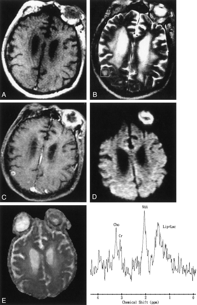
- Fig 1.
Images obtained in a 69-year-old man with Klebsiella pneumoniae bacteremia, renal abscess, brain abscess, and endophthalmitis of the left eye.
A, Axial T1-weighted image (500/30[TR/TE]) before administration of contrast material.
B, Axial T2-weighted image (4000/100). The 2 × 2 × 2 cm voxel (box) in the lesion, adjacent brain tissue, and neighboring fat represents the 1H-MRS volume of interest.
C, Axial contrast-enhanced T1-weighted MR image (500/30) shows a regular thin-walled ring-enhanced abscess approximately 9 mm in diameter in the superficial brain surface of right parietal region. Endophthalmitis in the left eye shows enhancement after contrast agent administration.
D, Axial diffusion-weighted (10,000/93; b = 1000 s/mm2) image shows marked hyperintensity in the abscess cavity and the left eye.
E, ADC map reveals slight hypointensity, representing restricted diffusion in the corresponding region.
F, In vivo 1H-MR spectrum (2000/270) was unacceptable because of contamination from neighboring fat and a very small lesion. The resonances of choline (Cho), creatine (Cr), and N-acetylaspartate (NAA) were interpreted to be caused by partial volume effects of the adjacent brain tissue. A hump resonance (1–1.5 ppm) was identified, and lactate (Lac) peak contaminated by neighboring fat with a lipid (Lip) peak were suggested. Multiple small peaks at various frequencies are present; these peaks may represent noise or unassigned metabolites.
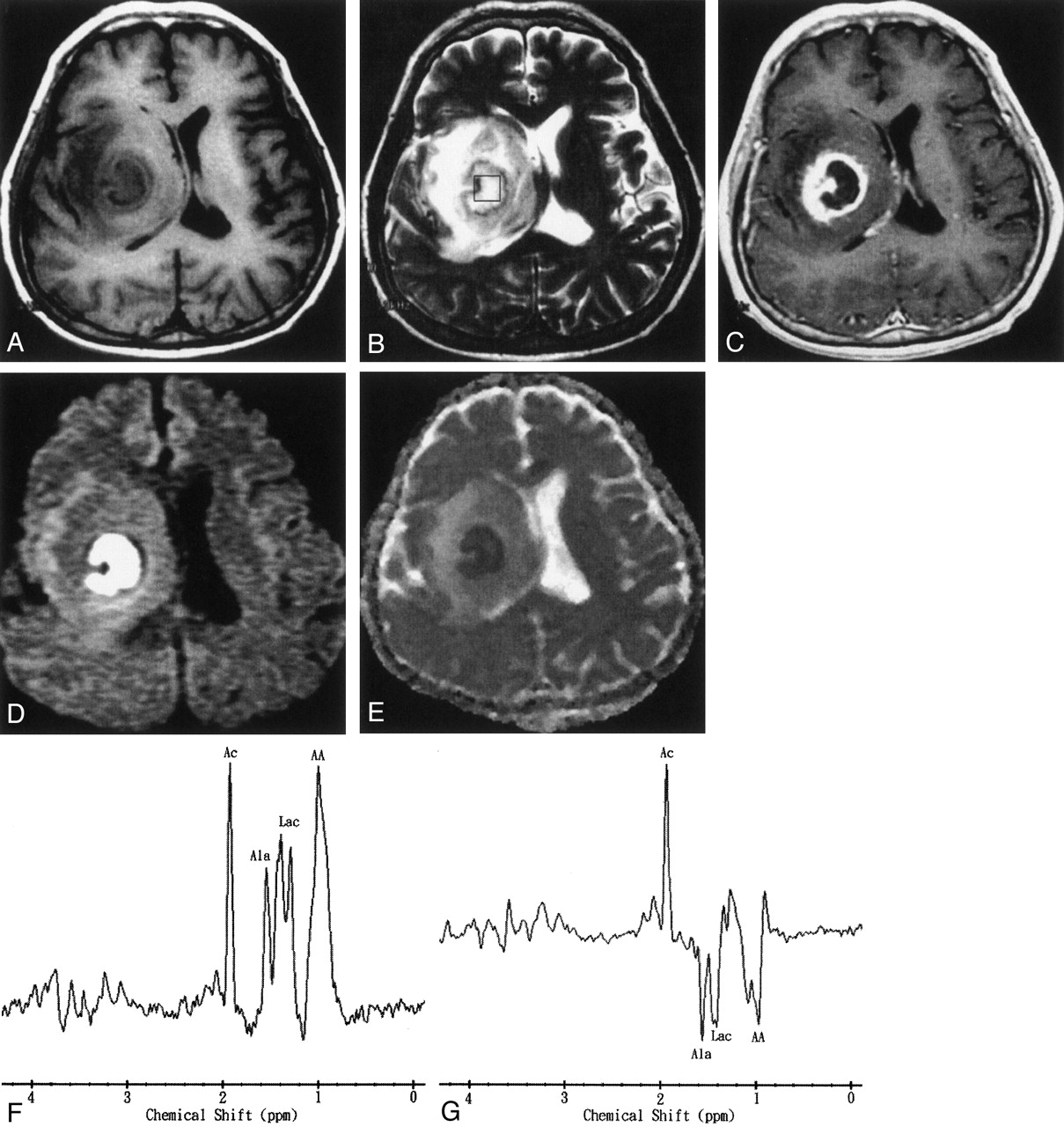
- Fig 2.
Images obtained in a 50-year-old man with surgically proven pyogenic brain abscess in the right basal ganglion.
A, Axial T1-weighted image (500/30) before administration of contrast material.
B, Axial T2-weighted image (4000/100). The 2 × 2 × 2 cm voxel (box) in the center of the lesion represents the 1H-MRS volume of interest.
C, Axial contrast-enhanced T1-weighted (500/30) MR image shows a ring-shaped cystic lesion and surrounding edema.
D, Axial diffusion-weighted (10,000/93; b = 1000 s/mm2) image shows marked hyperintensity in the abscess cavity and slight iso- to hypointensity surrounding the edema.
E, ADC map reveals hypointensity in the abscess cavity, representing restricted diffusion, and hyperintense areas surrounding the edema.
F and G, In vivo 1H spectra (2000/270 and 135) from the abscess cavity show resonances representing acetate (Ac), alanine (Ala), lactate (Lac), and amino acids (AA). At a TE of 135 (G), the phase reversal resonances are well depicted at 1.5, 1.3, and 0.9 ppm, which confirms the assignment to alanine, lactate, and amino acids, respectively.
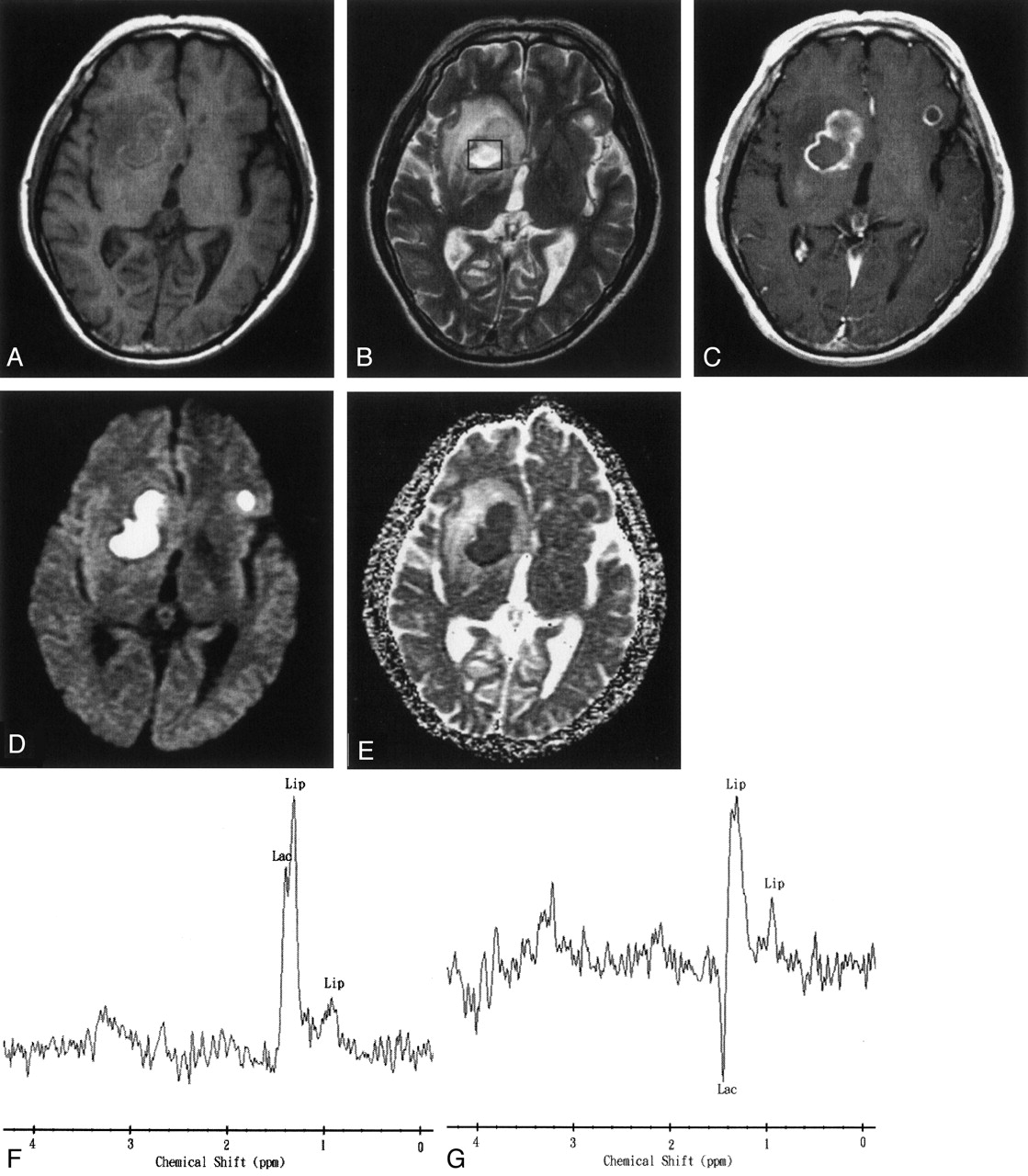
- Fig 3.
Images obtained 35 days after the start of initial antibiotic treatment in a 45-year-old man with multiple pyogenic brain abscesses.
A, Axial T1-weighted image (500/30) before administration of contrast material.
B, Axial T2-weighted image (4000/100). The 2 × 2 × 2 cm voxel (box) represents 1H-MRS volume of interest.
C, Axial contrast-enhanced T1-weighted (500/30) MR image shows two ring-shaped enhanced lesions in the right basal ganglion and left frontal lobe.
D, Axial diffusion-weighted (10,000/93; b = 1000 s/mm2) image shows markedly high signal intensity in the abscess cavity and slightly iso- to hypointense surrounding edema.
E, ADC map reveals low signal intensity in the abscess cavity, representing restricted diffusion, and hyperintense areas surrounding the edema.
F and G, In vivo 1H spectra (2000/270 and 135) from the abscess cavity show a lactate (Lac) peak (1.3 ppm) that is inverted at a TE of 135 and a lipid (Lip) peak (0.8–1.2 ppm). Note the similarity of this spectral pattern to that of a necrotic brain tumor.
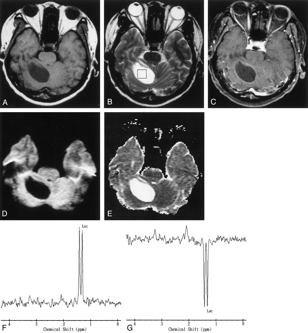
- Fig 4.
Images obtained in a 67-year-old man with a pathologically proven right cerebellar metastasis from primary lung adenocarcinoma.
A, Axial T1-weighted image (500/30) before administration of contrast material.
B, Axial T2-weighted image (4000/100). The 2 × 2 × 2 cm voxel (box) in the center of the lesion represents the 1H-MRS volume of interest.
C, Axial contrast-enhanced T1-weighted (500/30) MR image shows a ring-enhanced lesion in the right cerebellum.
D, Axial diffusion-weighted (10,000/93; b = 1000 s/mm2) image shows markedly low signal intensity in the necrotic part of the tumor.
E, ADC map reveals high signal intensity in the necrotic part of the tumor that is similar to that of CSF, reflecting marked diffusion.
F and G, In vivo 1H spectra (2000/270 and 135) from the necrotic center of the tumor show a lactate (Lac) peak at 1.3 ppm that is inverted at a TE of 135.


Tables
- TABLE 1:
1H-MRS and diffusion-weighted imaging findings in 14 patients with brain abscess or tumor
Case No. Age (y)/Sex Diagnosis Metabolites Detected Signal Intensity on Diffusion-Weighted Image 1 50/M Brain abscess Ace(+++), Ala(++), AA(+++), Lac(+++) Markedly high 2 44/M Brain abscess Ace(+++), Succ(++), Ala(++), AA(++), Lac(++), NAA(+), Cho(+), Cr/PCr(+) Markedly high 3 58/F Brain abscess Ace(+++), Ala(++), AA(+), Lac(++) Markedly high 4 26/F Brain abscess Ace(+++), Ala(++), AA(+), Lip(+), Lac(++) Markedly high 5 69/M Brain abscess * Markedly high 6 48/M Brain stem abscess ** Markedly high 7 45/M Brain abscess Lip(++), Lac(+) Markedly high 8 63/M Glioblastoma Lac(+++) Markedly low 9 74/M Glioblastoma Lac(++), NAA(+), Cho(+++), Cr/PCr(+) Markedly low 10 58/F Glioblastoma Lip(+), Lac(++) Slightly low 11 61/M Anaplastic astrocytoma Lac(+++), NAA(+), Cho(+++), Cr/PCr(+) Markedly low 12 52/M Metastatic brain tumor Lip(+), Lac(++) Markedly low 13 55/F Metastatic brain tumor Lip(+), Lac(++), NAA(+), Cho(+++), Cr/PCr(+) Markedly low 14 67/M Metastatic brain tumor Lac(+++) Markedly low Note.—M indicates male; F, female; +, small peak; ++, moderate peak; +++, large peak; Ace, acetate; Ala, alanine; AA, amino acids; Lac, lactate; Succ, succinate; NAA, N-acetylaspartate; Cho, choline; Cr, creatine; PCr, phosphocreatine; Lip, lipids; *, unacceptable because of contamination from neighboring fat; **, unacceptable because of poor shimming conditions.
In this issue


Jump to section
Related Articles
Cited By...
- Multiparametric imaging in the evaluation of intracerebral abscesses
- Multiparametric imaging in the evaluation of intracerebral abscesses
- Hypertensive emergency and seizures during haemodialysis
- MR Imaging Features of Acute Mastoiditis and Their Clinical Relevance
- A weak leg
- Proton MR Spectroscopy of Brain Abscesses
- Role of imaging in the diagnosis of acute bacterial meningitis and its complications
- In Vivo Proton MR Spectroscopy Evaluation of Pyogenic Brain Abscesses: A Report of 194 Cases
- Unusual findings in cerebral abscess: report of two cases.
- Glioblastoma multiforme with atypical diffusion-weighted MR findings