Advertisement

Main menu

  • Home
  • Content
    • Current Issue
    • Accepted Manuscripts
    • Article Preview
    • Past Issue Archive
    • AJNR Case Collection
    • Case of the Week Archive
    • Classic Case Archive
    • Case of the Month Archive
  • Special Collections
    • Spinal CSF Leak Articles (Jan 2020-June 2024)
    • 2024 AJNR Journal Awards
    • Most Impactful AJNR Articles
  • Multimedia
    • AJNR Podcast
    • AJNR Scantastics
    • Video Articles
  • For Authors
    • Submit a Manuscript
    • Author Policies
    • Fast publishing of Accepted Manuscripts
    • Graphical Abstract Preparation
    • Manuscript Submission Guidelines
    • Imaging Protocol Submission
    • Submit a Case for the Case Collection
  • About Us
    • About AJNR
    • Editorial Board
  • More
    • Become a Reviewer/Academy of Reviewers
    • Subscribers
    • Permissions
    • Alerts
    • Feedback
    • Advertisers
    • ASNR Home
  • Other Publications
    • ajnr

User menu

  • Alerts
  • Log in

Search

  • Advanced search
American Journal of Neuroradiology

American Journal of Neuroradiology

ASHNR American Society of Functional Neuroradiology ASHNR American Society of Pediatric Neuroradiology ASSR
  • Alerts
  • Log in

Advanced Search

  • Home
  • Content
    • Current Issue
    • Accepted Manuscripts
    • Article Preview
    • Past Issue Archive
    • AJNR Case Collection
    • Case of the Week Archive
    • Classic Case Archive
    • Case of the Month Archive
  • Special Collections
    • Spinal CSF Leak Articles (Jan 2020-June 2024)
    • 2024 AJNR Journal Awards
    • Most Impactful AJNR Articles
  • Multimedia
    • AJNR Podcast
    • AJNR Scantastics
    • Video Articles
  • For Authors
    • Submit a Manuscript
    • Author Policies
    • Fast publishing of Accepted Manuscripts
    • Graphical Abstract Preparation
    • Manuscript Submission Guidelines
    • Imaging Protocol Submission
    • Submit a Case for the Case Collection
  • About Us
    • About AJNR
    • Editorial Board
  • More
    • Become a Reviewer/Academy of Reviewers
    • Subscribers
    • Permissions
    • Alerts
    • Feedback
    • Advertisers
    • ASNR Home
  • Follow AJNR on Twitter
  • Visit AJNR on Facebook
  • Follow AJNR on Instagram
  • Join AJNR on LinkedIn
  • RSS Feeds

Welcome to the new AJNR, Updated Hall of Fame, and more. Read the full announcements.


AJNR is seeking candidates for the position of Associate Section Editor, AJNR Case Collection. Read the full announcement.


Research ArticleBRAIN

Proton MR Spectroscopy of Tumefactive Demyelinating Lesions

Amit M. Saindane, Soonmee Cha, Meng Law, Xiaonan Xue, Edmond A. Knopp and David Zagzag
American Journal of Neuroradiology September 2002, 23 (8) 1378-1386;
Amit M. Saindane
  • Find this author on Google Scholar
  • Find this author on PubMed
  • Search for this author on this site
Soonmee Cha
  • Find this author on Google Scholar
  • Find this author on PubMed
  • Search for this author on this site
Meng Law
  • Find this author on Google Scholar
  • Find this author on PubMed
  • Search for this author on this site
Xiaonan Xue
  • Find this author on Google Scholar
  • Find this author on PubMed
  • Search for this author on this site
Edmond A. Knopp
  • Find this author on Google Scholar
  • Find this author on PubMed
  • Search for this author on this site
David Zagzag
  • Find this author on Google Scholar
  • Find this author on PubMed
  • Search for this author on this site
  • Article
  • Figures & Data
  • Info & Metrics
  • Responses
  • References
  • PDF
Loading

Article Figures & Data

Figures

  • Tables
  • Fig 1.
    • Download figure
    • Open in new tab
    • Download powerpoint
    Fig 1.

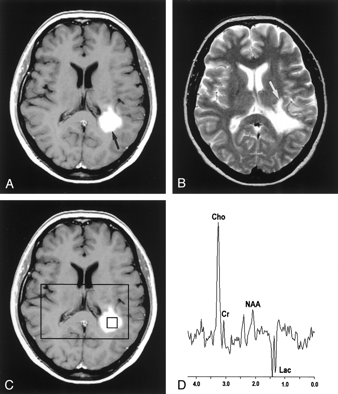
    Images in a 50-year-old woman with biopsy-proven tumefactive demyelinating lesion.

    A, Contrast-enhanced axial T1-weighted image (600/14/1) demonstrates an ill-defined enhancing mass (arrow) in the left frontoparietal periventricular white matter.

    B, Axial T2-weighted image (3400/119/1) shows increased signal intensity (arrow) around the lesion.

    C, Localizing image (600/14/1) for proton MR spectroscopy displays a voxel in the central portion of the lesion.

    D, Proton MR spectrum obtained by using PRESS (1500/144) demonstrates an elevated Cho value, a decreased NAA value, and a Lac doublet.

  • Fig 2.
    • Download figure
    • Open in new tab
    • Download powerpoint
    Fig 2.

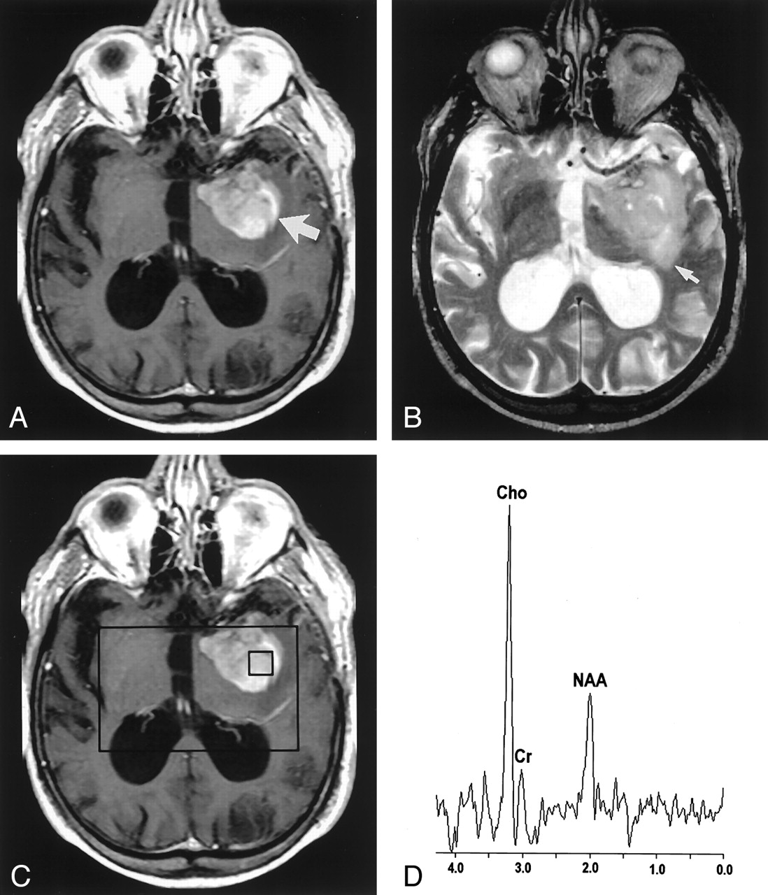
    Images in an 81-year-old man with a high-grade mixed glioma.

    A, Contrast-enhanced axial T1-weighted image (600/14/1) demonstrates an ill-defined enhancing mass (arrow) in the left temporal region.

    B, Axial T2-weighted image (3400/119/1) shows increased signal intensity (arrow) around the lesion.

    C, Localizing image (600/14/1) for proton MR spectroscopy displays a voxel in the central portion of the lesion.

    D, Proton MR spectrum obtained by using PRESS (1500/144) demonstrates an elevated Cho value and a decreased NAA value.

  • Fig 3.
    • Download figure
    • Open in new tab
    • Download powerpoint
    Fig 3.

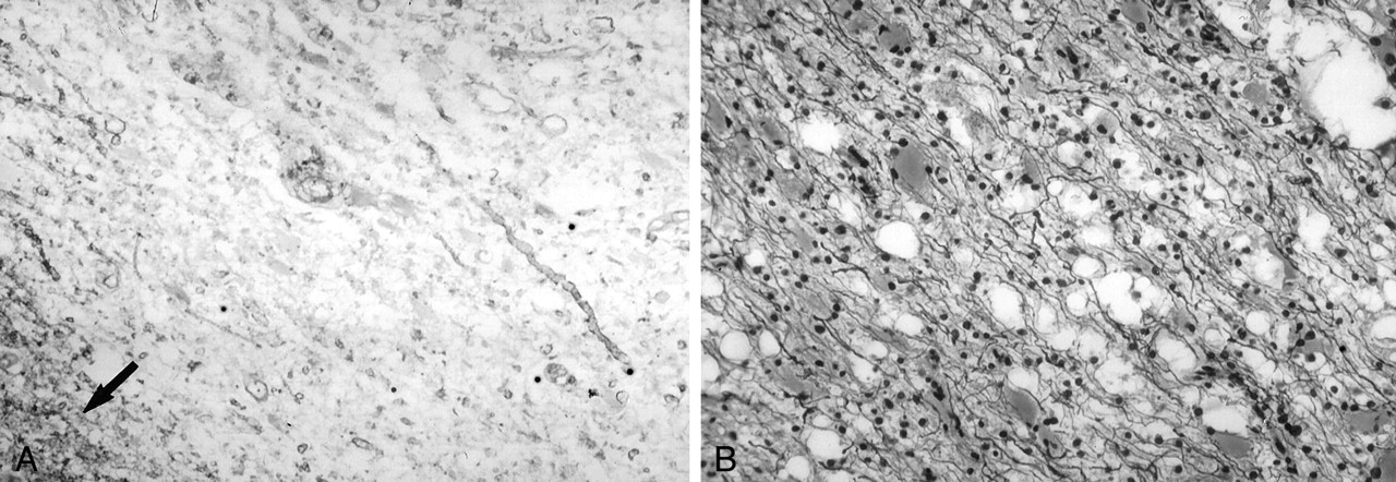
    Photomicrographs of a biopsy specimen from a tumefactive demyelinating lesion in a 50-year-old woman. Left, A Luxol fast blue stain demonstrates extensive myelin loss. The lower left corner (arrow) shows a relatively normal pattern of myelination. Right, Silver stain demonstrates relative preservation of axis cylinders.

Tables

  • Figures
    • View popup
    TABLE 1:

    MR imaging characteristics of tumefactive demyelinating lesions and glioma patients

    Patient No.Age (y)/SexLesionLesion Size (cm)EnhancementEdemaMass EffectNecrosis/Cystic DegenerationLesion DelineationHeterogeneityAdditional Lesions
    150/FTDL2.950%++++YesIllYesNo
    246/FTDL1.730%−−NoWellNoYes
    325/MTDL1.810%++YesIllYesYes
    457/MTDL1.2<5%+−YesIllYesYes
    541/FTDL2.8<5%++NoIllNoNo
    649/FTDL2.130%++YesWellNoNo
    751/MAA2.750%++++YesIntermediateYesYes
    839/MAA1.490%++NoIllYesYes
    944/MAO4.350%++++++YesIllYesNo
    1028/MAA1.990%++NoIllNoYes
    1138/MAMG1.490%+++NoIntermediateNoNo
    1238/MAA1.710%++YesIllYesYes
    1380/MAMG3.580%+++YesWellYesNo
    1425/FAA4.850%++++YesIllYesNo
    1560/MGBM2.380%++YesIllYesYes
    1634/MGBM5.250%++++++YesIllYesYes
    • Note.—TDL indicates tumefactive demyelinating lesion; AA, anaplastic astrocytoma; AO, anaplastic oligodendroglioma; AMG, anaplastic mixed glioma; GBM, glioblastoma multiforme.

    • View popup
    TABLE 2:

    MR spectroscopy characteristics of tumefactive demyelinating lesions and glioma patients

    Patient No.LesionEnhancing RegionCentral RegionPerilesional T2 RegionContralateral Normal Brain
    Cho/CrNAA/CrLacCho/CrNAA/CrLacCho/CrNAA/CrLacCho/CrNAA/CrLac
    1TDL4.61.0++3.51.0++1.71.2+0.51.6−
    2TDL1.81.9+3.03.2++1.62.9+0.91.6−
    3TDL1.70.7−4.92.3+1.91.2−0.71.6−
    4TDL2.41.3−3.32.7−1.50.3−0.82.0−
    5TDL2.81.9−1.62.5−1.42.6−1.43.0−
    6TDL1.71.7+2.31.4−1.11.7−0.91.9−
    7AA3.31.2−1.51.2−1.82.0−0.51.7−
    8AA3.30.7−9.01.1−1.00.9−0.71.2−
    9AO0.90.7−2.11.1−1.30.9+0.91.1−
    10AA4.30.9−3.80.8−2.01.4−0.71.7−
    11AMG2.50.6−2.50.6−2.01.3−1.63.6−
    12AA1.30.6−1.60.5−1.20.9−0.91.4−
    13AMG10.30.8+11.31.6−1.22.1+0.82.0−
    14AA12.70.6−4.70.8+1.51.3−1.11.5−
    15GBM1.80.7−2.20.9−1.51.2−1.01.5−
    16GBM10.01.0−4.40.8−2.31.0−1.22.1−
    • Note.—TDL indicates tumefactive demyelinating lesion; AA, anaplastic astrocytoma; AO, anaplastic oligodendroglioma; AMG, anaplastic mixed glioma; GBM, glioblastoma multiforme.

    • View popup
    TABLE 3:

    Comparison of spectroscopic characteristics of tumefactive demyelinating lesions and gliomas

    RegionTDL (n = 6)Glioma (n = 10)P Value
    MedianRangeMedianRange
    Enhancing RegionCho/Cr2.11.7–4.63.30.9–12.7.328
    NAA/Cr1.50.7–1.90.70.6–1.2.014
    Central RegionCho/Cr3.21.6–4.93.21.5–11.3.914
    NAA/Cr2.41.0–3.20.90.5–1.6.008
    Perilesional T2 RegionCho/Cr1.61.1–1.91.51.0–2.3.956
    NAA/Cr1.50.3–2.91.30.9–2.1.478
    Contralateral Normal BrainCho/Cr0.90.5–1.40.90.5–1.6.621
    NAA/Cr1.81.6–3.01.61.1–3.6.383
    • Note.—Using the Wilcoxon rank-sum test and a Bonferroni correction for multiple comparisons, P < .0125 is considered statistically significant.

    • View popup
    TABLE 4:

    Comparison of TDLs and gliomas with contralateral normal-appearing brain

    RegionCho/Cr P ValueNAA/Cr P Value
    TDL (n = 6)Enhancing Region.031.094
    Central Region.031.400
    Perilesional T2 Region.045.293
    Glioma (n = 10)Enhancing Region.007.006
    Central Region.006.007
    Perilesional T2 Region.006.041
PreviousNext
Back to top

In this issue

American Journal of Neuroradiology: 23 (8)
American Journal of Neuroradiology
Vol. 23, Issue 8
1 Sep 2002
  • Table of Contents
  • Index by author
Advertisement
Print
Download PDF
Email Article

Thank you for your interest in spreading the word on American Journal of Neuroradiology.

NOTE: We only request your email address so that the person you are recommending the page to knows that you wanted them to see it, and that it is not junk mail. We do not capture any email address.

Enter multiple addresses on separate lines or separate them with commas.
Proton MR Spectroscopy of Tumefactive Demyelinating Lesions
(Your Name) has sent you a message from American Journal of Neuroradiology
(Your Name) thought you would like to see the American Journal of Neuroradiology web site.
CAPTCHA
This question is for testing whether or not you are a human visitor and to prevent automated spam submissions.
Cite this article
Amit M. Saindane, Soonmee Cha, Meng Law, Xiaonan Xue, Edmond A. Knopp, David Zagzag
Proton MR Spectroscopy of Tumefactive Demyelinating Lesions
American Journal of Neuroradiology Sep 2002, 23 (8) 1378-1386;

Citation Manager Formats

  • BibTeX
  • Bookends
  • EasyBib
  • EndNote (tagged)
  • EndNote 8 (xml)
  • Medlars
  • Mendeley
  • Papers
  • RefWorks Tagged
  • Ref Manager
  • RIS
  • Zotero
0 Responses
Respond to this article
Share
Bookmark this article
Proton MR Spectroscopy of Tumefactive Demyelinating Lesions
Amit M. Saindane, Soonmee Cha, Meng Law, Xiaonan Xue, Edmond A. Knopp, David Zagzag
American Journal of Neuroradiology Sep 2002, 23 (8) 1378-1386;
del.icio.us logo Twitter logo Facebook logo Mendeley logo
  • Tweet Widget
  • Facebook Like
  • Google Plus One

Jump to section

  • Article
    • Abstract
    • Methods
    • Results
    • Discussion
    • Conclusion
    • Acknowledgments
    • References
  • Figures & Data
  • Info & Metrics
  • Responses
  • References
  • PDF

Related Articles

  • No related articles found.
  • PubMed
  • Google Scholar

Cited By...

  • Proton magnetic resonance spectroscopy of N-acetyl aspartate in first depressive episode and chronic major depressive disorder: a systematic review and meta-analysis
  • MR Imaging of Neoplastic Central Nervous System Lesions: Review and Recommendations for Current Practice
  • Tumefactive demyelinating lesions: a diagnostic challenge
  • Brain: non-infective and non-neoplastic manifestations of HIV
  • Proton MR Spectroscopy Improves Discrimination between Tumor and Pseudotumoral Lesion in Solid Brain Masses
  • Crossref
  • Google Scholar

This article has not yet been cited by articles in journals that are participating in Crossref Cited-by Linking.

More in this TOC Section

  • Progression of Microstructural Damage in Spinocerebellar Ataxia Type 2: A Longitudinal DTI Study
  • Usefulness of Quantitative Susceptibility Mapping for the Diagnosis of Parkinson Disease
  • Evaluating the Effects of White Matter Multiple Sclerosis Lesions on the Volume Estimation of 6 Brain Tissue Segmentation Methods
Show more Brain

Similar Articles

Advertisement

Indexed Content

  • Current Issue
  • Accepted Manuscripts
  • Article Preview
  • Past Issues
  • Editorials
  • Editors Choice
  • Fellow Journal Club
  • Letters to the Editor

Cases

  • Case Collection
  • Archive - Case of the Week
  • Archive - Case of the Month
  • Archive - Classic Case

Special Collections

  • Special Collections

Resources

  • News and Updates
  • Turn around Times
  • Submit a Manuscript
  • Author Policies
  • Manuscript Submission Guidelines
  • Evidence-Based Medicine Level Guide
  • Publishing Checklists
  • Graphical Abstract Preparation
  • Imaging Protocol Submission
  • Submit a Case
  • Become a Reviewer/Academy of Reviewers
  • Get Peer Review Credit from Publons

Multimedia

  • AJNR Podcast
  • AJNR SCANtastic
  • Video Articles

About Us

  • About AJNR
  • Editorial Board
  • Not an AJNR Subscriber? Join Now
  • Alerts
  • Feedback
  • Advertise with us
  • Librarian Resources
  • Permissions
  • Terms and Conditions

American Society of Neuroradiology

  • Not an ASNR Member? Join Now

© 2025 by the American Society of Neuroradiology All rights, including for text and data mining, AI training, and similar technologies, are reserved.
Print ISSN: 0195-6108 Online ISSN: 1936-959X

Powered by HighWire