Advertisement

Main menu

  • Home
  • Content
    • Current Issue
    • Accepted Manuscripts
    • Article Preview
    • Past Issue Archive
    • AJNR Case Collection
    • Case of the Week Archive
    • Classic Case Archive
    • Case of the Month Archive
  • Special Collections
    • Spinal CSF Leak Articles (Jan 2020-June 2024)
    • 2024 AJNR Journal Awards
    • Most Impactful AJNR Articles
  • Multimedia
    • AJNR Podcast
    • AJNR Scantastics
    • Video Articles
  • For Authors
    • Submit a Manuscript
    • Author Policies
    • Fast publishing of Accepted Manuscripts
    • Graphical Abstract Preparation
    • Manuscript Submission Guidelines
    • Imaging Protocol Submission
    • Submit a Case for the Case Collection
  • About Us
    • About AJNR
    • Editorial Board
  • More
    • Become a Reviewer/Academy of Reviewers
    • Subscribers
    • Permissions
    • Alerts
    • Feedback
    • Advertisers
    • ASNR Home
  • Other Publications
    • ajnr

User menu

  • Alerts
  • Log in

Search

  • Advanced search
American Journal of Neuroradiology

American Journal of Neuroradiology

ASHNR American Society of Functional Neuroradiology ASHNR American Society of Pediatric Neuroradiology ASSR
  • Alerts
  • Log in

Advanced Search

  • Home
  • Content
    • Current Issue
    • Accepted Manuscripts
    • Article Preview
    • Past Issue Archive
    • AJNR Case Collection
    • Case of the Week Archive
    • Classic Case Archive
    • Case of the Month Archive
  • Special Collections
    • Spinal CSF Leak Articles (Jan 2020-June 2024)
    • 2024 AJNR Journal Awards
    • Most Impactful AJNR Articles
  • Multimedia
    • AJNR Podcast
    • AJNR Scantastics
    • Video Articles
  • For Authors
    • Submit a Manuscript
    • Author Policies
    • Fast publishing of Accepted Manuscripts
    • Graphical Abstract Preparation
    • Manuscript Submission Guidelines
    • Imaging Protocol Submission
    • Submit a Case for the Case Collection
  • About Us
    • About AJNR
    • Editorial Board
  • More
    • Become a Reviewer/Academy of Reviewers
    • Subscribers
    • Permissions
    • Alerts
    • Feedback
    • Advertisers
    • ASNR Home
  • Follow AJNR on Twitter
  • Visit AJNR on Facebook
  • Follow AJNR on Instagram
  • Join AJNR on LinkedIn
  • RSS Feeds

Welcome to the new AJNR, Updated Hall of Fame, and more. Read the full announcements.


AJNR is seeking candidates for the position of Associate Section Editor, AJNR Case Collection. Read the full announcement.


Research ArticleBRAIN

Proton Chemical Shift Imaging in Pick Complex

Osamu Kizu, Kei Yamada and Tsunehiko Nishimura
American Journal of Neuroradiology September 2002, 23 (8) 1387-1392;
Osamu Kizu
  • Find this author on Google Scholar
  • Find this author on PubMed
  • Search for this author on this site
Kei Yamada
  • Find this author on Google Scholar
  • Find this author on PubMed
  • Search for this author on this site
Tsunehiko Nishimura
  • Find this author on Google Scholar
  • Find this author on PubMed
  • Search for this author on this site
  • Article
  • Figures & Data
  • Info & Metrics
  • Responses
  • References
  • PDF
Loading

Article Figures & Data

Figures

  • Tables
  • Fig 1.
    • Download figure
    • Open in new tab
    • Download powerpoint
    Fig 1.

    Proton MR spectra from bilateral basal ganglia and perisylvian regions in a case of FTD. Arrows indicate the chosen voxels. A T1-weighted MR image with VOI, which is outlined by a box, can be seen as a reference. In this case, NAA at 2.0 ppm was observed in the left basal ganglia.

  • Fig 2.
    • Download figure
    • Open in new tab
    • Download powerpoint
    Fig 2.

    Proton MR spectra from the bilateral basal ganglia and perisylvian regions in a case of CBD. Arrows indicate the chosen voxels. In this case, reduced NAA was observed in bilateral basal ganglia.

  • Fig 3.
    • Download figure
    • Open in new tab
    • Download powerpoint
    Fig 3.

    Proton MR spectra from bilateral basal ganglia and perisylvian regions in a case of PPA. Arrows indicate the chosen voxels. Note reduced NAA in the left basal ganglia and left perisylvian region.
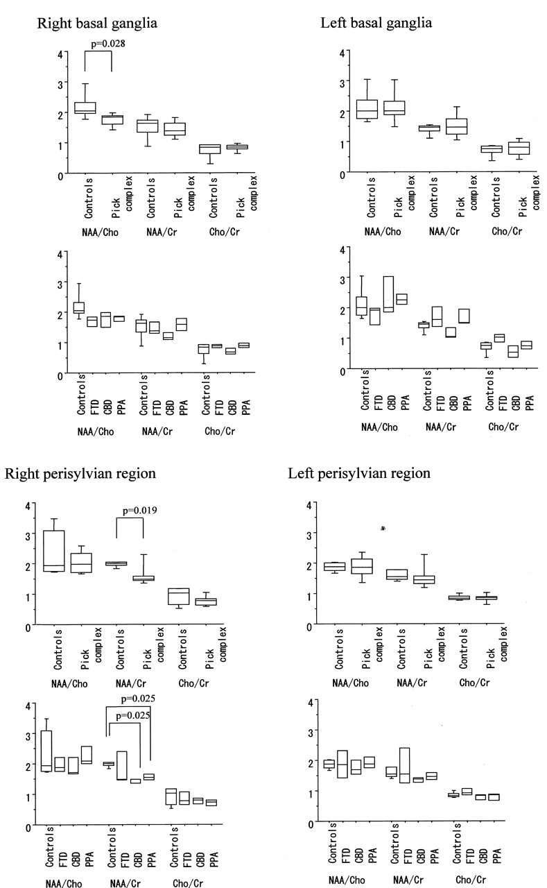
  • Fig 4.
    • Download figure
    • Open in new tab
    • Download powerpoint
    Fig 4.

    Box plots of peak ratios (NAA/Cho, NAA/Cr, and Cho/Cr) among controls subjects and patients with PC, FTD, CBD, and PPA. The box stretches from the lower hinge (defined as the 25th percentile) to the upper hinge (defined as the 75th percentile), and the median is shown as a line across the box. Outside the box, 10th and 90th percentile lines are shown. A statistically significant decrease in NAA/Cho was found in the right basal ganglia among patients with PC (P = .028). NAA/Cr ratio from the right perisylvian regions in patients with PPA and CBD were lower than that in control subjects (P = .025 and P = .025, respectively). No statistically significant difference in peak ratio was found in the left perisylvian regions or the left basal ganglia.

Tables

  • Figures
  • Summary of peak ratios in proton chemical shift imaging

    Right Basal GangliaLeft Basal Ganglia
    NAA/ChoNAA/CrCho/CrNAA/ChoNAA/CrCho/Cr
    Control subjects n = 52.18 ± 0.441.51 ± 0.390.74 ± 0.262.11 ± 0.551.40 ± 0.180.70 ± 0.20
    Pick complex n = 91.74 ± 0.21*1.41 ± 0.260.82 ± 0.132.13 ± 0.591.51 ± 0.400.76 ± 0.26
     FTD (n = 3)1.68 ± 0.221.47 ± 0.260.87 ± 0.701.73 ± 0.401.69 ± 0.440.98 ± 0.17
     CBD (n = 3)1.75 ± 0.341.19 ± 0.160.69 ± 0.132.39 ± 0.861.17 ± 0.230.54 ± 0.25
     PPA (n = 3)1.78 ± 0.121.58 ± 0.260.88 ± 0.092.27 ± 0.231.68 ± 0.350.75 ± 0.18
    Right Perisylvian RegionLeft Perisylvian Region
    NAA/ChoNAA/CrCho/CrNAA/ChoNAA/CrCho/Cr
    Control subjects n = 52.37 ± 0.791.97 ± 0.090.91 ± 0.291.87 ± 0.151.60 ± 0.170.85 ± 0.08
    Pick complex n = 92.05 ± 0.371.62 ± 0.42*0.77 ± 0.181.84 ± 0.371.54 ± 0.460.84 ± 0.14
     FTD (n = 3)1.97 ± 0.321.88 ± 0.720.84 ± 0.291.85 ± 0.601.80 ± 0.800.95 ± 0.17
     CBD (n = 3)1.91 ± 0.411.42 ± 0.10*0.76 ± 0.131.76 ± 0.321.35 ± 0.110.78 ± 0.11
     PPA (n = 3)2.26 ± 0.411.56 ± 0.13*0.70 ± 0.131.91 ± 0.241.47 ± 0.150.78 ± 0.15
    • Note.—Values are presented as the mean ± SD. FTD indicates frontotemporal dementia; CBD, corticobasal degeneration; PPA, primary progressive aphasia; NAA, N-acetylaspartate; Cho, choline; Cr, creatine.

    • * P < .05.

PreviousNext
Back to top

In this issue

American Journal of Neuroradiology: 23 (8)
American Journal of Neuroradiology
Vol. 23, Issue 8
1 Sep 2002
  • Table of Contents
  • Index by author
Advertisement
Print
Download PDF
Email Article

Thank you for your interest in spreading the word on American Journal of Neuroradiology.

NOTE: We only request your email address so that the person you are recommending the page to knows that you wanted them to see it, and that it is not junk mail. We do not capture any email address.

Enter multiple addresses on separate lines or separate them with commas.
Proton Chemical Shift Imaging in Pick Complex
(Your Name) has sent you a message from American Journal of Neuroradiology
(Your Name) thought you would like to see the American Journal of Neuroradiology web site.
CAPTCHA
This question is for testing whether or not you are a human visitor and to prevent automated spam submissions.
Cite this article
Osamu Kizu, Kei Yamada, Tsunehiko Nishimura
Proton Chemical Shift Imaging in Pick Complex
American Journal of Neuroradiology Sep 2002, 23 (8) 1387-1392;

Citation Manager Formats

  • BibTeX
  • Bookends
  • EasyBib
  • EndNote (tagged)
  • EndNote 8 (xml)
  • Medlars
  • Mendeley
  • Papers
  • RefWorks Tagged
  • Ref Manager
  • RIS
  • Zotero
0 Responses
Respond to this article
Share
Bookmark this article
Proton Chemical Shift Imaging in Pick Complex
Osamu Kizu, Kei Yamada, Tsunehiko Nishimura
American Journal of Neuroradiology Sep 2002, 23 (8) 1387-1392;
del.icio.us logo Twitter logo Facebook logo Mendeley logo
  • Tweet Widget
  • Facebook Like
  • Google Plus One

Jump to section

  • Article
    • Abstract
    • Methods
    • Results
    • Discussion
    • Conclusion
    • References
  • Figures & Data
  • Info & Metrics
  • Responses
  • References
  • PDF

Related Articles

  • No related articles found.
  • PubMed
  • Google Scholar

Cited By...

  • No citing articles found.
  • Crossref
  • Google Scholar

This article has not yet been cited by articles in journals that are participating in Crossref Cited-by Linking.

More in this TOC Section

  • Progression of Microstructural Damage in Spinocerebellar Ataxia Type 2: A Longitudinal DTI Study
  • Usefulness of Quantitative Susceptibility Mapping for the Diagnosis of Parkinson Disease
  • Evaluating the Effects of White Matter Multiple Sclerosis Lesions on the Volume Estimation of 6 Brain Tissue Segmentation Methods
Show more Brain

Similar Articles

Advertisement

Indexed Content

  • Current Issue
  • Accepted Manuscripts
  • Article Preview
  • Past Issues
  • Editorials
  • Editors Choice
  • Fellow Journal Club
  • Letters to the Editor

Cases

  • Case Collection
  • Archive - Case of the Week
  • Archive - Case of the Month
  • Archive - Classic Case

Special Collections

  • Special Collections

Resources

  • News and Updates
  • Turn around Times
  • Submit a Manuscript
  • Author Policies
  • Manuscript Submission Guidelines
  • Evidence-Based Medicine Level Guide
  • Publishing Checklists
  • Graphical Abstract Preparation
  • Imaging Protocol Submission
  • Submit a Case
  • Become a Reviewer/Academy of Reviewers
  • Get Peer Review Credit from Publons

Multimedia

  • AJNR Podcast
  • AJNR SCANtastic
  • Video Articles

About Us

  • About AJNR
  • Editorial Board
  • Not an AJNR Subscriber? Join Now
  • Alerts
  • Feedback
  • Advertise with us
  • Librarian Resources
  • Permissions
  • Terms and Conditions

American Society of Neuroradiology

  • Not an ASNR Member? Join Now

© 2025 by the American Society of Neuroradiology All rights, including for text and data mining, AI training, and similar technologies, are reserved.
Print ISSN: 0195-6108 Online ISSN: 1936-959X

Powered by HighWire