
Article Figures & Data
Figures
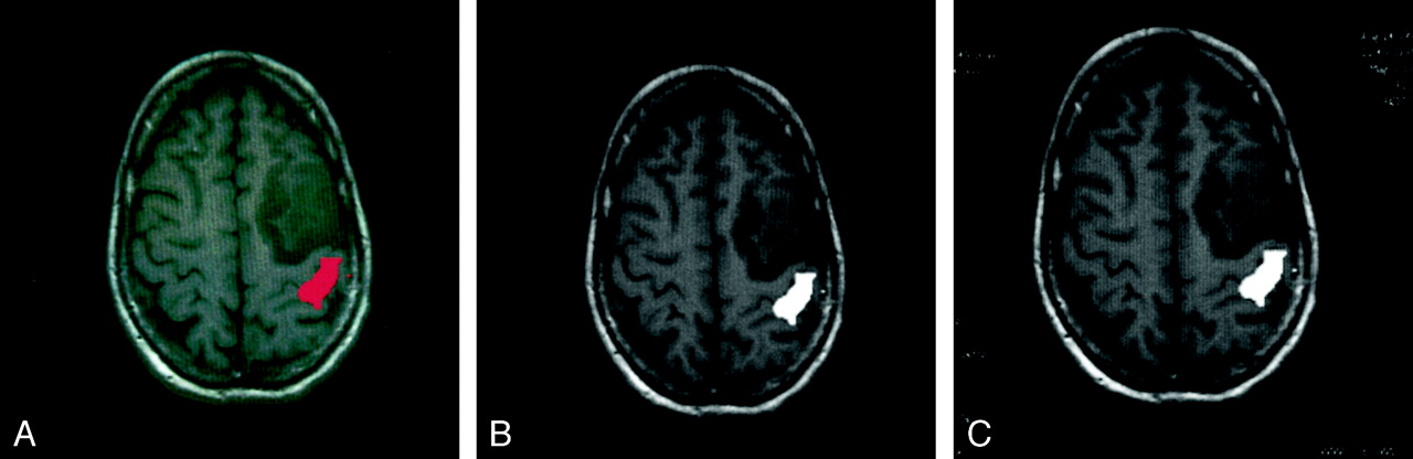
- Fig 1.
fMRIs on an off-line workstation and on a PACS image review station.
A, fMRI from a unilateral hand motor task study displayed on an off-line workstation demonstrates activation of the primary motor cortex (red) posterior to a brain tumor.
B, Same image as in A, after DICOM conversion, displayed on an off-line workstation. Activated area is displayed in white by using a gray-scale color table. Otherwise, the images in A and B appear identical.
C, Same DICOM image as in B, after transfer to PACS image review station (Images are identical). In addition, the PACS image review workstation displays modified header fields, including the study date and image acquisition parameters (patient specific information was not filmed).
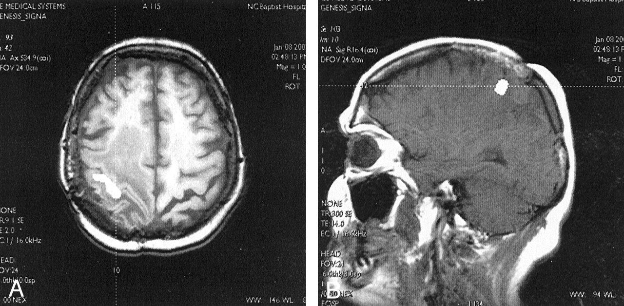
- Fig 2.
fMRIs with axial and sagittal overlays displayed on a PACS image review station with cross-referencing.
A, fMRI overlaid on axial T1-weighted image demonstrates activation of the primary motor cortex (white area) with surrounding tumor and edema. A cross-reference tool (dotted line) on the PACS image review station was applied to the activated region in the right motor cortex.
B, Images from the same fMRI study overlaid on a sagittal T1-weighted MR image. A cross-reference tool from the axial image in A cuts through the motor cortex activation cluster on the right side of the sagittal volume (dotted line). Patient-specific information (ie, name, examination number, accession number) was not filmed.


Tables
Fields Modified in the DICOM Header
Field Group Element SOP instance UID 0008 0018 Study date 0008 0020 Series date 0008 0021 Image date 0008 0023 Study time 0008 0030 Accession number 0008 0050 Institution name 0008 0080 Study description 0008 1030 Series description 0008 103e Patient name 0010 0010 Patient identification 0010 0020 Patient birthdate 0010 0030 Patient sex 0010 0040 Patient age 0010 1010 Patient weight 0010 1030 Additional patient history 0010 21b0 Study instance UID 0020 000d Series instance UID 0020 000e Series number 0020 0011 Image number 0020 0013 Image position patient 0020 0032 Image orientation patient 0020 0037 Frame of reference UID 0020 0052 Images in acquisition 0020 1002 Section location 0020 1041 Section thickness 0018 0050 Spacing between sections 0018 0088 Repetition time 0018 0080 Echo time 0018 0081 Magnetic field strength 0018 0087 Flip angle 0018 1314 Patient position 0018 5100 Photometric interpretation 0028 0004 Rows 0028 0010 Columns 0028 0011 Pixel spacing 0028 0030
In this issue


Jump to section
Related Articles
Cited By...
- No citing articles found.