Advertisement

Main menu

  • Home
  • Content
    • Current Issue
    • Accepted Manuscripts
    • Article Preview
    • Past Issue Archive
    • AJNR Case Collection
    • Case of the Week Archive
    • Classic Case Archive
    • Case of the Month Archive
  • Special Collections
    • Spinal CSF Leak Articles (Jan 2020-June 2024)
    • 2024 AJNR Journal Awards
    • Most Impactful AJNR Articles
  • Multimedia
    • AJNR Podcast
    • AJNR Scantastics
    • Video Articles
  • For Authors
    • Submit a Manuscript
    • Author Policies
    • Fast publishing of Accepted Manuscripts
    • Graphical Abstract Preparation
    • Manuscript Submission Guidelines
    • Imaging Protocol Submission
    • Submit a Case for the Case Collection
  • About Us
    • About AJNR
    • Editorial Board
  • More
    • Become a Reviewer/Academy of Reviewers
    • Subscribers
    • Permissions
    • Alerts
    • Feedback
    • Advertisers
    • ASNR Home
  • Other Publications
    • ajnr

User menu

  • Alerts
  • Log in

Search

  • Advanced search
American Journal of Neuroradiology

American Journal of Neuroradiology

ASHNR American Society of Functional Neuroradiology ASHNR American Society of Pediatric Neuroradiology ASSR
  • Alerts
  • Log in

Advanced Search

  • Home
  • Content
    • Current Issue
    • Accepted Manuscripts
    • Article Preview
    • Past Issue Archive
    • AJNR Case Collection
    • Case of the Week Archive
    • Classic Case Archive
    • Case of the Month Archive
  • Special Collections
    • Spinal CSF Leak Articles (Jan 2020-June 2024)
    • 2024 AJNR Journal Awards
    • Most Impactful AJNR Articles
  • Multimedia
    • AJNR Podcast
    • AJNR Scantastics
    • Video Articles
  • For Authors
    • Submit a Manuscript
    • Author Policies
    • Fast publishing of Accepted Manuscripts
    • Graphical Abstract Preparation
    • Manuscript Submission Guidelines
    • Imaging Protocol Submission
    • Submit a Case for the Case Collection
  • About Us
    • About AJNR
    • Editorial Board
  • More
    • Become a Reviewer/Academy of Reviewers
    • Subscribers
    • Permissions
    • Alerts
    • Feedback
    • Advertisers
    • ASNR Home
  • Follow AJNR on Twitter
  • Visit AJNR on Facebook
  • Follow AJNR on Instagram
  • Join AJNR on LinkedIn
  • RSS Feeds

Welcome to the new AJNR, Updated Hall of Fame, and more. Read the full announcements.


AJNR is seeking candidates for the position of Associate Section Editor, AJNR Case Collection. Read the full announcement.


OtherBRAIN

Visualization of Microvascularity in Glioblastoma Multiforme With 8-T High-Spatial-Resolution MR Imaging

Gregory A. Christoforidis, John C. Grecula, Herbert B. Newton, Allahyar Kangarlu, Amir M. Abduljalil, Petra Schmalbrock and Donald W Chakeres
American Journal of Neuroradiology October 2002, 23 (9) 1553-1556;
Gregory A. Christoforidis
  • Find this author on Google Scholar
  • Find this author on PubMed
  • Search for this author on this site
John C. Grecula
  • Find this author on Google Scholar
  • Find this author on PubMed
  • Search for this author on this site
Herbert B. Newton
  • Find this author on Google Scholar
  • Find this author on PubMed
  • Search for this author on this site
Allahyar Kangarlu
  • Find this author on Google Scholar
  • Find this author on PubMed
  • Search for this author on this site
Amir M. Abduljalil
  • Find this author on Google Scholar
  • Find this author on PubMed
  • Search for this author on this site
Petra Schmalbrock
  • Find this author on Google Scholar
  • Find this author on PubMed
  • Search for this author on this site
Donald W Chakeres
  • Find this author on Google Scholar
  • Find this author on PubMed
  • Search for this author on this site
  • Article
  • Figures & Data
  • Info & Metrics
  • Responses
  • References
  • PDF
Loading

Article Figures & Data

Figures

  • Fig 1.
    • Download figure
    • Open in new tab
    • Download powerpoint
    Fig 1.

    Comparison of MR images obtained at the inferior border of a glioblastoma multiforme. Although the arterial structures in the sylvian fissure (arrowheads) are identified at both 1.5- and 8-T imaging, the smaller venous structures (unlabeled arrows) are depicted only on the 8-T image. These venous structures represent draining veins from the tumoral bed. A indicates anterior; CP, choroid plexus within the trigone of the left lateral ventricle; L, lateral; M, medial; P, posterior; VOG, vein of Galen; and 3V, third ventricle.

    A, Image obtained at 8 T (714/10, 23° flip angle, 900 × 900 matrix, 2-mm section thickness, 20-cm FOV).

    B, FSE T2-weighted image obtained at 1.5 T (5650/104, 512 × 512 matrix, 3-mm section thickness, 20-cm FOV).

    C, Gadolinium-enhanced magnetization-transfer image obtained at 1.5 T (616/20, 256 × 256 matrix, 5-mm section thickness).

  • Fig 2.
    • Download figure
    • Open in new tab
    • Download powerpoint
    Fig 2.

    Axial images obtained through the superior aspect in patient with a glioblastoma multiforme demonstrate the ability to identify the fine angioarchitecture associated with the tumor at 8 T but not at 1.5 T. Note the enlargement and tortuosity of the transmedullary veins coursing over the tumor bed (long arrows), as compared with those in a healthy patient. The signal-intensity voids in the tumoral bed connect to the transmedullary veins (short arrows). Also note the area of overall decreased signal intensity that is associated with haphazardly arranged vessels, which are thought to represent neovascularity (arrowheads). A indicates anterior; CS, centrum semiovale; L, lateral; M, medial; P, posterior; SDV, subependymal draining vein.

    A, Image obtained at 8 T (714/10, 23° flip angle, 900 × 900 matrix, 2-mm section thickness, 20-cm FOV).

    B, FSE T2-weighted 1.5-T image (5650/104, 512 × 512 matrix, 3-mm section thickness, 20-cm FOV).

    C, Image obtained at 8 T at one section superior to that in B (714/10, 23° flip angle, 900 × 900 matrix, 2-mm section thickness, 20-cm FOV).

    D, Gadolinium-enhanced T1-weighted magnetization transfer images (616/20, 256 × 256 matrix, 5-mm section thickness).
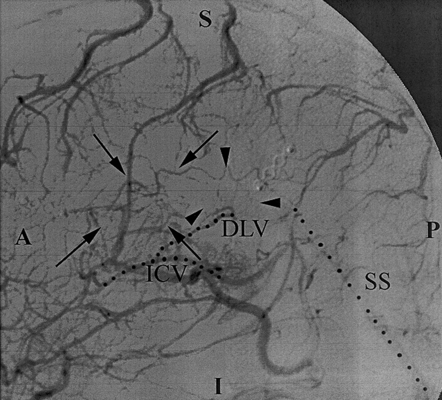
  • Fig 3.
    • Download figure
    • Open in new tab
    • Download powerpoint
    Fig 3.

    Lateral arteriogram obtained with a left carotid injection demonstrates tortuous vascularity (arrows and arrowheads) in the tumor bed within the glioblastoma multiforme. These findings were also identified on the 8-T images (Figs 1 and 2). However, the normal transmedullary veins and the smaller vessels are less conspicuous on DSA images than on 8-T images. A indicates anterior; DLV, direct lateral vein; I, inferior; ICV, internal cerebral vein; P, posterior; and SS, straight sinus.

  • Fig 4.
    • Download figure
    • Open in new tab
    • Download powerpoint
    Fig 4.

    Axial gradient-echo 8-T MR images (600/12, 23° flip angle, 2-mm section thickness, 1024 × 1024 matrix, 20-cm FOV) display the medullary veins in the head of a healthy volunteer. The image is centered at the centrum semiovale and shows many transmedullary veins (black arrowheads) that drain into the subependymal veins (arrows), which are depicted as linear signal intensity voids converging on the surface of the lateral ventricles. Also note the cortical penetrating veins (white arrowheads). A indicates anterior; L, lateral; M, medial; and P, posterior.

PreviousNext
Back to top

In this issue

American Journal of Neuroradiology: 23 (9)
American Journal of Neuroradiology
Vol. 23, Issue 9
1 Oct 2002
  • Table of Contents
  • Index by author
Advertisement
Print
Download PDF
Email Article

Thank you for your interest in spreading the word on American Journal of Neuroradiology.

NOTE: We only request your email address so that the person you are recommending the page to knows that you wanted them to see it, and that it is not junk mail. We do not capture any email address.

Enter multiple addresses on separate lines or separate them with commas.
Visualization of Microvascularity in Glioblastoma Multiforme With 8-T High-Spatial-Resolution MR Imaging
(Your Name) has sent you a message from American Journal of Neuroradiology
(Your Name) thought you would like to see the American Journal of Neuroradiology web site.
CAPTCHA
This question is for testing whether or not you are a human visitor and to prevent automated spam submissions.
Cite this article
Gregory A. Christoforidis, John C. Grecula, Herbert B. Newton, Allahyar Kangarlu, Amir M. Abduljalil, Petra Schmalbrock, Donald W Chakeres
Visualization of Microvascularity in Glioblastoma Multiforme With 8-T High-Spatial-Resolution MR Imaging
American Journal of Neuroradiology Oct 2002, 23 (9) 1553-1556;

Citation Manager Formats

  • BibTeX
  • Bookends
  • EasyBib
  • EndNote (tagged)
  • EndNote 8 (xml)
  • Medlars
  • Mendeley
  • Papers
  • RefWorks Tagged
  • Ref Manager
  • RIS
  • Zotero
0 Responses
Respond to this article
Share
Bookmark this article
Visualization of Microvascularity in Glioblastoma Multiforme With 8-T High-Spatial-Resolution MR Imaging
Gregory A. Christoforidis, John C. Grecula, Herbert B. Newton, Allahyar Kangarlu, Amir M. Abduljalil, Petra Schmalbrock, Donald W Chakeres
American Journal of Neuroradiology Oct 2002, 23 (9) 1553-1556;
del.icio.us logo Twitter logo Facebook logo Mendeley logo
  • Tweet Widget
  • Facebook Like
  • Google Plus One

Jump to section

  • Article
    • Abstract
    • Description of the Technique and Results
    • Discussion
    • Acknowledgments
    • Footnotes
    • References
  • Figures & Data
  • Info & Metrics
  • Responses
  • References
  • PDF

Related Articles

  • No related articles found.
  • PubMed
  • Google Scholar

Cited By...

  • No citing articles found.
  • Crossref
  • Google Scholar

This article has not yet been cited by articles in journals that are participating in Crossref Cited-by Linking.

More in this TOC Section

  • Progression of Microstructural Damage in Spinocerebellar Ataxia Type 2: A Longitudinal DTI Study
  • Usefulness of Quantitative Susceptibility Mapping for the Diagnosis of Parkinson Disease
  • Evaluating the Effects of White Matter Multiple Sclerosis Lesions on the Volume Estimation of 6 Brain Tissue Segmentation Methods
Show more Brain

Similar Articles

Advertisement

Indexed Content

  • Current Issue
  • Accepted Manuscripts
  • Article Preview
  • Past Issues
  • Editorials
  • Editors Choice
  • Fellow Journal Club
  • Letters to the Editor

Cases

  • Case Collection
  • Archive - Case of the Week
  • Archive - Case of the Month
  • Archive - Classic Case

Special Collections

  • Special Collections

Resources

  • News and Updates
  • Turn around Times
  • Submit a Manuscript
  • Author Policies
  • Manuscript Submission Guidelines
  • Evidence-Based Medicine Level Guide
  • Publishing Checklists
  • Graphical Abstract Preparation
  • Imaging Protocol Submission
  • Submit a Case
  • Become a Reviewer/Academy of Reviewers
  • Get Peer Review Credit from Publons

Multimedia

  • AJNR Podcast
  • AJNR SCANtastic
  • Video Articles

About Us

  • About AJNR
  • Editorial Board
  • Not an AJNR Subscriber? Join Now
  • Alerts
  • Feedback
  • Advertise with us
  • Librarian Resources
  • Permissions
  • Terms and Conditions

American Society of Neuroradiology

  • Not an ASNR Member? Join Now

© 2025 by the American Society of Neuroradiology All rights, including for text and data mining, AI training, and similar technologies, are reserved.
Print ISSN: 0195-6108 Online ISSN: 1936-959X

Powered by HighWire