
Article Figures & Data
Figures
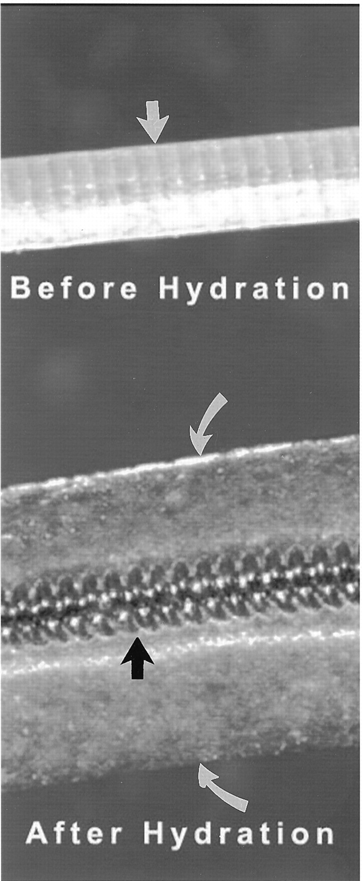
- Fig 1.
Hybrid hydrogel-platinum coil device. Top, prehydration image shows initial profile of the device. Highly compact hydrogel material is wrapped around a platinum coil. Indentations between winds of the underlying platinum coil can be seen through the compact hydrogel material (straight white arrow). The outer diameter of the coil is 0.008 inch; the thickness of the hydrogel is approximately 0.001 inch, such that the outer diameter of the device is 0.010 inch. Bottom, post-hydration image of the device shows marked expansion of the hydrogel material, which has become translucent. The outer edges of the hydrogel are denoted by the curved white arrows. The platinum coil is denoted by the black arrow. The radial thickness of the expanded hydrogel is approximately 0.013 inch, such that the total outer diameter of the hydrated device is 0.035 inch.
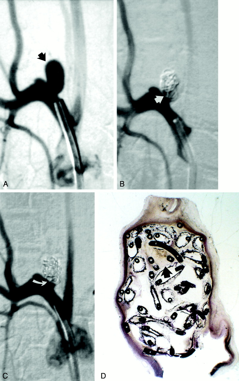
- Fig 2.
Study animal 4, 2-week implant.
A, Anteroposterior digital subtraction angiogram of the brachiocephalic artery shows a narrow-necked 5.4-mm-wide 7.8-mm-high aneurysm (black arrow).
B, Anteroposterior digital subtraction angiogram obtained immediately after embolization with a single complex coil and three hydrogel devices shows dense packing of the aneurysm dome, with persistent flow at the aneurysm neck (straight white arrow).
C, Anteroposterior digital subtraction angiogram obtained 2 weeks after embolization shows progressive occlusion of the aneurysm cavity, with the neck now occluded (curved white arrow).
D, Hematoxylin and eosin stain; original magnification, ×6.8. Coronal section obtained through the aneurysm cavity. The aneurysm cavity is filled with a mixture of unorganized thrombus (long straight arrow) and expanded hydrogel (short straight arrow). The hydrogel stains as faint, violet-colored, reticular material. There is no substantial inflammation. A thin fibrin membrane traverses the neck (curved arrow). Artifactual separation between the aneurysm wall and the coils and hydrogel occurred during processing (open arrow).
- Fig 3.
Study animal 6, 1-month implant.
A, Anteroposterior digital subtraction angiogram of the brachiocephalic artery shows a narrow-necked 4-mm-wide 7-mm-high aneurysm (black arrow).
B, Anteroposterior digital subtraction angiogram obtained immediately after embolization with a single complex coil and two hydrogel devices shows dense packing of the aneurysm dome, with persistent flow at the aneurysm neck (straight white arrow).
C, Anteroposterior digital subtraction angiogram obtained 1 month after embolization shows progressive occlusion of the aneurysm cavity, with the neck now occluded (curved white arrow).
D, Hematoxylin and eosin stain; original magnification, ×2.5. Coronal section obtained through the aneurysm cavity. The sac is nearly completely filled with hydrogel, which stains as faint, violet-colored, reticular material. There is a small amount of organizing thrombus near the dome (arrow).
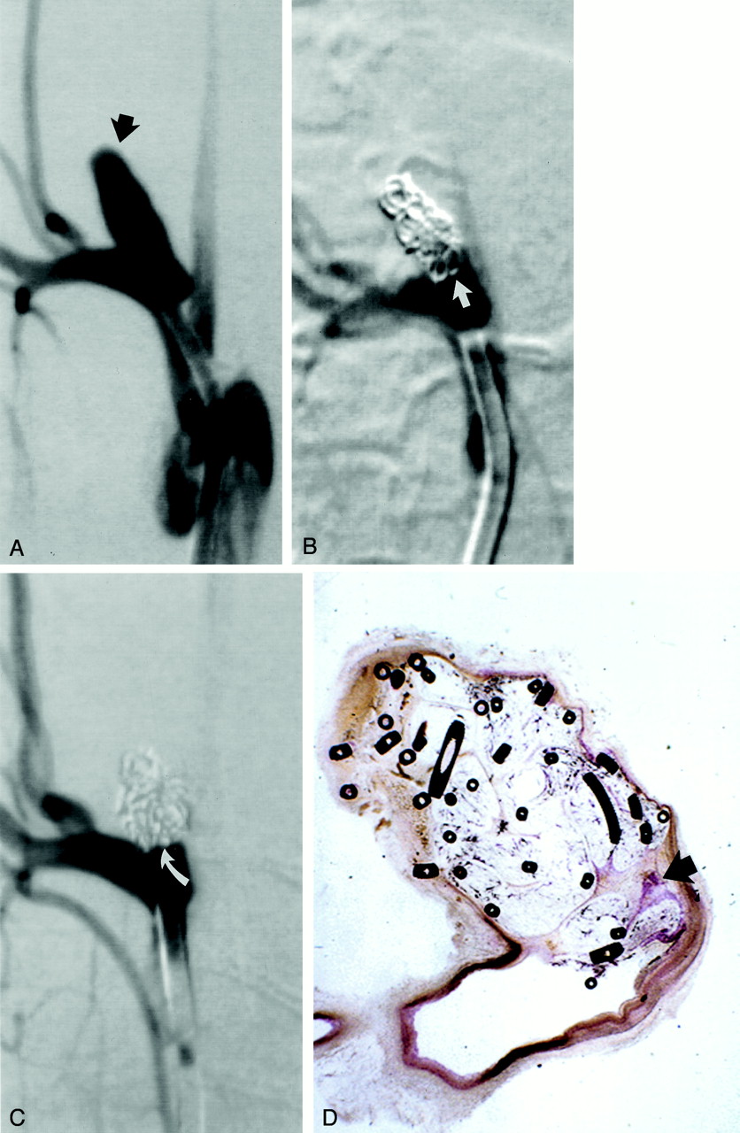
- Fig 4.
Study animal 10, 3-month implant.
A, Anteroposterior digital subtraction angiogram of the brachiocephalic artery shows a wide-necked 4-mm-wide 11-mm-high aneurysm (black arrow).
B, Anteroposterior digital subtraction angiogram obtained immediately after embolization with a single complex coil and a single hydrogel device shows dense packing of the aneurysm dome, with persistent flow at the aneurysm neck (straight white arrow).
C, Anteroposterior digital subtraction angiogram obtained 3 months after embolization shows progressive occlusion of the aneurysm cavity, with the neck now occluded (curved white arrow).
D, Hematoxylin and eosin stain; original magnification, ×6.3. Coronal section obtained through the aneurysm cavity. The sac is nearly completely filled with hydrogel, which stains as faint, violet-colored, reticular material. There is a small amount of organized tissue near the neck (arrow).
- Fig 5.
Study animal 16, 6-month implant.
A, Anteroposterior digital subtraction angiogram of the brachiocephalic artery shows a wide-necked 5.5-mm-wide 8.5-mm-high aneurysm (black arrow).
B, Anteroposterior digital subtraction angiogram obtained immediately after embolization with a single complex coil and a single hydrogel device shows dense packing of the aneurysm dome, with persistent flow at the aneurysm neck (straight white arrow).
C, Anteroposterior digital subtraction angiogram obtained 6 months after embolization shows progressive occlusion of the aneurysm cavity, with the neck now occluded (curved white arrow).
D, Hematoxylin and eosin stain; original magnification, ×6.8. Coronal section obtained through the aneurysm cavity. The sac is completely filled with hydrogel, which stains as faint, violet-colored, reticular material. A thin layer of organized tissue traverses the neck of the aneurysm (arrow).


In this issue


Jump to section
Related Articles
Cited By...
- Hydrogel Coils versus Bare Platinum Coils for the Treatment of Ruptured and Unruptured Aneurysms: An Updated Systematic Review and Meta-Analysis of Randomized Controlled Trials
- Endovascular Repair of Thoracic Aortic Pseudoaneurysms in Children
- An injectable shear-thinning biomaterial for endovascular embolization
- HydroCoils Are Associated with Lower Angiographic Recurrence Rates Than Are Bare Platinum Coils in Treatment of "Difficult-to-Treat" Aneurysms: A Post Hoc Subgroup Analysis of the HELPS Trial
- Mechanisms of Healing in Coiled Intracranial Aneurysms: A Review of the Literature
- HydroCoils Reduce Recurrence Rates in Recently Ruptured Medium-Sized Intracranial Aneurysms: A Subgroup Analysis of the HELPS Trial
- Reactive tissue proliferation and damage of elastic lamina caused by hydrogel coated coils in experimental rat aneurysms
- Initial experience with an extremely soft bare platinum coil, ED coil-10 Extra Soft, for endovascular treatment of cerebral aneurysms
- Vessel occlusion using a single long oversized coil in vertebral artery dissection: a technical note
- A multicenter registry of hydrocephalus following coil embolization of unruptured aneurysms: which patients are at risk and why it occurs
- The next generation HydroCoil: initial clinical experience with the HydroFill embolic coil
- Embolization of intracranial aneurysms with second-generation Matrix-2 detachable coils: mid-term and long-term results
- Embolization of Intracranial Aneurysms with HydroSoft Coils: Results of the Korean Multicenter Study
- The Woven EndoBridge: A New Aneurysm Occlusion Device
- Creation of Large Elastase-Induced Aneurysms: Presurgical Arterial Remodeling Using Arteriovenous Fistulas
- HydroCoils, Occlusion Rates, and Outcomes: A Large Single-Center Study
- Five-Year Follow-Up in Elastase-Induced Aneurysms in Rabbits
- Intrinsic Pathway-Mediated Apoptosis in Elastase-Induced Aneurysms in Rabbits
- Angiographic and Clinical Outcomes in 200 Consecutive Patients with Cerebral Aneurysm Treated with Hydrogel-Coated Coils
- Morbidity and Mortality Associated with Creation of Elastase-Induced Saccular Aneurysms in a Rabbit Model
- Molecular Indices of Apoptosis Activation in Elastase-Induced Aneurysms After Embolization With Platinum Coils
- Control of Aneurysm Volume by Adjusting the Position of Ligation During Creation of Elastase-Induced Aneurysms: A Prospective Study
- Embolization of Intracranial Aneurysms With Hydrogel-Coated Coils Versus Inert Platinum Coils: Effects on Packing Density, Coil Length and Quantity, Procedure Performance, Cost, Length of Hospital Stay, and Durability of Therapy
- Endovascular management of unruptured intracranial aneurysms
- Aneurysm Packing with HydroCoil Embolic System versus Platinum Coils: Initial Clinical Experience