Advertisement

Main menu

  • Home
  • Content
    • Current Issue
    • Accepted Manuscripts
    • Article Preview
    • Past Issue Archive
    • AJNR Case Collection
    • Case of the Week Archive
    • Classic Case Archive
    • Case of the Month Archive
  • Special Collections
    • Spinal CSF Leak Articles (Jan 2020-June 2024)
    • 2024 AJNR Journal Awards
    • Most Impactful AJNR Articles
  • Multimedia
    • AJNR Podcast
    • AJNR Scantastics
    • Video Articles
  • For Authors
    • Submit a Manuscript
    • Author Policies
    • Fast publishing of Accepted Manuscripts
    • Graphical Abstract Preparation
    • Manuscript Submission Guidelines
    • Imaging Protocol Submission
    • Submit a Case for the Case Collection
  • About Us
    • About AJNR
    • Editorial Board
  • More
    • Become a Reviewer/Academy of Reviewers
    • Subscribers
    • Permissions
    • Alerts
    • Feedback
    • Advertisers
    • ASNR Home
  • Other Publications
    • ajnr

User menu

  • Alerts
  • Log in

Search

  • Advanced search
American Journal of Neuroradiology

American Journal of Neuroradiology

ASHNR American Society of Functional Neuroradiology ASHNR American Society of Pediatric Neuroradiology ASSR
  • Alerts
  • Log in

Advanced Search

  • Home
  • Content
    • Current Issue
    • Accepted Manuscripts
    • Article Preview
    • Past Issue Archive
    • AJNR Case Collection
    • Case of the Week Archive
    • Classic Case Archive
    • Case of the Month Archive
  • Special Collections
    • Spinal CSF Leak Articles (Jan 2020-June 2024)
    • 2024 AJNR Journal Awards
    • Most Impactful AJNR Articles
  • Multimedia
    • AJNR Podcast
    • AJNR Scantastics
    • Video Articles
  • For Authors
    • Submit a Manuscript
    • Author Policies
    • Fast publishing of Accepted Manuscripts
    • Graphical Abstract Preparation
    • Manuscript Submission Guidelines
    • Imaging Protocol Submission
    • Submit a Case for the Case Collection
  • About Us
    • About AJNR
    • Editorial Board
  • More
    • Become a Reviewer/Academy of Reviewers
    • Subscribers
    • Permissions
    • Alerts
    • Feedback
    • Advertisers
    • ASNR Home
  • Follow AJNR on Twitter
  • Visit AJNR on Facebook
  • Follow AJNR on Instagram
  • Join AJNR on LinkedIn
  • RSS Feeds

Welcome to the new AJNR, Updated Hall of Fame, and more. Read the full announcements.


AJNR is seeking candidates for the position of Associate Section Editor, AJNR Case Collection. Read the full announcement.


LetterLetter

Multidetector-Row CT Angiographic Findings in the Bilateral Aortic Arch Origin of the Vertebral Arteries

Musturay Karcaaltincaba, James Strottman and Lacey Washington
American Journal of Neuroradiology January 2003, 24 (1) 157;
Musturay Karcaaltincaba
  • Find this author on Google Scholar
  • Find this author on PubMed
  • Search for this author on this site
James Strottman
  • Find this author on Google Scholar
  • Find this author on PubMed
  • Search for this author on this site
Lacey Washington
  • Find this author on Google Scholar
  • Find this author on PubMed
  • Search for this author on this site
  • Article
  • Figures & Data
  • Info & Metrics
  • Responses
  • References
  • PDF
Loading

We recently read the interesting article by Albayram et al (1), which concisely explained the anomalies of the vertebral arteries. We would like to present another rare case of bilateral vertebral arteries originating from the aortic arch. We incidentally diagnosed this condition on the basis of findings on a chest CT scan obtained in a patient with trauma. Our trauma chest CT protocol uses 8 × 2.5-mm detector configuration with a pitch of 1.35, allowing us to scan the chest in under 10 seconds. After the study, we reconstruct 1.25-mm-thick images at 1.25-mm intervals.

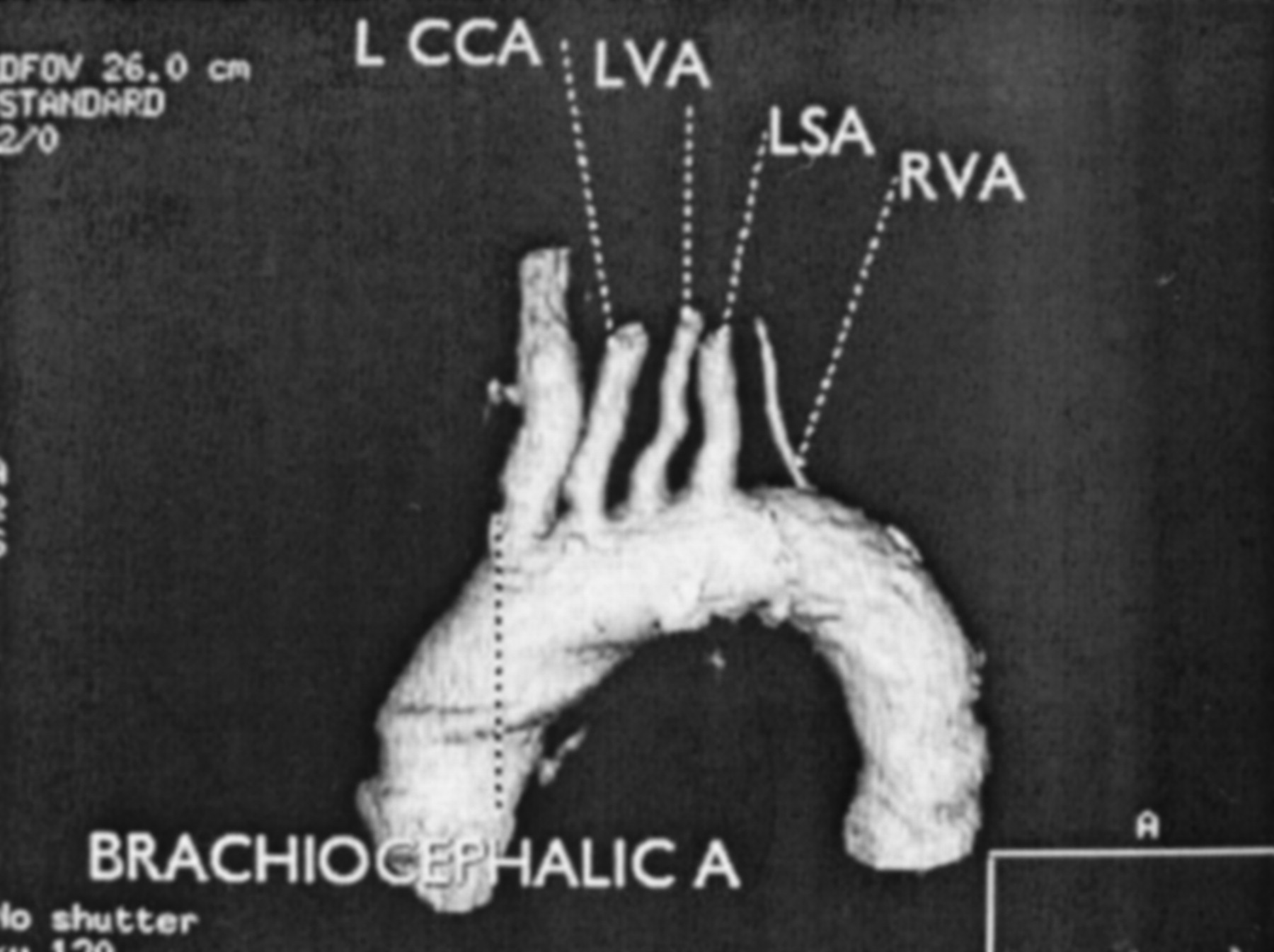
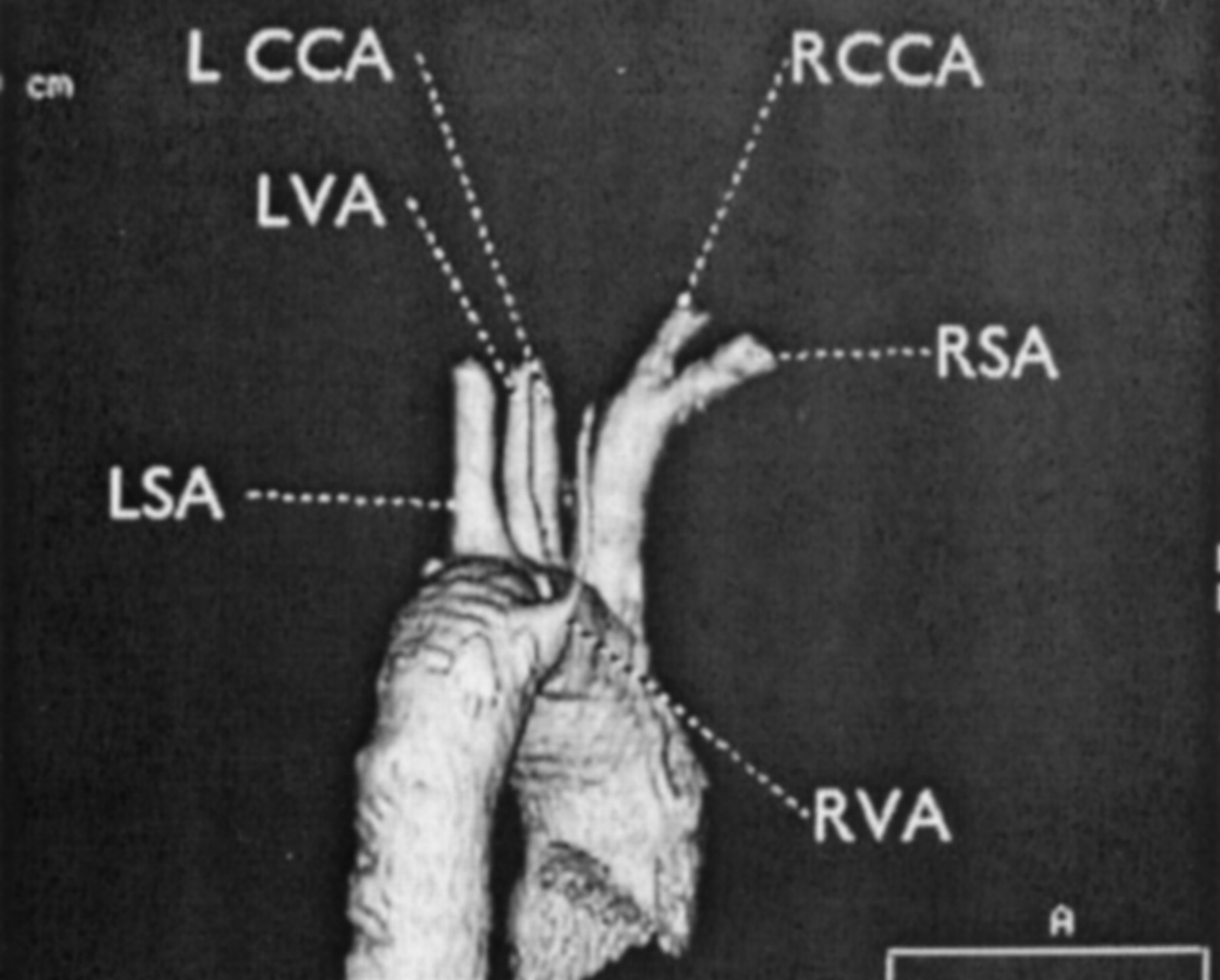
Both vertebral arteries originate from aortic arch (Figs 1 and 2). The left vertebral artery arises between the origins of the left common carotid and left subclavian arteries. An aberrant right vertebral artery arises from the descending aorta distal to the origin of the left subclavian artery, with a dilatation at the origin consistent with a Kommerel diverticulum (Figs 2 and 3), and it passes posterior to the esophagus (Fig 4). An aberrant right vertebral artery is a rare anomaly, and it can easily be distinguished from an aberrant right subclavian artery by the presence of the normal right subclavian artery (Fig 2) (2).

High-resolution multidetector-row CT angiography enables the display of images from different views. These can be useful as a roadmap in interventional and surgical therapeutic decision making, especially in patients with complex aortic arch anatomy.

Fig 1.
  • Download figure
  • Open in new tab
  • Download powerpoint
Fig 1.

CT angiogram, left anterior oblique projection. Three-dimensional volume-rendered image demonstrates the bilateral origin of the vertebral arteries.

Fig 2.
  • Download figure
  • Open in new tab
  • Download powerpoint
Fig 2.

CT angiography, posterior projection. Three-dimensional volume-rendered image shows the posterior arch origin of an aberrant right vertebral artery. Note the Kommerel diverticulum at the origin.

Fig 3.
  • Download figure
  • Open in new tab
  • Download powerpoint
Fig 3.

Curved planar reformatted image outlines the course of the aberrant right vertebral artery.

Fig 4.
  • Download figure
  • Open in new tab
  • Download powerpoint
Fig 4.

Axial image at the level of left brachiocephalic vein shows the retroesophageal location of an aberrant right vertebral artery.

References

  1. ↵
    Albayram S, Gailloud P, Wasserman BA. Bilateral arch origin of the vertebral arteries. AJNR Am J Neuroradiol 2002;23:455–458
    Abstract/FREE Full Text
  2. ↵
    Lemke AJ, Benndorf G, Liebig T, Felix R. Anomalous origin of the right vertebral artery: review of the literature and case report of the right vertebral artery origin distal to the left subclavian artery. AJNR Am J Neuroradiol 1999;20:1318–1321
    Abstract/FREE Full Text
  • Copyright © American Society of Neuroradiology
Back to top

In this issue

American Journal of Neuroradiology: 24 (1)
American Journal of Neuroradiology
Vol. 24, Issue 1
1 Jan 2003
  • Table of Contents
  • Index by author
Advertisement
Print
Download PDF
Email Article

Thank you for your interest in spreading the word on American Journal of Neuroradiology.

NOTE: We only request your email address so that the person you are recommending the page to knows that you wanted them to see it, and that it is not junk mail. We do not capture any email address.

Enter multiple addresses on separate lines or separate them with commas.
Multidetector-Row CT Angiographic Findings in the Bilateral Aortic Arch Origin of the Vertebral Arteries
(Your Name) has sent you a message from American Journal of Neuroradiology
(Your Name) thought you would like to see the American Journal of Neuroradiology web site.
CAPTCHA
This question is for testing whether or not you are a human visitor and to prevent automated spam submissions.
Cite this article
Musturay Karcaaltincaba, James Strottman, Lacey Washington
Multidetector-Row CT Angiographic Findings in the Bilateral Aortic Arch Origin of the Vertebral Arteries
American Journal of Neuroradiology Jan 2003, 24 (1) 157;

Citation Manager Formats

  • BibTeX
  • Bookends
  • EasyBib
  • EndNote (tagged)
  • EndNote 8 (xml)
  • Medlars
  • Mendeley
  • Papers
  • RefWorks Tagged
  • Ref Manager
  • RIS
  • Zotero
0 Responses
Respond to this article
Share
Bookmark this article
Multidetector-Row CT Angiographic Findings in the Bilateral Aortic Arch Origin of the Vertebral Arteries
Musturay Karcaaltincaba, James Strottman, Lacey Washington
American Journal of Neuroradiology Jan 2003, 24 (1) 157;
del.icio.us logo Twitter logo Facebook logo Mendeley logo
  • Tweet Widget
  • Facebook Like
  • Google Plus One

Jump to section

  • Article
    • References
  • Figures & Data
  • Info & Metrics
  • Responses
  • References
  • PDF

Related Articles

  • No related articles found.
  • PubMed
  • Google Scholar

Cited By...

  • Anomalous vertebral artery origins: the first and second reports of two variants
  • Non-invasive imaging of aberrant right subclavian artery pathologies and aberrant right vertebral artery
  • Crossref
  • Google Scholar

This article has not yet been cited by articles in journals that are participating in Crossref Cited-by Linking.

More in this TOC Section

  • Brain AVM’s Nidus: What if We Hadn’t Understood Anything?
  • Letter to the Editor regarding “Automated Volumetric Software in Dementia: Help or Hindrance to the Neuroradiologist?”
  • Reply:
Show more Letters

Similar Articles

Advertisement

Indexed Content

  • Current Issue
  • Accepted Manuscripts
  • Article Preview
  • Past Issues
  • Editorials
  • Editors Choice
  • Fellow Journal Club
  • Letters to the Editor

Cases

  • Case Collection
  • Archive - Case of the Week
  • Archive - Case of the Month
  • Archive - Classic Case

Special Collections

  • Special Collections

Resources

  • News and Updates
  • Turn around Times
  • Submit a Manuscript
  • Author Policies
  • Manuscript Submission Guidelines
  • Evidence-Based Medicine Level Guide
  • Publishing Checklists
  • Graphical Abstract Preparation
  • Imaging Protocol Submission
  • Submit a Case
  • Become a Reviewer/Academy of Reviewers
  • Get Peer Review Credit from Publons

Multimedia

  • AJNR Podcast
  • AJNR SCANtastic
  • Video Articles

About Us

  • About AJNR
  • Editorial Board
  • Not an AJNR Subscriber? Join Now
  • Alerts
  • Feedback
  • Advertise with us
  • Librarian Resources
  • Permissions
  • Terms and Conditions

American Society of Neuroradiology

  • Not an ASNR Member? Join Now

© 2025 by the American Society of Neuroradiology All rights, including for text and data mining, AI training, and similar technologies, are reserved.
Print ISSN: 0195-6108 Online ISSN: 1936-959X

Powered by HighWire