
Article Figures & Data
Figures
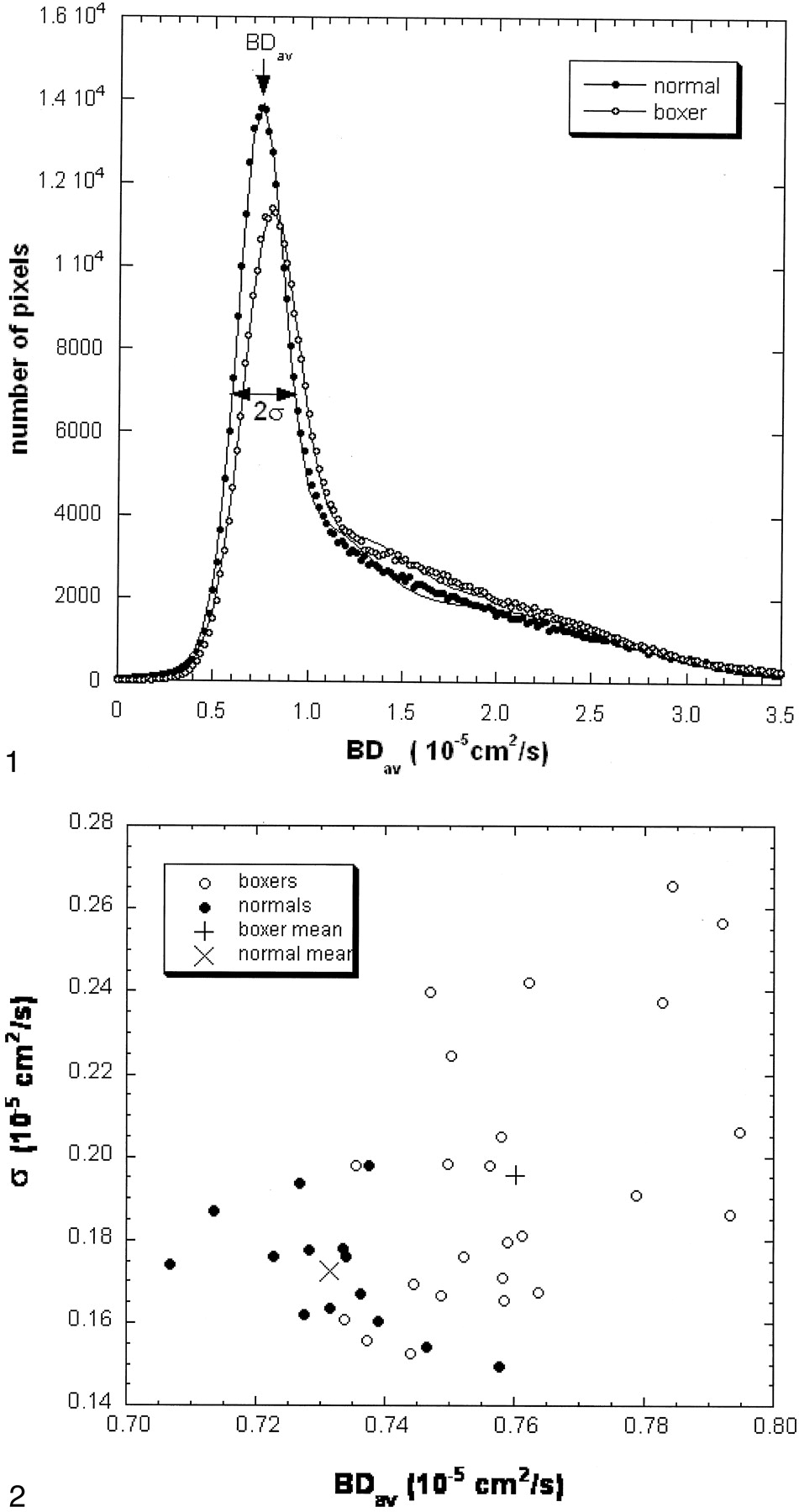
- Fig 1.
Normalized brain diffusion distribution histograms in a control subject and a boxer (case 15). The areas under the two curves are the same. The Dav data (dots and circles) are fitted with a triple Gaussian function to represent the two-compartment nature and the mixing between the two compartments (lines). The narrow peak represents the distribution of the brain tissue about its mean. The second and the third compartments have a broader distribution. The mean of the brain tissue pixel distribution is recognized as a mean diffusion constant for the entire brain (BDav). The distribution width (σ) of the brain tissue compartment is also recorded. The fitted curve of the boxer (circles) shifts to the right as compared with the curve of the control subject (dots). The second compartment level of the boxer’s curve is higher than that of the control subject.
- Fig 2.
BDav versus σ for boxers and control subjects: Overall, the boxer group shows elevated BDav and σ.
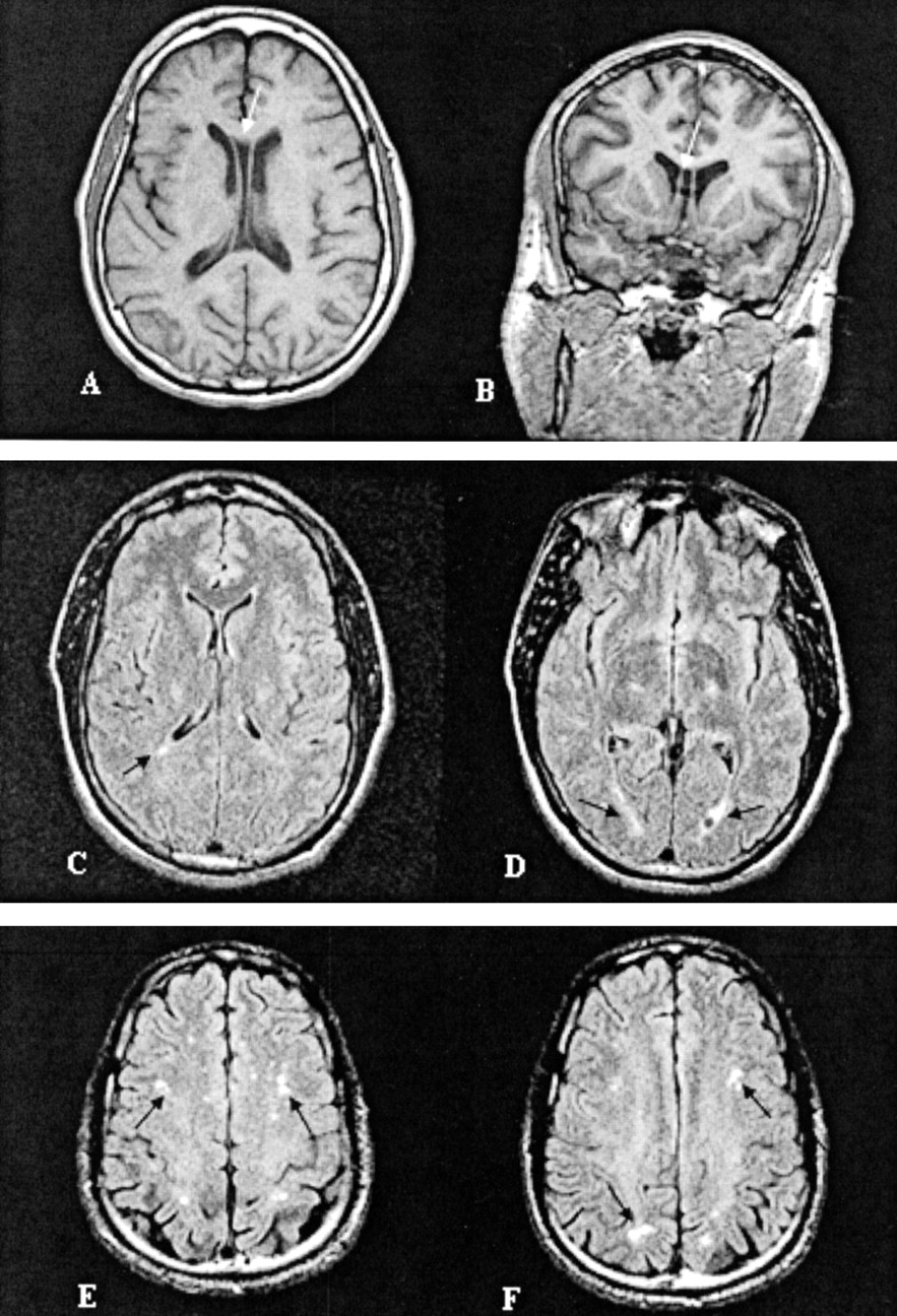
- Fig 3.
Representative images of MR findings in boxers: A, Cavum septum pellucidum (case 14); B, nonspecific periventricular white matter disease (case 22); and C, mild subcortical white matter demyelination (case 21).


Tables
Case (No.) Age (y) BDav σ MR Findings 1 20.84 0.7562 0.1980 Normal 2 22.52 0.7502 0.2245 Left minimal hippocampal atrophy, otherwise normal 3 24.68 0.7933 0.1861 Volume loss, CSP 4 26.00 0.7637 0.1672 CSP, mild atrophy, non-specific SWM 5 26.51 0.7622 0.2419 Normal 6 27.04 0.7372 0.1558 Normal 7 27.09 0.7590 0.1795 Normal 8 27.71 0.7613 0.1812 Normal 9 27.72 0.7486 0.1667 Normal 10 30.00 0.7580 0.2048 Normal 11 30.07 0.7497 0.1984 CSP, nonspecific SWM 12 30.17 0.7586 0.1655 Nonspecific SWM in left frontal lobe 13 32.29 0.7920 0.2564 Mild volume loss inappropriate to age 14 33.64 0.7828 0.2376 CSP, SWM, mild volume loss 15 33.72 0.7947 0.2061 Normal 16 34.13 0.7842 0.2655 CSP 17 35.14 0.7444 0.1695 Normal 18 36.10 0.7439 0.1526 Normal 19 36.12 0.7337 0.1606 Cerebellar atrophy, mild non-specific PWMD 20 38.05 0.7584 0.1711 Atrophy in left inferior cerebella, mild dilatation of sulci 21 38.52 0.7522 0.1759 Nonspecific SWM, atrophy inappropriate to age 22 40.00 0.7469 0.2397 PWMD 23 42.92 0.7355 0.1979 Normal 24 53.09 0.7786 0.1909 Normal Note.—CSP indicates cavum septum pellucidum; PWMD, nonspecific periventricular white matter disease; and SWM, subcortical white matter disease.
Premature Volume Loss CSP PWMD SWM Normal Number (n) 8 5 2 4 13 Percentage 33.3% 20.8% 8.3% 16.7% 54.2% Note.—Some boxers had more than one positive finding. CSP indicates cavum septum pellucidum; PWMD, nonspecific periventricular white matter disease; and SWM, subcortical white matter disease.
In this issue


Jump to section
Related Articles
Cited By...
- Towards Understanding Comprehensive Morphometric Changes and Its Correlation with Cognition and Exposure to Fighting in Active Professional Boxers
- Diffusion Measures Indicate Fight Exposure-Related Damage to Cerebral White Matter in Boxers and Mixed Martial Arts Fighters
- Cerebral Microhemorrhages Detected by Susceptibility-Weighted Imaging in Amateur Boxers
- Clinics in neurology and neurosurgery-extradural and subdural haematoma
- No cumulative effects for one or two previous concussions
- A review of structural magnetic resonance neuroimaging