
Article Figures & Data
Figures
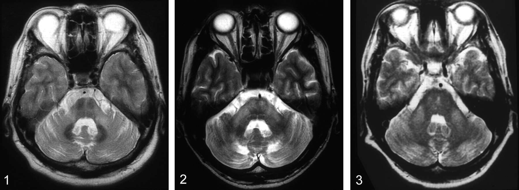
- Fig 1.
A 48-year-old woman with spinocerebellar ataxia (SCA6). T2-weighted fast spin-echo (FSE) MR image (3,600/102/2) shows bilateral symmetrical hyperintensity of atrophic MCPs. The pons with the “cross sign” and cerebellum are also atrophic. These MR findings are identical to that of sOPCA.
- Fig 2.
A 38-year-old man with ALD. T2-weighted FSE MR image (3,600/102/2) shows bilateral symmetrical hyperintensity of both MCPs, pyramidal tracts in the pons, and cerebellar white matter. The cerebellum is atrophic, and the fourth ventricle is slightly dilated.
- Fig 3.
A 34-year-old woman with Wilson disease. T2-weighted FSE MR image (3,600/102/2) shows symmetrical bilateral hyperintensity in both MCPs. There are several small hyperintensities in the pons. Mild atrophy of the cerebellum is seen.
- Fig 4.
A 60-year-old woman with hypoglycemic coma. A, T2-weighted FSE MR image (3,600/102/2) on admission shows subtle symmetrical hyperintensity in both MCPs. B, Isotropic diffusion-weighted MR image (b = 1,000 s/mm2) obtained at the same time shows markedly hyperintense signal intensity in both MCPs. In addition, small symmetrical hyperintensities along the pyramidal tracts are demonstrated (arrows). These hyperintensities disappeared completely on repeated MR imaging performed the next day (not shown).
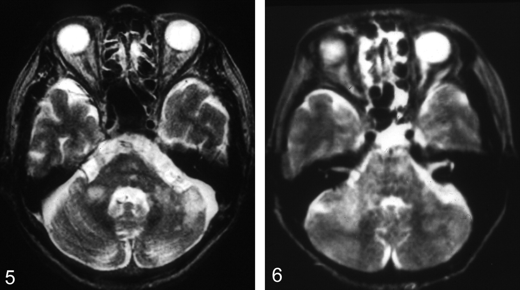
- Fig 5.
A 57-year-old man with bilateral MCP infarction. T2-weighted FSE MR image (3,600/102/2) shows bilateral hyperintensity in both MCPs. Also demonstrated are additional cerebellar infarction in the left AICA distribution and lacunar infarctions in the pons.
- Fig 6.
A 55-year-old woman with diffuse infiltrating B-cell malignant lymphoma. T2-weighted SE image (3,000/90/1) shows a heterogeneous hyperintensity extending from the pons into the cerebellar white matter through the MCPs. The hyperintensity is asymmetrical. Enlargement of the pons and right MCP is demonstrated.
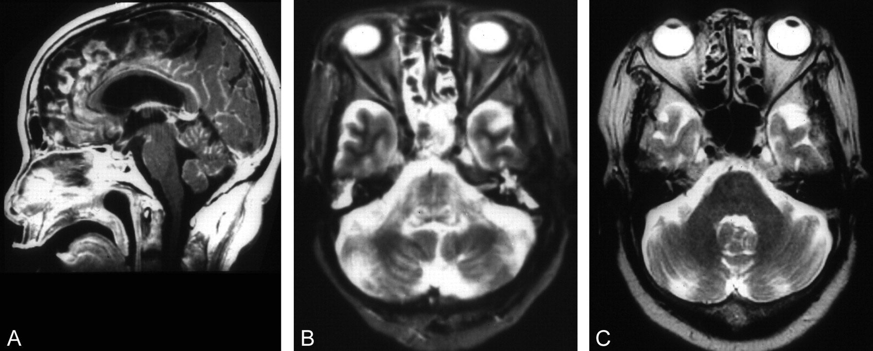
- Fig 7.
A 77-year-old woman with meningeal carcinomatosis. A, Postcontrast T1-weighted SE midsagittal image (600/15/1) shows enhancement in the cerebellar fissures and cortical sulci as well as the anterior margin of the midbrain and upper pons. B, T2-weighted FSE MR image (3,600/102/2) shows symmetrical hyperintensities of MCPs, upper cerebellar peduncles, lateral and dorsal pons. C, T2-weighted FSE MR image (3,600/102/2) obtained 5 months earlier showed no hyperintensities at the same level of that shown in B.


Tables
Diseases involving bilateral middle cerebellar peduncles
Diagnosis (no.) Age/sex Pontine Lesions Cbll Lesions Other Lesions Degenerative diseases (11) sOPCA (8) 47, 50, 53, 55/M,61, 68, 71, 72/F Cross sign, atrophy Atrophy Atrophy of basal ganglia with linear hyperintensity at lateral margins (3) SCA2 (1) 42/M Cross sign, atrophy Atrophy (-) SCA6 (1) 48/F Cross sign, atrophy Atrophy (-) Shy-Drager synd (1) 57/M Linear lesion on midline (-) (-) Metabolic diseases (6) Adrenoleukodystrophy (2) 4, 38/M Lesions along pyramidal tracts (1), heterogeneous lesions (1), WML (2) Large symmetrical parieto-occipial WML (1), lesions along pyramidal tracts (1) Wilson disease (2) 24/M, 34/F Several small lesions (2), atrophy (1) Mild atrophy (1) Midbrain (tegmentum) and basal ggl lesions (2) and thalamic lesions (1) Alcoholic liver cirrhosis (1) 51/M (-) Mild atrophy T1 hyperintensity at globi pallidi Hypoglycemic coma (1) 52/F Lesions along pyramidal tracts on DWI (-) Hyperintensity lesions along pyramidal tracts on T2WI and DWI Neoplasms (3) Diffuse infiltrating ML (1) 55/F Heterogeneous lesions, enlargement Diffuse WML Whole corpus callosum and diffuse cbr WML Brain stem glioma (1) 74/M Ring-enhancement, enlargement (-) Tumor extension into medulla oblongata and midbrain Meningeal carcinomatosis (1) 77/F Multiple lacunar infarcts Enhancement in fissures Leptomeningeal enhancement, multiple lacunar infarction, mild hydrocephalus Cerebrovascular disease, hypertensive and its related encephalopathies (3) AICA infarction (1) 57/M Lacunar infarcts Infarction in AICA distribution Multiple lacunar infarction Hypertensive encephalopathy (1) 51/F Heterogeneous lesion, enlargement Diffuse WML, swelling Midbrain, basal ganglia, thalamus, bilateral hippocampus, periventricular WML Cyclosporin-A encephalopathy (1) 16/F A few punctate lesions Diffuse swelling Small parietal WML Inflammatory & demyelinating diseases (4) Multiple sclerosis (1) 17/F Small tegmental lesions Mild atrophy Small lesions in corpus callosum, small oval periventricular WML ADEM (1) 38/M Heterogeneous lesions (-) Multiple subcortical WML Behcet disease (1) 66/F Subtle heterogeneous lesions (-) Non-specific punctate WML HIV encephalopathy (1) 31/M Diffuse hyperintensity (-) Frontal WML, mild brain atropohy Total (27) mean age, 48.5 y Note.—ADEM, acute disseminated encephalomyelitis; cbll, cerebellar; cbr, cerebral; DWI, diffusion-weighted imaging; ggl, ganglia; HIV, human immunodeficiency virus; ML, malignant lymphoma; SCA, spinocerebellar ataxia; sOPCA, sporadic olivopontocerebellar atrophy; T2WI, T2-weighted imaging; WML, white matter lesions.
In this issue


Jump to section
Related Articles
Cited By...
- The Inferior Cerebellar Peduncle Sign: A Novel Imaging Marker for Differentiating Multiple System Atrophy Cerebellar Type from Spinocerebellar Ataxia
- Suspecting unwitnessed hypoglycaemia
- Imaging Patterns Characterizing Mitochondrial Leukodystrophies
- MR Imaging Features of the Cerebellum in Adult-Onset Neuronal Intranuclear Inclusion Disease: 8 Cases
- Reverse 'hot cross bun', 'Mercedes-Benz', 'face of the giant panda and her cub' signs with pontine infarcts: a radiological pandora
- Neuroimaging of Rapidly Progressive Dementias, Part 1: Neurodegenerative Etiologies
- Neuroimaging and clinical features in type II (late-onset) Alexander disease
- Unusual MRI findings in a case of Marchiafava Bignami disease
- Pearls & Oy-sters: A distinctive watershed area in the vertebrobasilar territory
- Tuberculosis Involving Bilateral Middle Cerebellar Peduncles
- Serial diffusion and perfusion-weighted MR in transient hypoglycemia