
Article Figures & Data
Figures
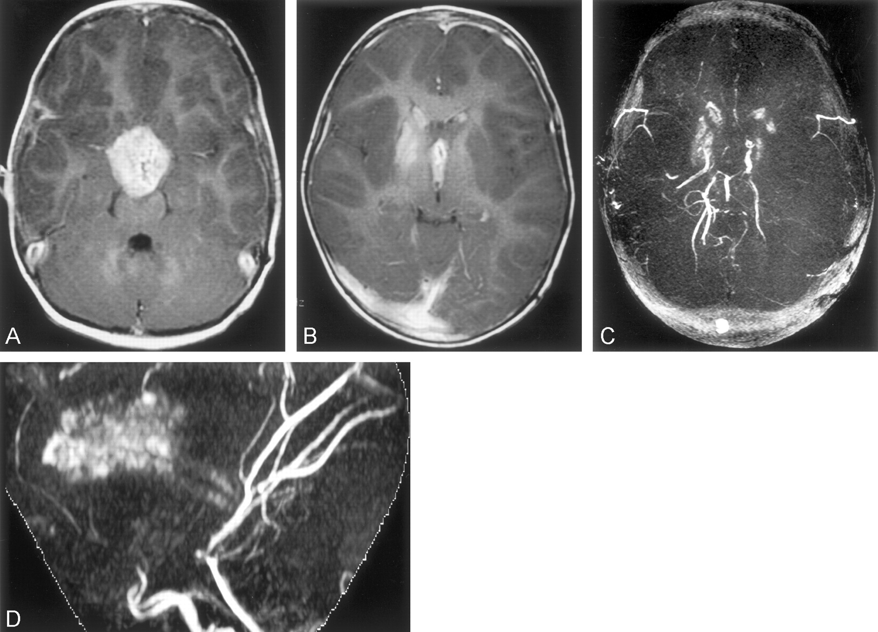
- Fig 1.
Case 1. This 2.5-year-old female patient was unresponsive.
A and B, Axial T1-weighted contrast-enhanced images (350/12/2 [TR/TE/NEX]) show a necrotic enhancing suprasellar mass encasing the distal internal carotid arteries.
C, Composite image.
D, Maximum intensity projection image from 3D time-of-flight MR angiogram, No blood flow is visualized beyond the supraclinoid portions of internal carotid arteries.
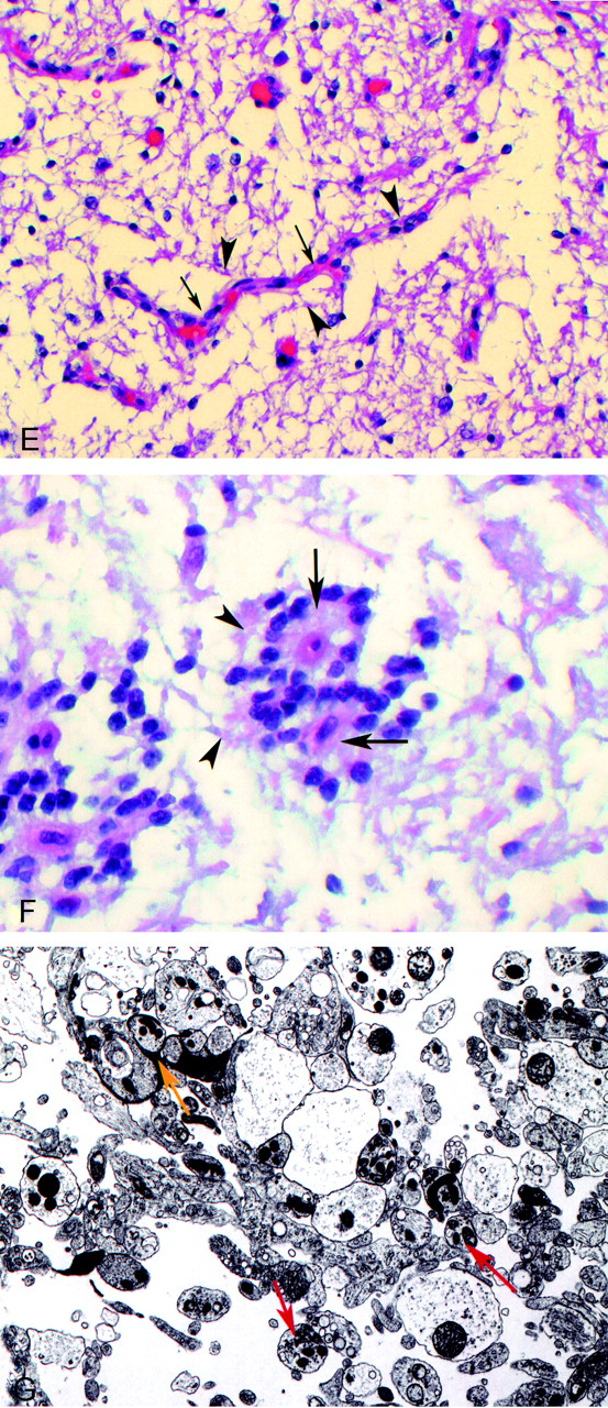
- Fig 2.
Case 2. Two-year-old male patient with diplopia and a decrease in left eye vision.
A, Axial T2-weighted image (3200/84/1) shows a hyperintense suprasellar mass encasing the anterior portion of the circle of Willis.
B, Axial T1-weighted image (433/16/2) shows a hypointense suprasellar mass encasing the circle of Willis.
C, Contrast-enhanced sagittal T1-weighted image (650/14/2) shows a solid-cystic enhancing hypothalamic-suprasellar mass.
D, Contrast-enhanced coronal view T1-weighted image (400/16/2).
E, Photomicrograph of hematoxylin and eosin-stained normal tanycytes (arrowheads) radiating to a blood vessel (arrows) in area postrema.
F, Photomicrograph of hematoxylin and eosin-stained neoplastic cells (arrowheads) radiating to blood vessel (arrows) in tanycytoma specimen.
G, Photomicrograph of electron microscopy of tumor specimen reveals tanycytes with long synaptic processes (red arrow) and synaptoid complexes (yellow arrow).
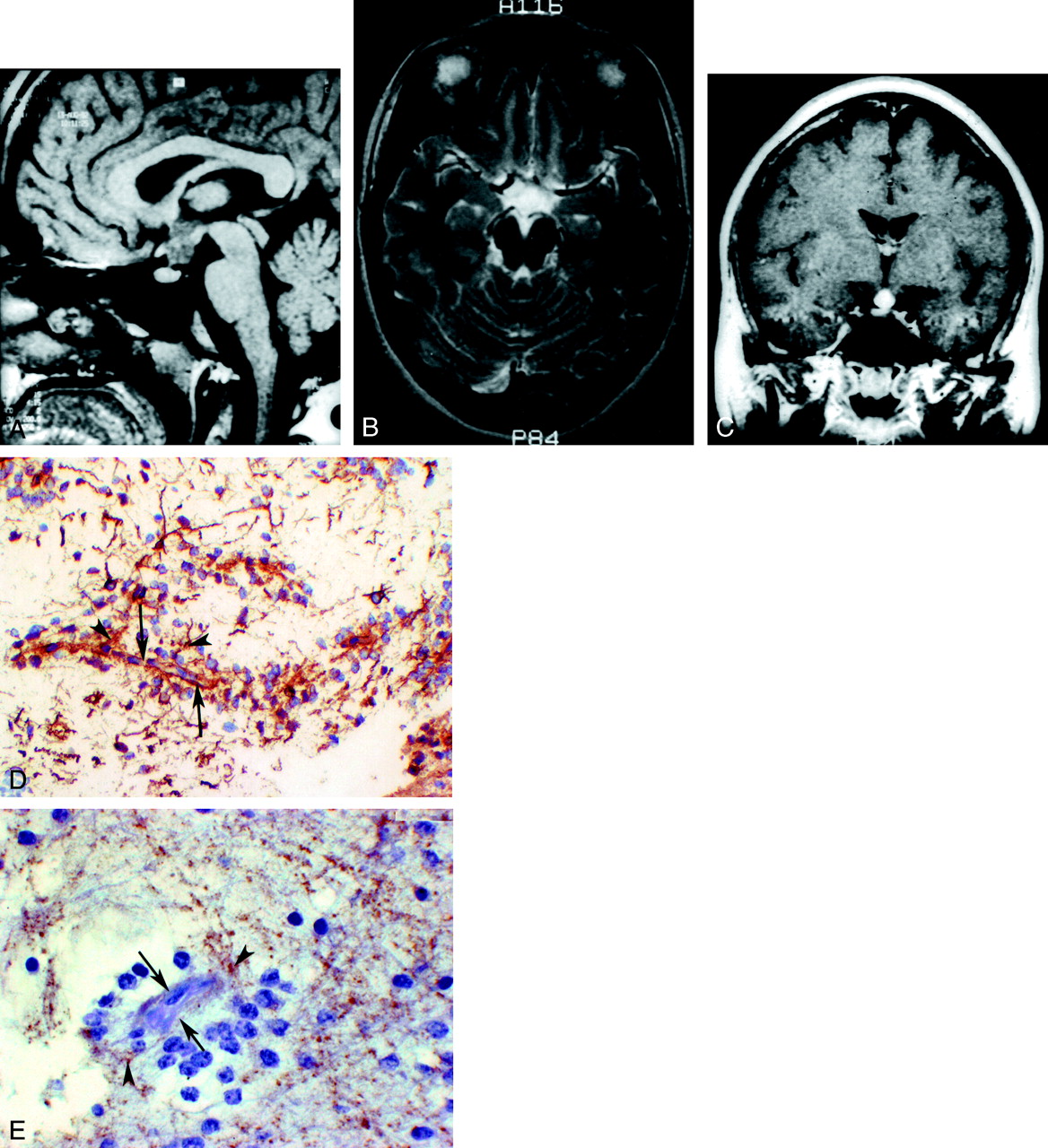
- Fig 3.
Case 4. This 26-year-old female patient presented with galactorrhea and hypothyroidism.
A, Sagittal view T1-weighted image (433/8/2) shows a hypointense hypothalamic mass.
B, Axial view T2-weighted image (3200/88/2) shows a hyperintense hypothalamic mass with suprasellar extension.
C, Coronal view T1-weighted contrast-enhanced image (400/9/2) shows a homogeneously enhancing hypothalamic-suprasellar mass.
D, Photomicrograph of tumor specimen stained with glial fibrillary acidic protein shows positive brown-stained glial processes (arrowheads) radiating to blood vessel (arrows).
E, Photomicrograph of tumor specimen stained with synaptophysin shows positive brown-stained glial processes (arrowheads) radiating to blood vessel (arrows).


Tables
Patient No. Age (y)/Sex Symptoms Location Imaging 1 2/F Headache, nausea, emesis Hypothalamus-suprasellar mass Large, partially cystic intensely enhancing mass; encases circle of Willis 2 2/M Diplopia, decrease in left eye vision Hypothalamus-suprasellar mass Large, intensely enhancing mass; encases circle of Willis; (Fig 2A–D) 3 3/M Headache, emesis Hypothalamus-suprasellar mass Large, partially cystic intensely enhancing mass; encases circle of Willis 4 26/F Galactorrhea, hypothyroidism Hypothalamus Homogeneous enhancing mass (Fig 3A–C) 5 56/F Headache Left lateral ventricle Minimally enhancing, well-defined mass Note.—F indicates female; M, male.
Patient No. Surgery Therapy Pathology Follow-up 1 Partial resection Radiation: 5012 cGy Tanycytes; occlusion of bilateral internal carotid arteries (Fig. 1A–D) Deceased <1 yr from initial diagnosis; bilateral, massive strokes chemo: carboplatin vincristine 2 Partial resection Radiation: 5040 cGy Tanycytes; (Fig. 2E–G) Decrease residual tumor chemo: carboplatin vincristine 3 Partial resection Radiation: 5400 cGy Tanycytes; encased circle of Willis Deceased <1 yr from initial diagnosis; cardiac arrest chemo: carboplatin vincristine 4 Complete resection Radiation: 5400 cGy Tanycytes; + GFAP stain, + synaptophysin stain (Fig. 3D and E) Lost to follow-up 5 Complete resection None Tanycytes No recurrence at 4 months Note.—Chemo indicates chemotherapy; GFAP, glial fibrillary acidic protein.
In this issue


Jump to section
Related Articles
Cited By...
- No citing articles found.