Advertisement

Main menu

  • Home
  • Content
    • Current Issue
    • Accepted Manuscripts
    • Article Preview
    • Past Issue Archive
    • AJNR Case Collection
    • Case of the Week Archive
    • Classic Case Archive
    • Case of the Month Archive
  • Special Collections
    • Spinal CSF Leak Articles (Jan 2020-June 2024)
    • 2024 AJNR Journal Awards
    • Most Impactful AJNR Articles
  • Multimedia
    • AJNR Podcast
    • AJNR Scantastics
    • Video Articles
  • For Authors
    • Submit a Manuscript
    • Author Policies
    • Fast publishing of Accepted Manuscripts
    • Graphical Abstract Preparation
    • Manuscript Submission Guidelines
    • Imaging Protocol Submission
    • Submit a Case for the Case Collection
  • About Us
    • About AJNR
    • Editorial Board
  • More
    • Become a Reviewer/Academy of Reviewers
    • Subscribers
    • Permissions
    • Alerts
    • Feedback
    • Advertisers
    • ASNR Home
  • Other Publications
    • ajnr

User menu

  • Alerts
  • Log in

Search

  • Advanced search
American Journal of Neuroradiology

American Journal of Neuroradiology

ASHNR American Society of Functional Neuroradiology ASHNR American Society of Pediatric Neuroradiology ASSR
  • Alerts
  • Log in

Advanced Search

  • Home
  • Content
    • Current Issue
    • Accepted Manuscripts
    • Article Preview
    • Past Issue Archive
    • AJNR Case Collection
    • Case of the Week Archive
    • Classic Case Archive
    • Case of the Month Archive
  • Special Collections
    • Spinal CSF Leak Articles (Jan 2020-June 2024)
    • 2024 AJNR Journal Awards
    • Most Impactful AJNR Articles
  • Multimedia
    • AJNR Podcast
    • AJNR Scantastics
    • Video Articles
  • For Authors
    • Submit a Manuscript
    • Author Policies
    • Fast publishing of Accepted Manuscripts
    • Graphical Abstract Preparation
    • Manuscript Submission Guidelines
    • Imaging Protocol Submission
    • Submit a Case for the Case Collection
  • About Us
    • About AJNR
    • Editorial Board
  • More
    • Become a Reviewer/Academy of Reviewers
    • Subscribers
    • Permissions
    • Alerts
    • Feedback
    • Advertisers
    • ASNR Home
  • Follow AJNR on Twitter
  • Visit AJNR on Facebook
  • Follow AJNR on Instagram
  • Join AJNR on LinkedIn
  • RSS Feeds

Welcome to the new AJNR, Updated Hall of Fame, and more. Read the full announcements.


AJNR is seeking candidates for the position of Associate Section Editor, AJNR Case Collection. Read the full announcement.


Getting new auth cookie, if you see this message a lot, tell someone!
Research ArticleSPINE

Refractory Occipital Neuralgia: Preoperative Assessment with CT-Guided Nerve Block Prior to Dorsal Cervical Rhizotomy

Vibhu Kapoor, William E. Rothfus, Stephen Z. Grahovac, Stephen Z. Amin Kassam and Michael B. Horowitz
American Journal of Neuroradiology November 2003, 24 (10) 2105-2110;
Vibhu Kapoor
  • Find this author on Google Scholar
  • Find this author on PubMed
  • Search for this author on this site
William E. Rothfus
  • Find this author on Google Scholar
  • Find this author on PubMed
  • Search for this author on this site
Stephen Z. Grahovac
  • Find this author on Google Scholar
  • Find this author on PubMed
  • Search for this author on this site
Stephen Z. Amin Kassam
  • Find this author on Google Scholar
  • Find this author on PubMed
  • Search for this author on this site
Michael B. Horowitz
  • Find this author on Google Scholar
  • Find this author on PubMed
  • Search for this author on this site
  • Article
  • Figures & Data
  • Info & Metrics
  • Responses
  • References
  • PDF
Loading

Article Figures & Data

Figures

  • Fig 1.
    • Download figure
    • Open in new tab
    • Download powerpoint
    Fig 1.

    Left C2 nerve root block. Axial CT at level of C2 vertebral body demonstrates final location of tip of 25-gauge spinal needle (arrow) posterior to lateral mass (open arrow) of C2 vertebra.

  • Fig 2.
    • Download figure
    • Open in new tab
    • Download powerpoint
    Fig 2.

    Three-dimensional illustration of left C2 nerve root block, posterosuperior view. Posterior arch of C1 has been removed for clarity. C2 nerve is depicted in yellow, vertebral artery in red and injected medication in green.
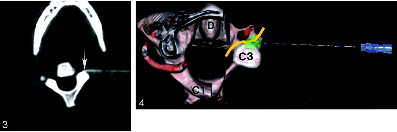
  • Fig 3.
    • Download figure
    • Open in new tab
    • Download powerpoint
    Fig 3.

    Left C3 nerve root block. Axial CT at level of C2–C3 intervertebral foramen demonstrates final location of tip of 25-gauge spinal needle (arrow) anterior to superior articular facet of C3.

  • Fig 4.
    • Download figure
    • Open in new tab
    • Download powerpoint
    Fig 4.

    Three-dimensional illustration of left C2 nerve root block viewed from above. Posterior arches of C1 and C2 have been removed for clarity. C3 nerve is depicted in yellow and injected medication in green. Note vertebral artery (red) just anterior to the nerve. D, dens.
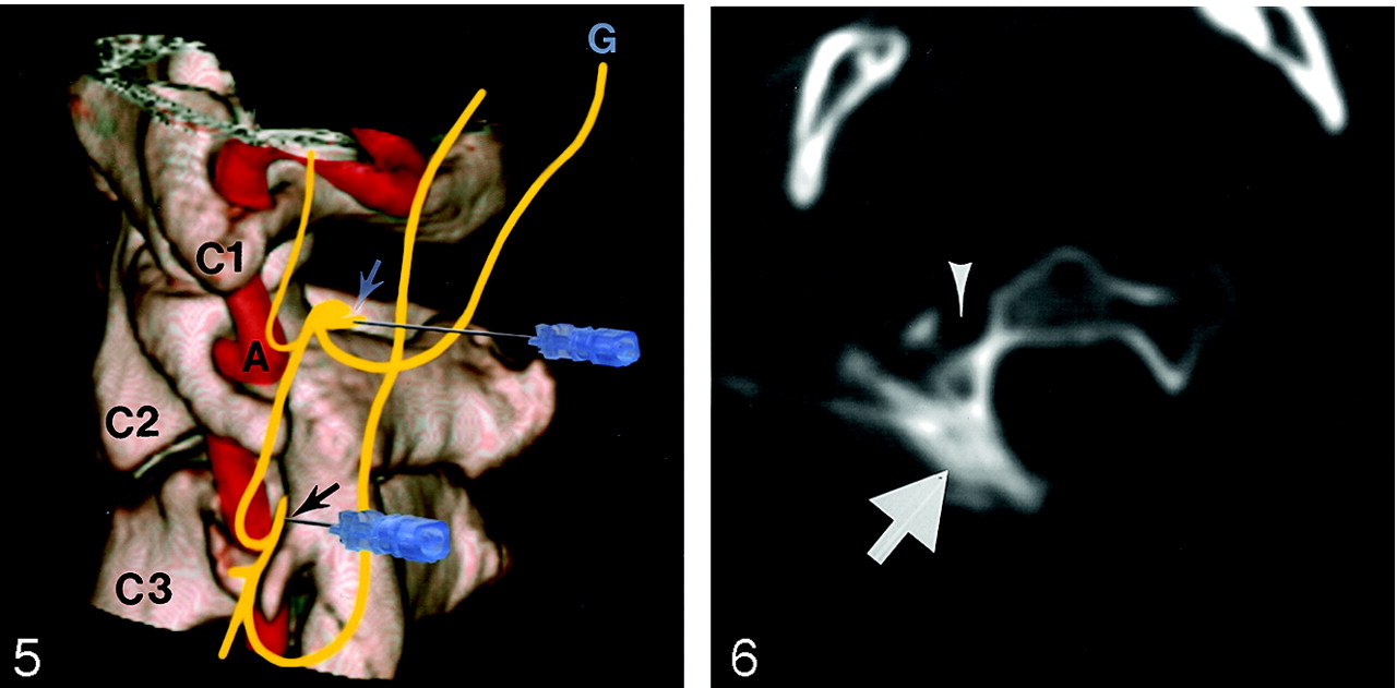
  • Fig 5.
    • Download figure
    • Open in new tab
    • Download powerpoint
    Fig 5.

    Composite 3D illustration of left C2 (white arrow) and C3 (black arrow) nerve root block viewed from left. Vertebral artery (A) runs close to C3 nerve. Note connections between C1, C2, and C3 nerves. G, greater occipital nerve.

  • Fig 6.
    • Download figure
    • Open in new tab
    • Download powerpoint
    Fig 6.

    Right C2 nerve root block. Iodinated contrast medium (arrow) is injected before injection of bupivacaine to confirm accurate placement of needle tip. Arrowhead marks foramina transversaria that transmits vertebral artery and veins.

  • Fig 7.
    • Download figure
    • Open in new tab
    • Download powerpoint
    Fig 7.

    Illustration of occipital region and upper cervical vertebrae viewed from behind showing course of greater occipital nerve (G) with possible sites of compression: F, due to atlantoaxial joint disease; M, as it penetrates tendinous portion of the trapezius muscles (Z); T, as occipital nerve pierces posterior atlantoaxial membrane (depicted in light semitransparent blue); and between posterior arches of C1 and C2 vertebrae. Note connections between greater occipital nerve (G) and C1 (i) and C3 (ii) nerves. O, occipital bone.

Back to top

In this issue

American Journal of Neuroradiology: 24 (10)
American Journal of Neuroradiology
Vol. 24, Issue 10
1 Nov 2003
  • Table of Contents
  • Index by author
Advertisement
Print
Download PDF
Email Article

Thank you for your interest in spreading the word on American Journal of Neuroradiology.

NOTE: We only request your email address so that the person you are recommending the page to knows that you wanted them to see it, and that it is not junk mail. We do not capture any email address.

Enter multiple addresses on separate lines or separate them with commas.
Refractory Occipital Neuralgia: Preoperative Assessment with CT-Guided Nerve Block Prior to Dorsal Cervical Rhizotomy
(Your Name) has sent you a message from American Journal of Neuroradiology
(Your Name) thought you would like to see the American Journal of Neuroradiology web site.
CAPTCHA
This question is for testing whether or not you are a human visitor and to prevent automated spam submissions.
Cite this article
Vibhu Kapoor, William E. Rothfus, Stephen Z. Grahovac, Stephen Z. Amin Kassam, Michael B. Horowitz
Refractory Occipital Neuralgia: Preoperative Assessment with CT-Guided Nerve Block Prior to Dorsal Cervical Rhizotomy
American Journal of Neuroradiology Nov 2003, 24 (10) 2105-2110;

Citation Manager Formats

  • BibTeX
  • Bookends
  • EasyBib
  • EndNote (tagged)
  • EndNote 8 (xml)
  • Medlars
  • Mendeley
  • Papers
  • RefWorks Tagged
  • Ref Manager
  • RIS
  • Zotero
0 Responses
Respond to this article
Share
Bookmark this article
Refractory Occipital Neuralgia: Preoperative Assessment with CT-Guided Nerve Block Prior to Dorsal Cervical Rhizotomy
Vibhu Kapoor, William E. Rothfus, Stephen Z. Grahovac, Stephen Z. Amin Kassam, Michael B. Horowitz
American Journal of Neuroradiology Nov 2003, 24 (10) 2105-2110;
del.icio.us logo Twitter logo Facebook logo Mendeley logo
  • Tweet Widget
  • Facebook Like
  • Google Plus One

Jump to section

  • Article
    • Abstract
    • Methods
    • Results
    • Discussion
    • Conclusion
    • Acknowledgments
    • Footnotes
    • References
  • Figures & Data
  • Info & Metrics
  • Responses
  • References
  • PDF

Related Articles

  • No related articles found.
  • PubMed
  • Google Scholar

Cited By...

  • Sonographic Evaluation of the Greater Occipital Nerve in Unilateral Occipital Neuralgia
  • Crossref
  • Google Scholar

This article has not yet been cited by articles in journals that are participating in Crossref Cited-by Linking.

More in this TOC Section

  • Bern Score Validity for SIH
  • MP2RAGE 7T in MS Lesions of the Cervical Spine
  • Resisted Inspiration for CSF-Venous Fistula
Show more Spine

Similar Articles

Advertisement

Indexed Content

  • Current Issue
  • Accepted Manuscripts
  • Article Preview
  • Past Issues
  • Editorials
  • Editors Choice
  • Fellow Journal Club
  • Letters to the Editor

Cases

  • Case Collection
  • Archive - Case of the Week
  • Archive - Case of the Month
  • Archive - Classic Case

Special Collections

  • Special Collections

Resources

  • News and Updates
  • Turn around Times
  • Submit a Manuscript
  • Author Policies
  • Manuscript Submission Guidelines
  • Evidence-Based Medicine Level Guide
  • Publishing Checklists
  • Graphical Abstract Preparation
  • Imaging Protocol Submission
  • Submit a Case
  • Become a Reviewer/Academy of Reviewers
  • Get Peer Review Credit from Publons

Multimedia

  • AJNR Podcast
  • AJNR SCANtastic
  • Video Articles

About Us

  • About AJNR
  • Editorial Board
  • Not an AJNR Subscriber? Join Now
  • Alerts
  • Feedback
  • Advertise with us
  • Librarian Resources
  • Permissions
  • Terms and Conditions

American Society of Neuroradiology

  • Not an ASNR Member? Join Now

© 2025 by the American Society of Neuroradiology All rights, including for text and data mining, AI training, and similar technologies, are reserved.
Print ISSN: 0195-6108 Online ISSN: 1936-959X

Powered by HighWire