Advertisement

Main menu

  • Home
  • Content
    • Current Issue
    • Accepted Manuscripts
    • Article Preview
    • Past Issue Archive
    • AJNR Case Collection
    • Case of the Week Archive
    • Classic Case Archive
    • Case of the Month Archive
  • Special Collections
    • Spinal CSF Leak Articles (Jan 2020-June 2024)
    • 2024 AJNR Journal Awards
    • Most Impactful AJNR Articles
  • Multimedia
    • AJNR Podcast
    • AJNR Scantastics
    • Video Articles
  • For Authors
    • Submit a Manuscript
    • Author Policies
    • Fast publishing of Accepted Manuscripts
    • Graphical Abstract Preparation
    • Manuscript Submission Guidelines
    • Imaging Protocol Submission
    • Submit a Case for the Case Collection
  • About Us
    • About AJNR
    • Editorial Board
  • More
    • Become a Reviewer/Academy of Reviewers
    • Subscribers
    • Permissions
    • Alerts
    • Feedback
    • Advertisers
    • ASNR Home
  • Other Publications
    • ajnr

User menu

  • Alerts
  • Log in

Search

  • Advanced search
American Journal of Neuroradiology

American Journal of Neuroradiology

ASHNR American Society of Functional Neuroradiology ASHNR American Society of Pediatric Neuroradiology ASSR
  • Alerts
  • Log in

Advanced Search

  • Home
  • Content
    • Current Issue
    • Accepted Manuscripts
    • Article Preview
    • Past Issue Archive
    • AJNR Case Collection
    • Case of the Week Archive
    • Classic Case Archive
    • Case of the Month Archive
  • Special Collections
    • Spinal CSF Leak Articles (Jan 2020-June 2024)
    • 2024 AJNR Journal Awards
    • Most Impactful AJNR Articles
  • Multimedia
    • AJNR Podcast
    • AJNR Scantastics
    • Video Articles
  • For Authors
    • Submit a Manuscript
    • Author Policies
    • Fast publishing of Accepted Manuscripts
    • Graphical Abstract Preparation
    • Manuscript Submission Guidelines
    • Imaging Protocol Submission
    • Submit a Case for the Case Collection
  • About Us
    • About AJNR
    • Editorial Board
  • More
    • Become a Reviewer/Academy of Reviewers
    • Subscribers
    • Permissions
    • Alerts
    • Feedback
    • Advertisers
    • ASNR Home
  • Follow AJNR on Twitter
  • Visit AJNR on Facebook
  • Follow AJNR on Instagram
  • Join AJNR on LinkedIn
  • RSS Feeds

Welcome to the new AJNR, Updated Hall of Fame, and more. Read the full announcements.


AJNR is seeking candidates for the position of Associate Section Editor, AJNR Case Collection. Read the full announcement.


OtherSPINE

Dumbbell-Shaped Epidural Capillary Hemangioma

Bruno Badinand, Christophe Morel, Nicolas Kopp, Van A. Tran Min and François Cotton
American Journal of Neuroradiology February 2003, 24 (2) 190-192;
Bruno Badinand
  • Find this author on Google Scholar
  • Find this author on PubMed
  • Search for this author on this site
Christophe Morel
  • Find this author on Google Scholar
  • Find this author on PubMed
  • Search for this author on this site
Nicolas Kopp
  • Find this author on Google Scholar
  • Find this author on PubMed
  • Search for this author on this site
Van A. Tran Min
  • Find this author on Google Scholar
  • Find this author on PubMed
  • Search for this author on this site
François Cotton
  • Find this author on Google Scholar
  • Find this author on PubMed
  • Search for this author on this site
  • Article
  • Figures & Data
  • Info & Metrics
  • Responses
  • References
  • PDF
Loading

Abstract

Summary: We report a case of a purely epidural capillary hemangioma of the thoracic spine with foraminal extension. Epidural hemangiomas are rare; only a few cases of dumbbell-shaped ones have been reported, and all were cavernous. MR imaging showed characteristic findings of a capillary hemangioma, which are also consistent with other epidural lesions such as neuromas or meningiomas.

Hemangiomas of the spine are usually lesions of the vertebral bodies, and purely epidural hemangiomas are rare. Few cases of epidural cavernous hemangiomas have been reported, and descriptions of capillary hemangiomas at this location are extremely rare in the literature. We report a case of a purely epidural capillary hemangioma with foraminal extension. The MR imaging findings are presented.

Case Report

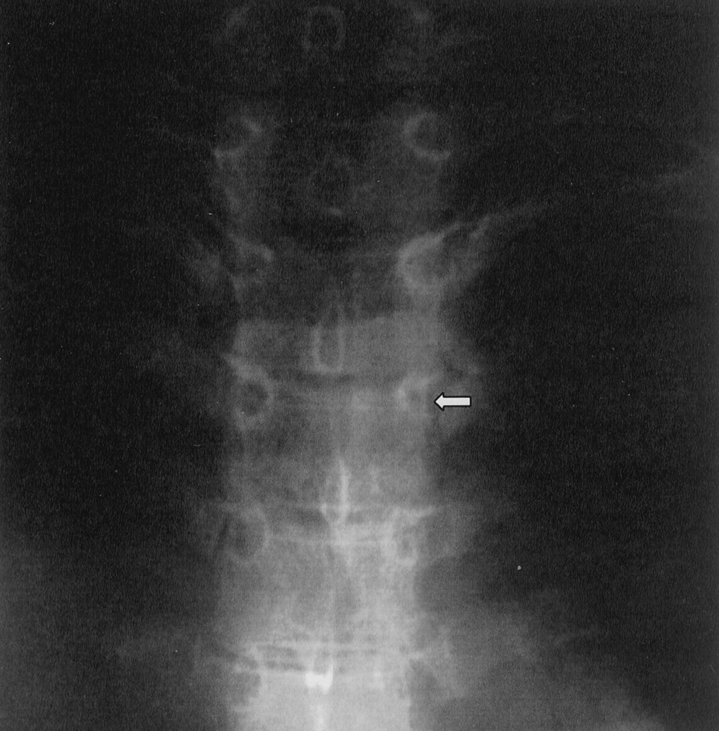
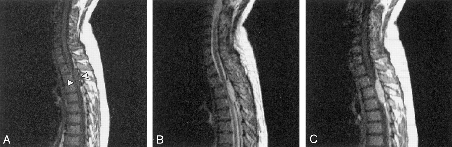
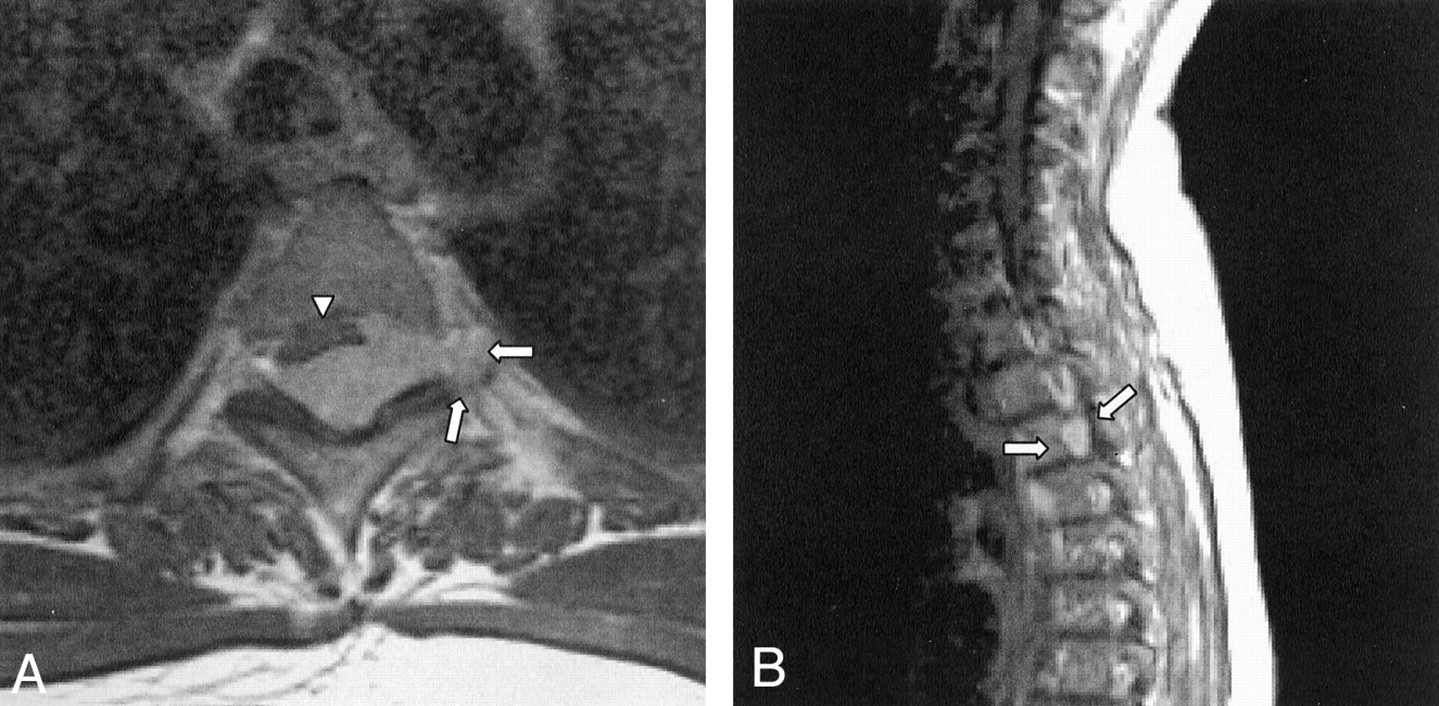
A 40-year-old woman presented with a 1-year history of progressive pain and muscular contracture of her lower limbs. The patient also had a recent onset of motor deficit in the lower limbs, which caused difficulties with walking and sphincteric problems. Her medical history was unremarkable except for a duodenal ulcer, which was not treated, and endometriosis. On physical examination, the patient’s motor strength in her lower limbs was decreased. This finding was associated with hypesthesia and a brisk osteotendinous reflex. Imaging of the dorsal spine showed thinning of the left pedicle at T4 on radiographs (Fig 1) and widening of the left foramen at T3-T4 on CT scans. On MR images, the lesion was a well-circumscribed and probably epidural mass that extended from T2 to T4, with extension to the left foramen at T3-T4, as shown on coronal images. The lesion was 15 mm thick, 30 mm wide (including foraminal extension), and 40 mm long. On T1-weighted (600/15) sagittal MR images, the mass had homogeneous intermediate signal intensity similar to that of the spinal cord (Fig 2A), and on T2-weighted (3000/120) sagittal images, it had homogeneous high signal intensity (Fig 2B). The mass was surrounded by a rim of low signal intensity on both images. The T1-weighted sagittal and coronal images obtained after the intravenous administration of gadolinium-based contrast agent showed mostly intense homogeneous enhancement of the mass (Fig 2C), as well as clear extension to the left foramen at T3-T4 (Fig 3). The preoperative differential diagnosis included neuroma and meningioma. T2–T4 laminectomy revealed a red-purple hemorrhagic epidural mass that was relatively well encapsulated. The mass was easily cleaved, and excision was easy except for the foraminal extension, which was not removed. Pathologic examination showed a vascular tumor composed of thin and irregular capillary vessels caught in low-attenuating fibrosis. This finding was consistent with a capillary hemangioma (Fig 4). Follow-up MR imaging performed 8 months later showed at last no signs of residual tumor.

Fig 1.
  • Download figure
  • Open in new tab
  • Download powerpoint
Fig 1.

Anteroposterior radiograph of the thoracic spine shows a thinning of the left pedicle at the T3-T4 level (arrow)

Fig 2.
  • Download figure
  • Open in new tab
  • Download powerpoint
Fig 2.

Sagittal MR images.

A, T1-weighted image (600/15) shows a mass, probably epidural, with homogeneous isointensity relative to the spinal cord. The mass is surrounded by a rim of low signal intensity (arrowheads) located at the T2–T4 level.

B, T2-weighted image (3000/120) shows the homogeneously hyperintense mass surrounded by a rim of low signal intensity.

C, Gadolinium-enhanced T1-weighted image shows mostly intense, homogeneous enhancement of the lesion.

Fig 3.
  • Download figure
  • Open in new tab
  • Download powerpoint
Fig 3.

Gadolinium-enhanced T1-weighted MR images clearly show extension to the left foramen at T3-T4 (arrows).

A, Axial image. The epidural mass compresses the anterior aspect of the thoracic spine (arrowhead).

B, Sagittal image.

Fig 4.
  • Download figure
  • Open in new tab
  • Download powerpoint
Fig 4.

Photomicrograph obtained at histologic examination shows a tumor composed of thin and irregular capillary vessels (arrowheads) caught in low-attenuating fibrosis (arrows) (hematoxylin and eosin, original magnification ×100).

Discussion

Hemangiomas are congenital vascular malformations that pathologists frequently consider to be hamartomatous malformations (1). They are classified by the predominant type of vascular channel (capillary, cavernous, arteriovenous or venous) observed at histologic examination. Nonvascular components may also be observed, particularly in cavernous hemangiomas. These components include fat, smooth muscle, fibrous tissue, and hemosiderin (2).

Usual locations are in the soft tissue (3), in cutaneous or subcutaneous tissues, or in osseous tissue, especially that in spinal column. The lesions have been detected in 11% of spines examined at autopsy (4). Purely vertebral hemangiomas are common, and epidural hemangiomas are actually considered to be an extension of vertebral lesions in the epidural space (1, 5). They are generally isolated. Purely epidural hemangiomas are rare.

Few groups have reported cases of purely epidural lesions (1, 5, 6). Some of these lesions were dumbbell-shaped and located in the thoracic spine (5), but all were a cavernous type of hemangioma. To our knowledge, only one publication has reported a case of a purely epidural capillary hemangioma of the spine (7). We did not find any other examples of foraminal extension of this specific type in the literature.

The MR imaging findings in this case are similar to those found in previously reported cases of extraosseous capillary hemangioma (3, 4, 7). They are closely connected at pathologic examination, which shows capillary vessels caught in low-attenuating fibrosis without fat or hemosiderin components. This finding explains the homogeneous aspect and the isointense signal relative to the spinal cord on T1-weighted images, as some authors have described (2). The rim of low signal intensity seen on T1- and T2-weighted MR images may correspond with the encapsulated aspect observed during surgery, and it could suggest the benignity of the tumor. On T1- and T2-weigthed sagittal images, the low signal intensity at the anterior part of the lesion may also correspond to the posterior longitudinal ligament. Therefore, it may be an important landmark for the epidural location of the lesion.

Nevertheless, these radiologic aspects are nonspecific. Well-circumscribed tumors and the dumbbell shape on MR images are also commonly found with other benign tumors such as neuroma or meningioma. This case reminds us that hemangioma should be considered in the differential diagnosis of epidural lesions.

References

  1. ↵
    Feider HK, Yuille DL. An epidural cavernous hemangioma of the spine. AJNR Am J Neuroradiol 1991;12:243–244
    FREE Full Text
  2. ↵
    Archives of the AFIP. Musculoskeletal angiomatous lesions: radiologic-pathologic correlation of hemangioma. RadioGraphics 1995;15:893–917
    PubMed
  3. ↵
    Shin JH, Lee HK, Jeon SR, Park SH. Spinal intradural capillary hemangioma: MR findings. AJNR Am J Neuroradiol 2000;21:954–956
    Abstract/FREE Full Text
  4. ↵
    Lee RR, Melhem E. Neoplasms of the spine. In: Orrison WW Jr, eds. Neuroimaging. 2 ed. Philadelphia: Saunders;2000;1334–1336
  5. ↵
    Haimes AB, Krol G. Dumbbell-shaped spinal cavernous hemangioma: a case report. AJNR Am J Neuroradiol 1991;12:1021–1022
    FREE Full Text
  6. ↵
    Padolecchia R, Acerbi G, Puglioli M, Collavoli PL, Ravelli V, Caciagli P. Epidural spinal cavernous hemangioma. Spine 1998 May 15;23:1136–1140
    CrossRefPubMed
  7. ↵
    Gupta S, Kumar S, Banerji D, Pandey R, Gujral R. Magnetic resonance imaging features of an epidural spinal hemangioma. Australas Radiol 1996 Aug;40:342–344
    PubMed
  • Received May 30, 2002.
  • Accepted after revision July 12, 2002.
  • Copyright © American Society of Neuroradiology
View Abstract
Back to top

In this issue

American Journal of Neuroradiology: 24 (2)
American Journal of Neuroradiology
Vol. 24, Issue 2
1 Feb 2003
  • Table of Contents
  • Index by author
Advertisement
Print
Download PDF
Email Article

Thank you for your interest in spreading the word on American Journal of Neuroradiology.

NOTE: We only request your email address so that the person you are recommending the page to knows that you wanted them to see it, and that it is not junk mail. We do not capture any email address.

Enter multiple addresses on separate lines or separate them with commas.
Dumbbell-Shaped Epidural Capillary Hemangioma
(Your Name) has sent you a message from American Journal of Neuroradiology
(Your Name) thought you would like to see the American Journal of Neuroradiology web site.
CAPTCHA
This question is for testing whether or not you are a human visitor and to prevent automated spam submissions.
Cite this article
Bruno Badinand, Christophe Morel, Nicolas Kopp, Van A. Tran Min, François Cotton
Dumbbell-Shaped Epidural Capillary Hemangioma
American Journal of Neuroradiology Feb 2003, 24 (2) 190-192;

Citation Manager Formats

  • BibTeX
  • Bookends
  • EasyBib
  • EndNote (tagged)
  • EndNote 8 (xml)
  • Medlars
  • Mendeley
  • Papers
  • RefWorks Tagged
  • Ref Manager
  • RIS
  • Zotero
0 Responses
Respond to this article
Share
Bookmark this article
Dumbbell-Shaped Epidural Capillary Hemangioma
Bruno Badinand, Christophe Morel, Nicolas Kopp, Van A. Tran Min, François Cotton
American Journal of Neuroradiology Feb 2003, 24 (2) 190-192;
del.icio.us logo Twitter logo Facebook logo Mendeley logo
  • Tweet Widget
  • Facebook Like
  • Google Plus One

Jump to section

  • Article
    • Abstract
    • Case Report
    • Discussion
    • References
  • Figures & Data
  • Info & Metrics
  • Responses
  • References
  • PDF

Related Articles

  • No related articles found.
  • PubMed
  • Google Scholar

Cited By...

  • Spinal extradural cavernous haemangioma in an elderly man
  • Crossref
  • Google Scholar

This article has not yet been cited by articles in journals that are participating in Crossref Cited-by Linking.

More in this TOC Section

  • Bern Score Validity for SIH
  • MP2RAGE 7T in MS Lesions of the Cervical Spine
  • Deep Learning for STIR Spine MRI Quality
Show more Spine

Similar Articles

Advertisement

Indexed Content

  • Current Issue
  • Accepted Manuscripts
  • Article Preview
  • Past Issues
  • Editorials
  • Editors Choice
  • Fellow Journal Club
  • Letters to the Editor

Cases

  • Case Collection
  • Archive - Case of the Week
  • Archive - Case of the Month
  • Archive - Classic Case

Special Collections

  • Special Collections

Resources

  • News and Updates
  • Turn around Times
  • Submit a Manuscript
  • Author Policies
  • Manuscript Submission Guidelines
  • Evidence-Based Medicine Level Guide
  • Publishing Checklists
  • Graphical Abstract Preparation
  • Imaging Protocol Submission
  • Submit a Case
  • Become a Reviewer/Academy of Reviewers
  • Get Peer Review Credit from Publons

Multimedia

  • AJNR Podcast
  • AJNR SCANtastic
  • Video Articles

About Us

  • About AJNR
  • Editorial Board
  • Not an AJNR Subscriber? Join Now
  • Alerts
  • Feedback
  • Advertise with us
  • Librarian Resources
  • Permissions
  • Terms and Conditions

American Society of Neuroradiology

  • Not an ASNR Member? Join Now

© 2025 by the American Society of Neuroradiology All rights, including for text and data mining, AI training, and similar technologies, are reserved.
Print ISSN: 0195-6108 Online ISSN: 1936-959X

Powered by HighWire