
Article Figures & Data
Figures
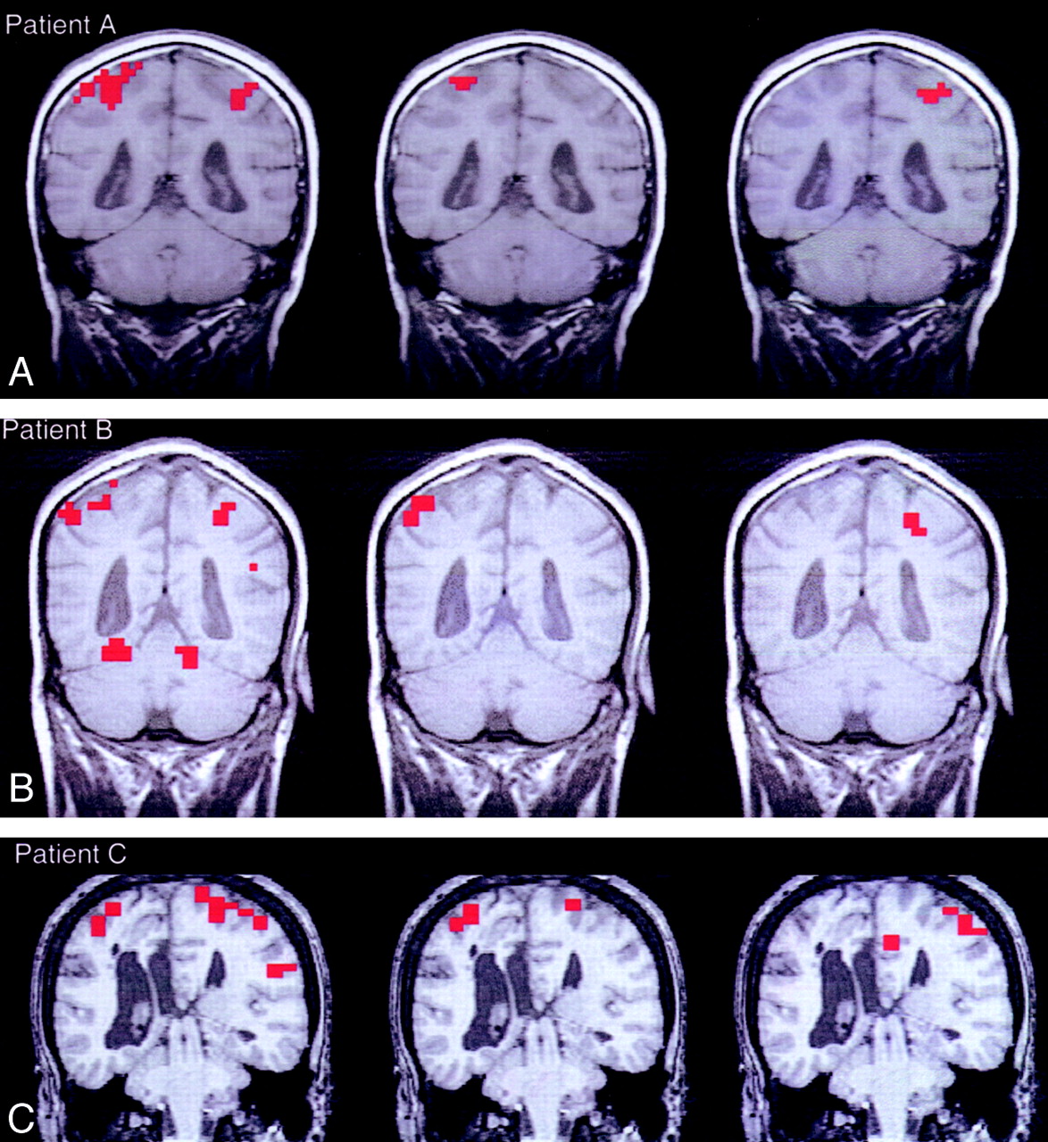
- Fig 1.
Functional connectivity and activation in the sensorimotor cortex of the three patients with agenesis of the corpus callosum. Left, Image displays the activation data from sensorimotor cortex identified by bilateral finger tapping. Middle, Image displays the functional connectivity data as the voxels functionally connected with a seed voxel cluster were chosen in the right sensorimotor cortex. Right, Image displays the functional connectivity data as voxels functionally connected to a seed voxel were selected in the left sensorimotor cortex. Note that with one exception (middle image in C), all functionally connected voxels are in the hemisphere ipsilateral to that of the seed voxel.
A, Patient A.
B, Patient B.
C, Patient C.
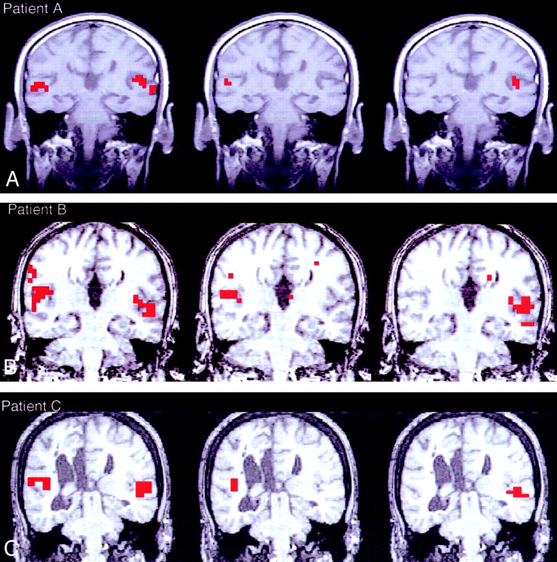
- Fig 2.
Functional connectivity in the auditory cortex of the three patients with agenesis of the corpus callosum. Left, Image displays the activation data from the auditory cortex during a text-listening task. Middle, Image displays the functional connectivity data as the voxels functionally connected to a seed voxel cluster were chosen in the right auditory cortex. Right, Image displays the functional connectivity data as voxels functionally connected to a seed voxel were selected in the left auditory cortex.
A, Patient A.
B, Patient B.
C, Patient C
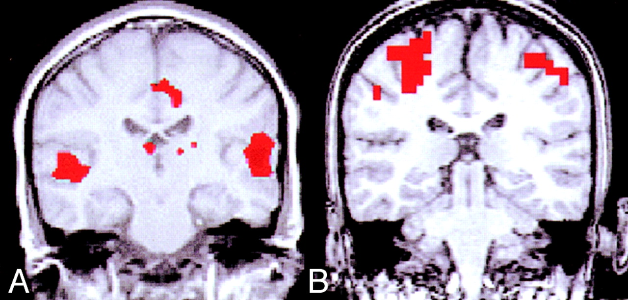
- Fig 3.
Functional connectivity maps from healthy control subjects with an intact corpus callosum. Notice the bilateral connectivity in both maps in contrast to the agenesis data shown in Figures 1 and 2.
A, Seed voxel chosen from the right auditory cortex.
B, Seed voxel chosen from the right sensorimotor cortex.


Tables
- TABLE:
Number of Functionally Connected Voxels in Each Hemisphere for a Seed Voxel in One Hemisphere in the Sensorimotor or Auditory Cortex
A: Pattern of functionally connected voxels in sensorimotor cortex Patient and Seed Voxel Cluster Seed Voxel in L Hemisphere Seed Voxel in R Hemisphere No. of Voxels in the LH No. of Voxels in the RH No. of Voxels in the LH No. of Voxels in the RH A 1 5 0 0 6 2 6 0 NA NA 3 8 0 NA NA 4 10 0 NA NA B 1 7 0 0 12 2 8 0 0 14 3 6 0 0 6 4 10 0 7 17 5 19 7 NA NA C 1 8 0 2 5 2 7 0 0 2 B: Pattern of functionally connected voxels in auditory cortex Patient and Seed Voxel Cluster Seed Voxel in L Hemisphere Seed Voxel in R Hemisphere No. of Voxels in the LH No. of Voxels in the RH No. of Voxels in the LH No. of Voxels in the RH A, 1 6 0 0 3 B 1 6 0 0 9 2 7 0 0 5 3 5 0 0 7 4 9 0 0 10 5 NA NA 0 9 C, 1 5 0 0 3 Note.—The seed voxels were correlated with every other voxel in the data set, and voxels exceeding a threshold of 0.4 (P < .01) were classified as functionally connected. LH indicates the left hemisphere; RH, right hemisphere; NA, not applicable (three patients had different number of clusters in each hemisphere).
In this issue


Jump to section
Related Articles
Cited By...
- Callosal Interhemispheric Communication in Mild Traumatic Brain Injury: A Mediation Analysis on WM Microstructure Effects
- Laminar-specific interhemispheric connectivity mapping with bilateral line-scanning fMRI
- Increased cognitive complexity reveals abnormal brain network activity in individuals with corpus callosum dysgenesis
- On the role of the corpus callosum in interhemispheric functional connectivity in humans
- Stable long-range interhemispheric coordination is supported by direct anatomical projections
- Causal effect of disconnection lesions on interhemispheric functional connectivity in rhesus monkeys
- Interhemispheric Functional Connectivity following Prenatal or Perinatal Brain Injury Predicts Receptive Language Outcome
- Impaired interhemispheric connectivity in medication-naive patients with major depressive disorder
- Intact Bilateral Resting-State Networks in the Absence of the Corpus Callosum
- Predicting human resting-state functional connectivity from structural connectivity
- Regional Variation in Interhemispheric Coordination of Intrinsic Hemodynamic Fluctuations