
Article Figures & Data
Figures
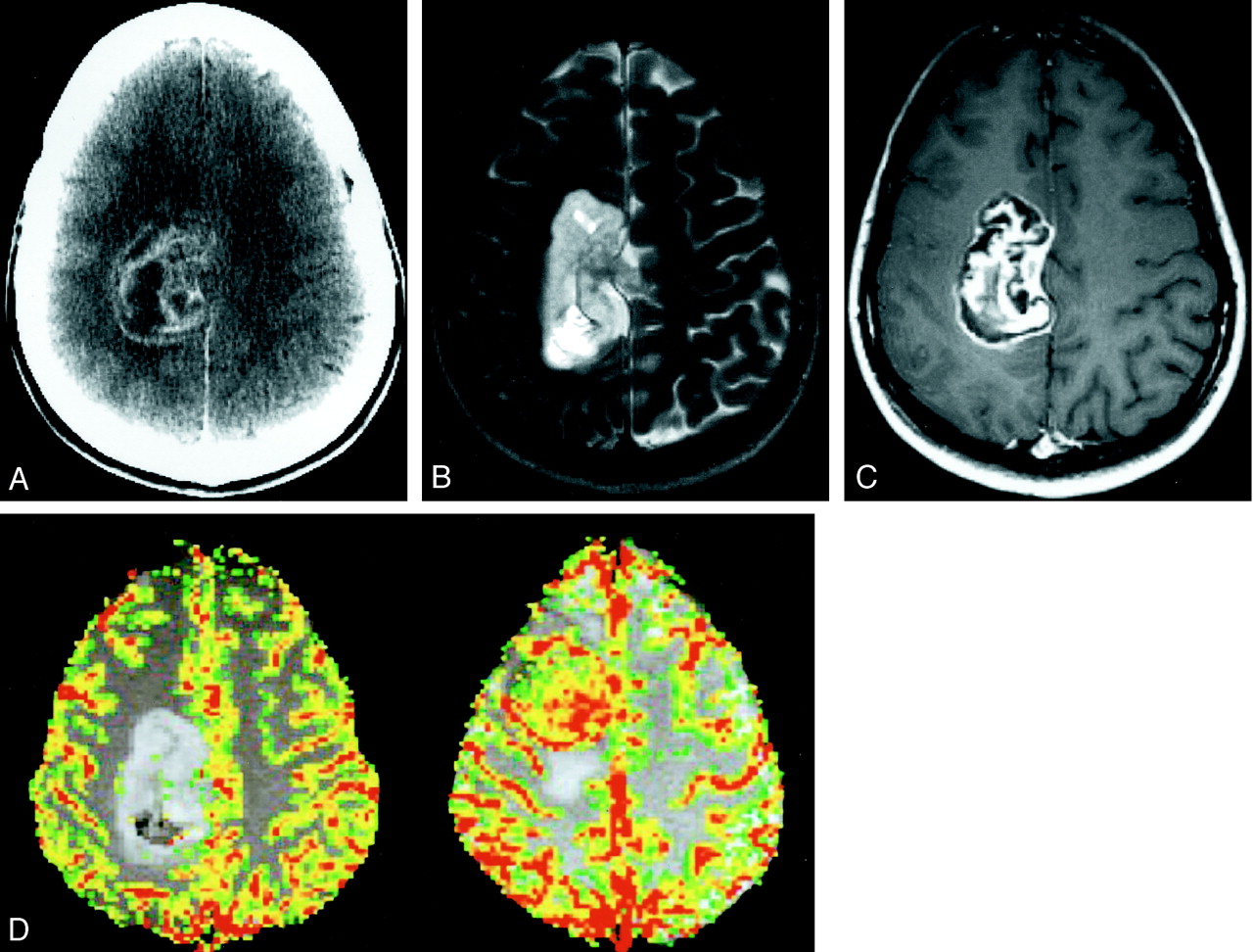
- Fig 1.
Images from the case of a 28-year-old woman with a 6-month history of headaches and left arm tingling and numbness that had gradually progressed to involve the left half of her face.
A, Axial contrast-enhanced CT scan shows a 6-cm, lobulated, heterogeneously enhancing mass in the right parafalcine parietal region, extending into the underlying brain parenchyma.
B, Axial T2-weighted image (3400/119/1 [TR/TE/number of excitations]) shows the mass to have high signal intensity relative to cortex, with scattered foci of low signal intensity. The surrounding brain parenchyma appears normal, with no evidence of vasogenic edema.
C, Axial contrast-enhanced T1-weighted image (600/14/1) shows enhancement with linear areas of extremely low signal intensity, consistent with islands of cartilaginous tissue.
D, Axial color overlay perfusion MR imaging map (1000/54). The image is a perfusion color overlay of a chondrosarcoma (left) that shows hypoperfusion in the region of the mass relative to normal white matter. The image on the right is a perfusion color overlay of a meningioma (right), which in comparison shows increased perfusion.
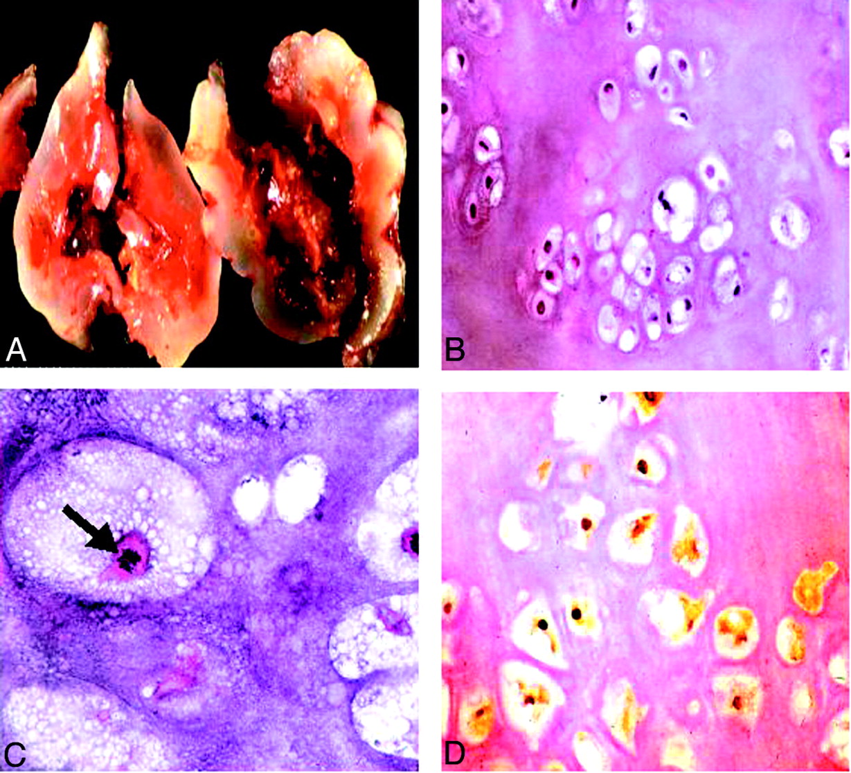
- Fig 2.
Histopathologic findings.
A, Gross specimen shows a bluish white, pearly, glistening lobulated tumor.
B, Low power slide shows a hyaline tumor of low to moderate cellularity with mild variation in size and shape of the tumor cells with mostly small and dark nuclei, showing mild nuclear pleomorphism (hematoxylin and eosin; original magnification, ×40).
C, High power slide reveals mitotic figures (arrow). Findings consistent with the appearance of a conventional chondrosarcoma (hematoxylin and eosin; original magnification, ×200).
D, Nuclear and, to a lesser degree, cytoplasmic immunoreactivity for S-100 protein are present (immunoperoxidase; original magnification, ×100), indicative of a cartilaginous tumor.


In this issue


Jump to section
Related Articles
Cited By...
- No citing articles found.