
Article Figures & Data
Figures
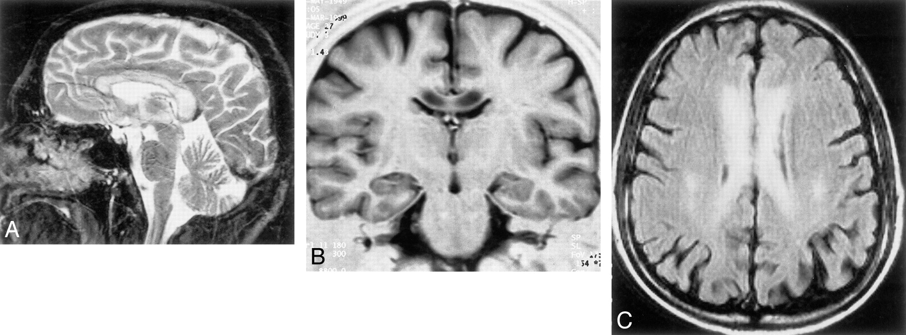
- Fig 1.
MR images obtained 1 month after the onset of symptoms.
A, Sagittal T2-weighted MR image (2600/14, 85/2 [TR/TE, second TE/NEX]). Hyperintensity of the entire corpus callosum is present.
B, Coronal inversion recovery image (9520/60/2 [TR/TI/NEX]). The corpus callosum appears hypointense, with sparing of fibers in the superficial and central portion.
C, Axial fluid-attenuated inversion recovery image (9999/105/1). Bilateral foci of hyperintensity in the white matter of the centrum semiovale.
- Fig 2.
MR images obtained 4 months after the onset of symptoms (A–C) and 11 months after onset (D and E).
A, Sagittal T2-weighted MR image (2600/14, 85/2). Slight reduction of abnormal signal intensity can be seen in the genu and anterior half of the body of the corpus callosum, which appears thinned.
B, Axial T2-weighted MR image (2600/14, 85/2).
C, Axial fluid-attenuated inversion recovery image (9999/105/1) shows the disappearance of foci of hyperintensity in the centrum semiovale.
D, Sagittal T2-weighted image (2600/14, 85/2). Slight hyperintensity persists in the splenium; the corpus callosum is moderately atrophic.
E, Axial T2-weighted image (2600/14, 85/2) shows slight hyperintensity of the splenium.
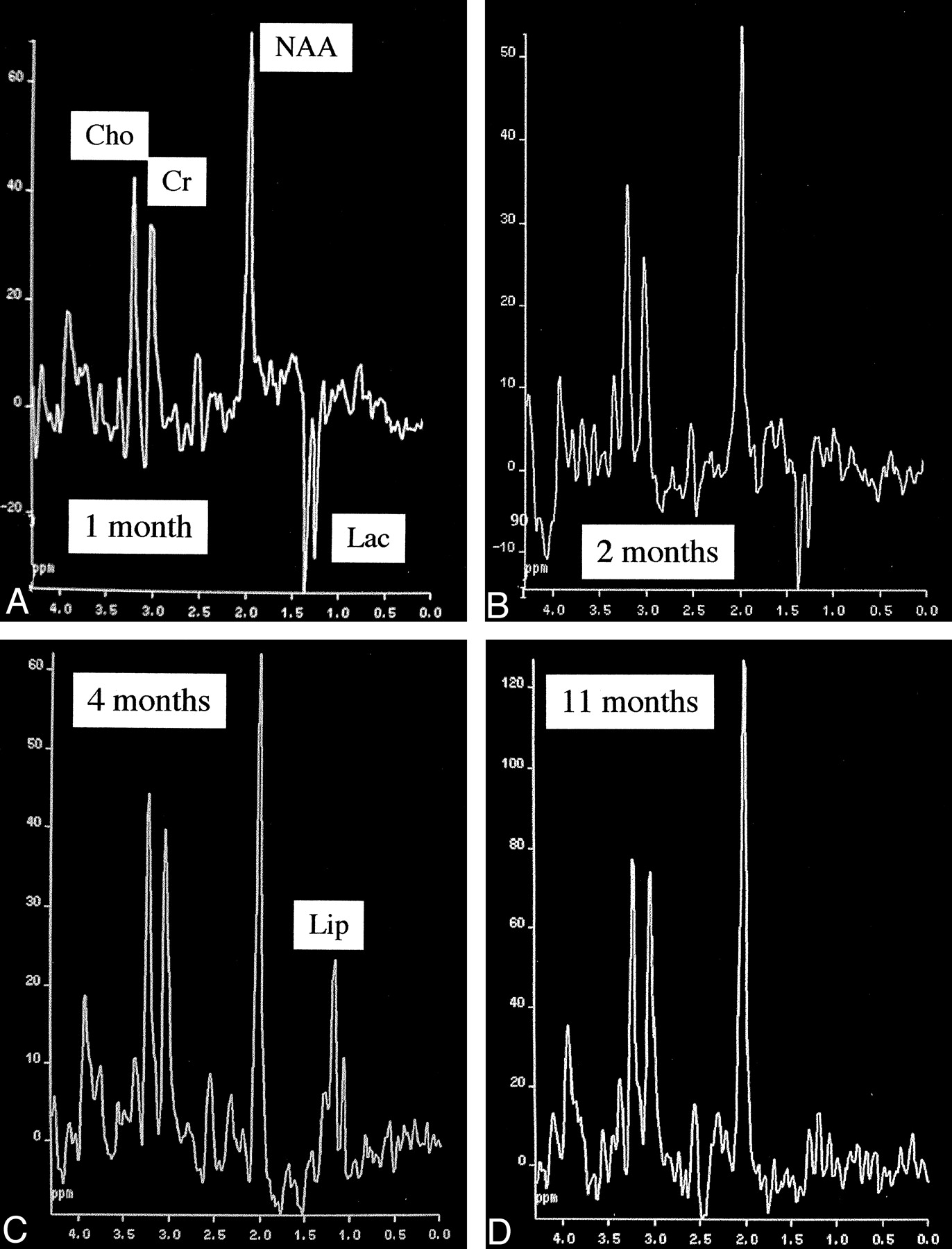
- Fig 3.
Evolution with time of metabolic profile of the splenium of the corpus callosum. Progressive normalization of the Cho/Cr ratio is initially slightly increased; progressive reduction of the NAA/Cr ratio, with a minimum at 4 months and a partial recovery at 11 months; presence of Lac at 1 and 2 months replaced by Lip at 4 months.
A, Metabolic profile at 1 month.
B, Metabolic profile at 2 months.
C, Metabolic profile at 4 months.
D, Metabolic profile at 11 months.


Tables
Main MR imaging signal changes and correspondent metabolic profile of MR spectroscopy during the evolution of the disease
Months after Onset MR Imaging Cho/Cr NAA/Cr Lactate Lipids 1 T2 hyperintensity of the entire corpus callosum; foci of in the centrum semiovale 1.35 1.98 ++ – 2 Unchanged 1.17 1.67 + – 4 Persistent hyperintensity in the splenium and posterior half of the body of the corpus callosum; corpus callosum slightly atrophic 1.06 1.26 – + 11 Slight hyperintensity of the splenium; moderate atrophy of the corpus callosum 0.87 1.45 – – Normal range 0.9–1.3 1.8–2.4 – – Note.—Cho indicates choline; Cr, creatine; NAA, N-acetylaspartate.


