
Article Figures & Data
Figures
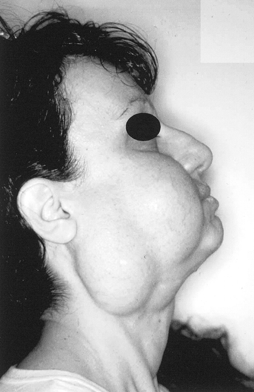
- Fig 1.
Photograph of a 37-year-old white female patient who presented with a 2-year history of slowly progressive swelling of the right cheek and parotidomasseteric and submandibular areas.
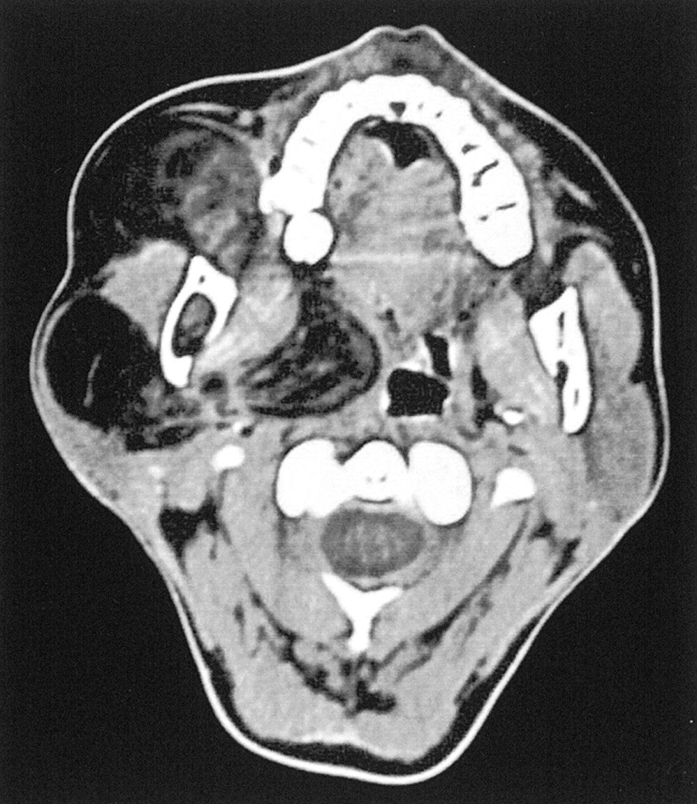
- Fig 2.
Axial contrast-enhanced CT scan shows an expansive, ill-defined, inhomogeneously hypoattenuated mass occupying the parotidomasseteric region and extending deeply toward the right parapharyngeal space. The parotid gland is compressed and displaced posteriorly. No contrast enhancement is evident. Linear densities within the lipomatous region are consistent with fibrous materials revealed by histologic examination.
- Fig 3.
MR images obtained with a 1.0-T superconductive imaging unit.
A, Coronal spin-echo T1-weighted MR image (512/17) shows an inhomogeneous hyperintense lesion located in the parotidomasseteric region and extending deeply toward the parapharyngeal space.
B, On spin-echo T2-weighted MR image without fat suppression (2300/80), decreasing signal intensity is appreciable and irregular linear regions of markedly hypointense signal are present.
C, Axial contrast-enhanced fat-suppressed spin-echo T1-weighted MR image (900/22) shows partial suppression of the lesion, consistent with fat. The lesion nevertheless shows ill-defined margins without a clear plane of cleavage between the mass and the adjacent muscular structures, especially the pterygoid and masseter muscles that appeared displaced and compressed. No evidence of contrast enhancement within the mass can be seen.
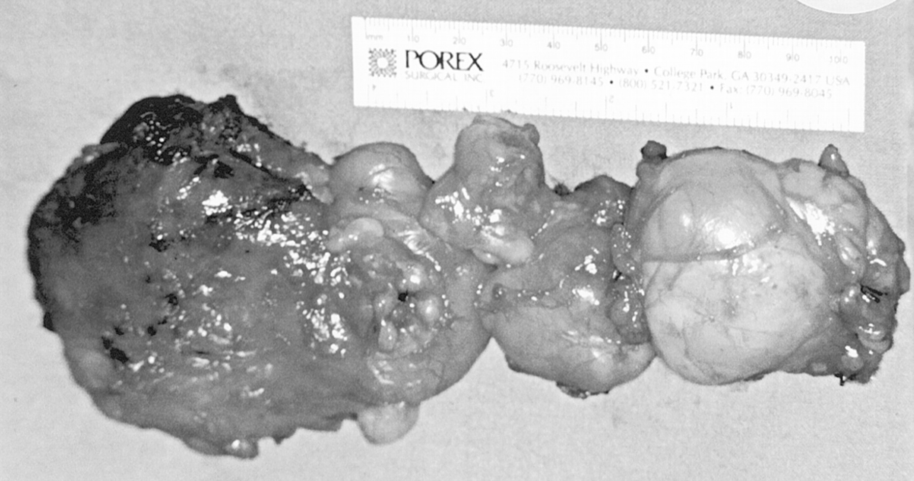
- Fig 4.
Photograph of surgical specimen. Removal of the mass was difficult, because the posterior part of the lesion was firmly attached to the surrounding muscles and the ramus of mandible. The gross appearance of the mass is that of an irregular, lobulated, firm fatty lesion with no evidence of encapsulation.
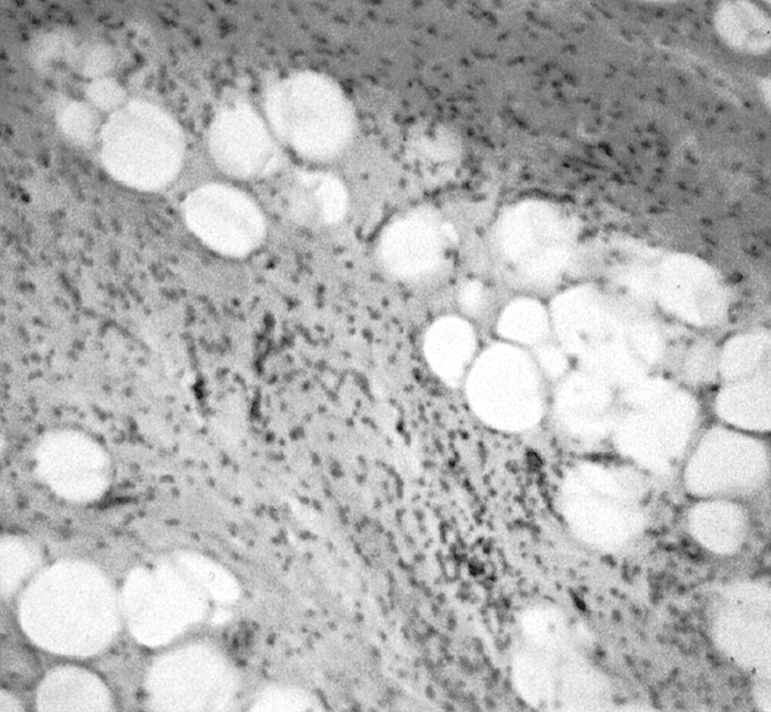
- Fig 5.
Pathologic specimen (hematoxylin and eosin; original magnification, ×400). The lesion is composed of uniform-appearing mature adipocytes, with the presence of myxoid hypocellular areas and infiltrates of inflammatory cells, especially lymphocytes. Diffuse infiltration of entrapped striated muscles can be seen; no evidence of lipoblasts, cellular atypism, or mitoses indicating liposarcoma is seen.


