
Article Figures & Data
Figures
- Fig 1.
Location of regions of interest. These are divided into six areas on the basis of the idealized standard concept of arterial distributions of the brain, as follows: territories of the ACA, MCA, PCA, anterior BZ (between the territories of the ACA and MCA), posterior BZ (between the territories of the MCA and PCA), and internal BZ (between the territories of the cortical branches and penetrators of the MCA).
- Fig 2.
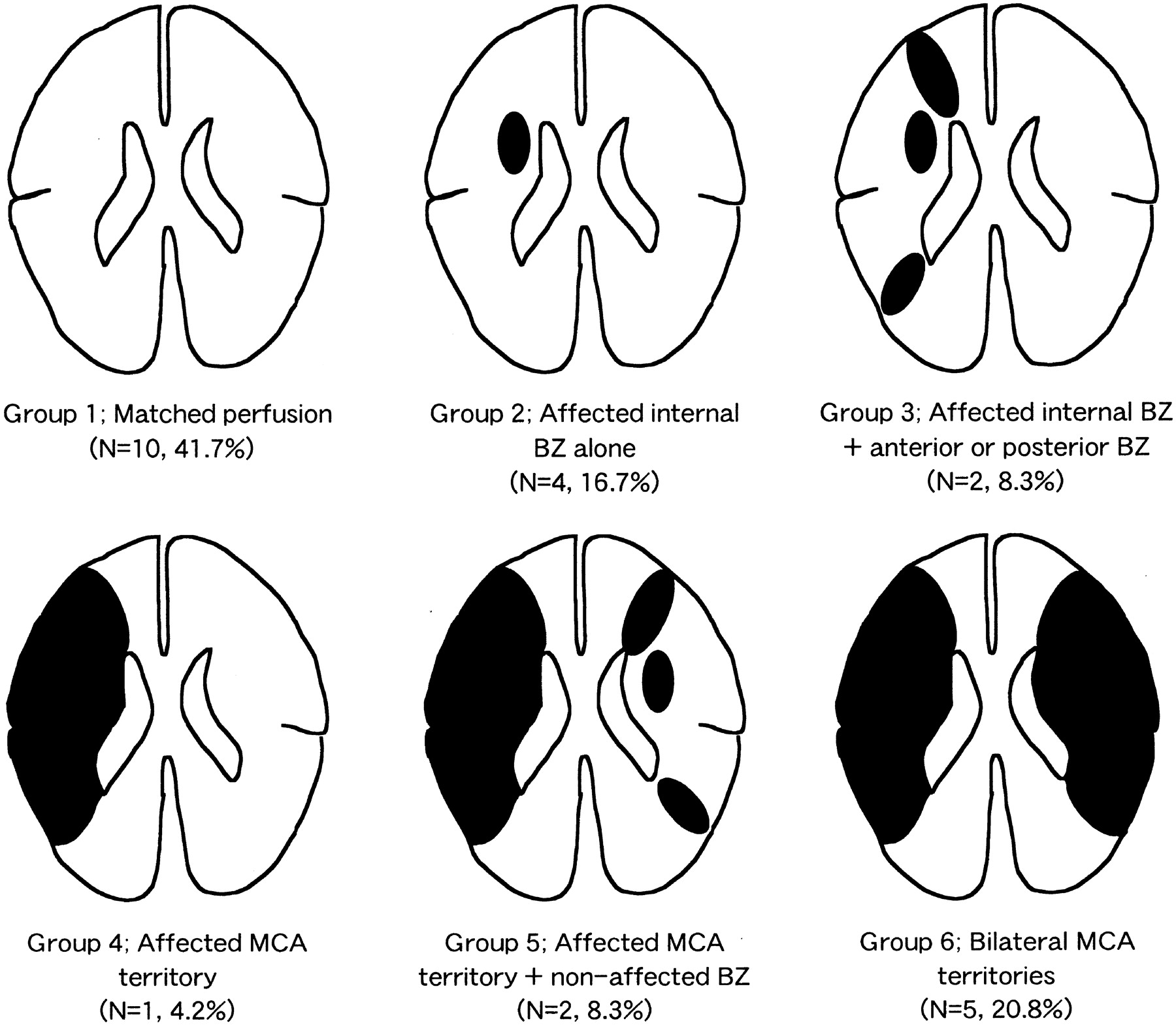
Distribution of MP in unilateral occlusive disease of the major cerebral arteries.


Tables
Case No. Age (y)/Sex Associated Conditions Type of Ischemia Mechanism Neurologic Deficits* CT/MRI Findings Angiographic Findings Main Flow to Affected MCA† Other Collateral‡ PET Findings, Distribution of MP§ 1 66/M HT, HLP Completed stroke Hemodynamic L sensory deficit Bil lacunar infarcts R ICS Antegrade flow None Matched perfusion (G1) 2 71/M HT Completed stroke A-to-A embolism L hemiparesis, L hemianopsia R small cortical infact R ICS Antegrade flow None Matched perfusion (G1) 3 62/M DM, HT, HLP, MI, ASO Completed stroke Unclassified R hemiparesis, motor aphasia L lacunar infarcts L ICS None None Affected IBZ and SBZ (G3) 4 66/M DM, HT, AP, ASO Completed stroke Unclassified R hemiparesis L lacunar infarct L ICS Antegrade flow Acom→L ACA Matched perfusion (G1) 5 78/M HT Completed stroke Unclassified L hemiparesis, L sensory deficit R small cortical infact R ICS Antegrade flow None Matched perfusion (G1) 6 81/F HT, ASO, af Completed stroke Hemodynamic R hemiparesis, R sensory deficit L lacunar infarct L ICS None None Bil MCAt including BZ (G6) 7 66/F HT Asymptomatic None R ICS None None Affected IBZ alone (G2) 8 63/F HT, HLP TIA A-to-A embolism R hemiparesis, motor aphasia Bil lacunar infarcts L ICS Antegrade flow Acom→L ACA Matched perfusion (G1) 9 68/M HT, HLP TIA Hemodynamic L hemiparesis Bil lacunar infarcts + R small cortical infact R ICS R opthalmic A→ Acom→R ACA Affected MCAt including BZ + non-affected BZ (G5) 10 68/M DM, HT, HLP Completed stroke A-to-A embolism R hemiparesis Bil lacunar infarcts + L small cortical infact L ICS Antegrade flow None Matched perfusion (G1) 11 62/M DM, HT, HLP Completed stroke Unclassified R hemiparesis L lacunar infarct L ICO Acom→ None Affected IBZ alone (G2) 12 52/F DM, HT Completed stroke Unclassified R hemiparesis, motor aphasia L lacunar infarct L ICO Acom→ L PCA→LM Affected IBZ and SBZ (G3) 13 70/M HT Completed stroke A-to-A embolism L hemiplegia, USN, anosgnosia R small cortical infact R ICO R ACA→LM A com→R ACA Affected MCAt including BZ + non-affected BZ (G5) 14 69/M HT TIA Hemodynamic L hemiparesis R small cortical infact R ICO R opthalmic A→ A com→R ACA Bil MCAt including BZ (G6) 15 75/M HT, AP Completed stroke A-to-A embolism L hemiparesis R small cortical infact R ICO None None Affected IBZ alone (G2) 16 69/M DM Completed stroke Unclassified Global aphasia L small cortical infact L ICO Pcom→ None Bil MCAt including BZ (G6) 17 58/M DM, ASO TIA Hemodynamic R hemiparesis, motor aphasia L lacunar infarct L MCS L ACA, PCA→LM A com→L ACA Bil MCAt including BZ (G6) 18 60/M DM, HT Completed stroke Unclassified R hemiparesis L lacunar infarct L MCO L ACA, PCA→LM None Affected MCAt including BZ (G4) 19 64/M HT Completed stroke Hemodynamic R hemiparesis L lacunar infarct L MCO None None Matched perfusion (G1) 20 56/F HT, HLP Completed stroke Unclassified L hemiparesis R lacunar infarcts R MCO R ACA→LM None Affected MCAt including BZ + non-affected BZ (G5) 21 67/M HT, HLP Completed stroke Unclassified R hemiparesis, R hemianopsia, anosognosia L small cortical infacts L MCO L ACA, PCA→LM None Matched perfusion (G1) 22 71/F HT, HLP Completed stroke Unclassified R hemiparesis None L MCO L ACA→LM A com→L ACA Affected IBZ alone (G2) 23 54/M DM, HT Asymptomatic Bil lacunar infarcts R MCO R ACA, PCA→LM None Matched perfusion (G1) 24 69/M Asymptomatic L small cortical infacts L MCO L ACA→LM None Matched perfusion (G1) Note.—Abbreviations are as follows: af indicates atrial fibrillation; AP, angina pectoris; ASO, arteriosclerosis obliterans; DM, diabetes mellitus; HLP, hyperlipidemia; IBZ, internal BZ; ICO, occlusion of the ICA; ICS, severe stenosis of the ICA; MCAt, MCA territory; MCO, occlusion of the M1 portion; MCS, severe stenosis of the M1 portion; HT, hypertension; MI, old myocardial infarction; SBZ, superficial BZ; USN, unilateral spatial neglect; Bil, bilateral.
* Defects at the onset in patients with TIA and at the time of hemodynamic studies in other patients.
† In patients who underwent conventional angiography or intra-arterial digital subtraction angiography.
‡ Number shows the type of the MP distribution.
- TABLE 2:
Values for regional CBF, CBV, CMRO2, OEF, and CBF/CBV ratio of patients and control subjects
Value Ipsilateral Side to the Vascular Lesion Contralateral Side to the Vascular Lesion Hemisphere Anterior BZ Posterior BZ Internal BZ MCA Territory Hemisphere Anterior BZ Posterior BZ Internal BZ Non-BZ CBF (mL/100 g/min) Control 47.5 ± 9.4 44.0 ± 8.1 45.9 ± 9.2 43.1 ± 12.9 53.6 ± 11.9 47.5 ± 9.4 44.0 ± 8.1 45.9 ± 9.2 43.1 ± 12.9 53.6 ± 11.9 ICA stenosis 35.1 ± 6.1* 29.7 ± 5.0* 32.5 ± 6.1* 28.6 ± 5.8* 38.4 ± 8.0* 37.8 ± 5.2* 32.3 ± 4.6* 34.9 ± 5.5* 30.4 ± 3.4* 43.2 ± 7.2 ICA occlusion 30.7 ± 5.5* 26.5 ± 6.3* 30.7 ± 4.5* 26.9 ± 5.7* 31.8 ± 7.0* 34.3 ± 4.4* 30.7 ± 4.7* 32.6 ± 7.0* 29.2 ± 3.1* 37.7 ± 5.9* MCA lesion 35.4 ± 3.4* 31.2 ± 4.0* 33.1 ± 3.3* 29.9 ± 4.8* 36.6 ± 4.6* 38.6 ± 4.6 33.5 ± 4.3* 36.5 ± 5.9 33.2 ± 7.0 41.7 ± 4.9 CBV (mL/100 g) Control 3.64 ± 0.41 3.06 ± 0.60 3.47 ± 0.40 2.45 ± 0.51 4.08 ± 0.53 3.64 ± 0.41 3.06 ± 0.60 3.47 ± 0.40 2.45 ± 0.51 4.08 ± 0.53 ICA stenosis 3.41 ± 0.46 2.67 ± 0.60 2.94 ± 0.76 2.51 ± 0.76 3.72 ± 0.62 3.35 ± 0.40 2.55 ± 0.42 3.16 ± 0.61 2.42 ± 0.55 3.79 ± 0.34 ICA occlusion 4.04 ± 0.91 3.24 ± 1.10 3.37 ± 0.70 3.34 ± 0.98 3.99 ± 0.88 3.37 ± 0.34 2.58 ± 0.71 3.31 ± 0.66 2.64 ± 0.66 3.32 ± 0.35* MCA lesion 3.65 ± 0.39 2.94 ± 0.52 3.33 ± 0.56 2.87 ± 1.28 3.93 ± 0.52 3.55 ± 0.65 3.00 ± 0.35 2.83 ± 0.95 2.78 ± 0.47 3.67 ± 0.39 CMRO2 (mL/100 g/min) Control 34.0 ± 0.46 31.5 ± 5.1 35.8 ± 4.9 29.8 ± 5.5 37.7 ± 4.3 34.0 ± 0.46 31.5 ± 5.1 35.8 ± 4.9 29.8 ± 5.5 37.7 ± 4.3 ICA stenosis 25.9 ± 2.6* 22.2 ± 2.9* 25.6 ± 4.4* 21.3 ± 3.3* 28.2 ± 3.0* 27.9 ± 3.3* 24.1 ± 3.0* 27.0 ± 4.4* 23.4 ± 3.3* 31.7 ± 4.6 ICA occlusion 27.3 ± 2.7* 22.8 ± 3.2* 29.0 ± 2.7* 23.3 ± 4.3 28.9 ± 3.4* 29.2 ± 1.8 26.3 ± 3.3 28.6 ± 3.4* 24.8 ± 1.4 32.2 ± 3.2 MCA lesion 27.2 ± 2.2* 23.5 ± 1.7* 26.7 ± 3.3* 22.4 ± 3.9* 28.9 ± 4.2* 29.2 ± 1.7* 24.3 ± 2.5* 28.8 ± 2.2* 24.2 ± 2.7* 33.4 ± 5.5 OEF Control 41.9 ± 6.4 42.0 ± 7.5 45.2 ± 5.8 41.0 ± 6.3 41.4 ± 6.7 41.9 ± 6.4 42.0 ± 7.5 45.2 ± 5.8 41.0 ± 6.3 41.4 ± 6.7 ICA stenosis 47.1 ± 6.5 48.6 ± 8.0 49.8 ± 7.9 48.5 ± 8.1 46.2 ± 6.1 46.4 ± 5.7 47.5 ± 7.0 48.0 ± 7.1 47.6 ± 5.4 45.7 ± 5.4 ICA occlusion 53.7 ± 5.4* 52.2 ± 4.8 56.5 ± 4.9* 52.6 ± 5.7* 54.4 ± 6.9* 51.1 ± 2.8* 51.1 ± 3.1 52.9 ± 4.7 50.3 ± 3.0 50.9 ± 2.9* MCA lesion 48.8 ± 5.1 49.0 ± 5.4 50.7 ± 5.7 48.7 ± 5.1 49.4 ± 5.4 47.3 ± 5.8 46.0 ± 6.3 50.8 ± 5.8 48.5 ± 7.4 46.8 ± 6.1 CBF/CBV ratio (/min) Control 0.078 ± 0.016 0.071 ± 0.016 0.078 ± 0.013 0.060 ± 0.014 0.078 ± 0.013 0.078 ± 0.016 0.071 ± 0.016 0.078 ± 0.013 0.060 ± 0.014 0.078 ± 0.013 ICA stenosis 0.101 ± 0.019 0.093 ± 0.019 0.098 ± 0.036 0.090 ± 0.026 0.101 ± 0.024 0.093 ± 0.022 0.084 ± 0.018 0.096 ± 0.026 0.080 ± 0.024 0.093 ± 0.025 ICA occlusion 0.135 ± 0.039* 0.130 ± 0.049* 0.111 ± 0.037 0.121 ± 0.036* 0.130 ± 0.032* 0.101 ± 0.018 0.088 ± 0.028 0.108 ± 0.025 0.088 ± 0.023 0.090 ± 0.015 MCA lesion 0.106 ± 0.016 0.099 ± 0.023 0.102 ± 0.021 0.100 ± 0.039 0.111 ± 0.020 0.095 ± 0.014 0.092 ± 0.014 0.080 ± 0.031 0.090 ± 0.020 0.091 ± 0.013 Note.—Values are the mean ± SD.
* P < .05, different from control as determined by means of ANOVA with the Scheffé F test.
- TABLE 3:
Standardized values for regional CBF, CBV, CMRO2, OEF, and CBF/CBV ratio of patients and control subjects
Value Ipsilateral Side to the Vascular Lesion Contralateral Side to the Vascular Lesion ABZ/MCA PBZ/MCA IBZ/MCA ABZ/MCA PBZ/MCA IBZ/MCA CBF Control 0.83 ± 0.06 0.86 ± 0.09 0.80 ± 0.07 0.83 ± 0.06 0.86 ± 0.09 0.80 ± 0.07 ICA stenosis 0.78 ± 0.10 0.86 ± 0.11 0.75 ± 0.10 0.76 ± 0.07 0.82 ± 0.13 0.71 ± 0.08 ICA occlusion 0.83 ± 0.09 0.98 ± 0.09 0.83 ± 0.08 0.82 ± 0.05 0.86 ± 0.08 0.79 ± 0.12 MCA lesion 0.86 ± 0.11 0.91 ± 0.09 0.83 ± 0.15 0.80 ± 0.06 0.87 ± 0.08 0.79 ± 0.11 CBV Control 0.75 ± 0.10 0.86 ± 0.12 0.60 ± 0.09 0.75 ± 0.10 0.86 ± 0.12 0.60 ± 0.09 ICA stenosis 0.73 ± 0.19 0.81 ± 0.22 0.70 ± 0.24 0.68 ± 0.12 0.84 ± 0.17 0.64 ± 0.15 ICA occlusion 0.81 ± 0.13 0.86 ± 0.19 0.81 ± 0.30 0.78 ± 0.22 1.04 ± 0.27 0.79 ± 0.18 MCA lesion 0.75 ± 0.13 0.85 ± 0.14 0.72 ± 0.30 0.82 ± 0.11 0.77 ± 0.25 0.76 ± 0.13 CMRO2 Control 0.83 ± 0.06 0.95 ± 0.07 0.79 ± 0.10 0.83 ± 0.06 0.95 ± 0.07 0.79 ± 0.10 ICA stenosis 0.79 ± 0.09 0.91 ± 0.11 0.76 ± 0.09 0.77 ± 0.09 0.86 ± 0.13 0.74 ± 0.06 ICA occlusion 0.79 ± 0.07 1.01 ± 0.05 0.78 ± 0.09 0.82 ± 0.05 0.90 ± 0.09 0.78 ± 0.10 MCA lesion 0.83 ± 0.14 0.93 ± 0.08 0.78 ± 0.10 0.74 ± 0.11 0.88 ± 0.15 0.74 ± 0.13 OEF Control 1.01 ± 0.03 1.10 ± 0.08 0.99 ± 0.07 1.01 ± 0.03 1.10 ± 0.08 0.99 ± 0.07 ICA stenosis 1.05 ± 0.09 1.08 ± 0.04 1.05 ± 0.09 1.04 ± 0.07 1.05 ± 0.06 1.04 ± 0.06 ICA occlusion 0.97 ± 0.05 1.05 ± 0.09 1.00 ± 0.13 1.00 ± 0.03 1.04 ± 0.06 0.99 ± 0.05 MCA lesion 0.99 ± 0.06 1.03 ± 0.04 0.99 ± 0.12 0.98 ± 0.03 1.09 ± 0.03 1.04 ± 0.06 CBF/CBV ratio Control 0.91 ± 0.09 1.00 ± 0.07 0.76 ± 0.11 0.91 ± 0.09 1.00 ± 0.07 0.76 ± 0.11 ICA stenosis 0.96 ± 0.30 0.98 ± 0.33 0.94 ± 0.36 0.93 ± 0.19 1.08 ± 0.34 0.90 ± 0.36 ICA occlusion 0.98 ± 0.22 0.86 ± 0.15 0.95 ± 0.38 0.97 ± 0.28 1.20 ± 0.26 1.00 ± 0.37 MCA lesion 0.91 ± 0.21 0.92 ± 0.10 0.88 ± 0.26 1.02 ± 0.21 0.88 ± 0.35 0.99 ± 0.14 Note.—ABZ indicates the anterior BZ; IBZ, internal BZ; PBZ, posterior BZ. No significant differences were observed between the four groups (Kruskal-Wallis test).
Group ICA Stenosis ICA Occlusion MCA Lesion* Total 1 6 0 4 10 2 1 2 1 4 3 1 1 0 2 4 0 0 1 1 5 0 1 1 2 6 2 2 1 5 Total 10 6 8 24 * MCA lesions include severe stenosis or occlusion of the trunk of the MCA.
Group Main Route of Blood Flow to the Affected MCA Territory Total Antegrade Acom Lept OA/Pcom 1 5 0 4 0 9 2 0 1 1 0 2 3 0 1 0 0 1 4 0 0 1 0 1 5 0 0 2 0 2 6 0 0 1 3 4 Total 5 2 9 3 19 Note.—Lept indicates leptomeningeal anastomosis; OA, ophthalmic artery.
In this issue


Jump to section
Related Articles
Cited By...
- Detecting Misery Perfusion in Unilateral Steno-Occlusive Disease of the Internal Carotid Artery or Middle Cerebral Artery by MR Imaging
- The Pathophysiology of Watershed Infarction in Internal Carotid Artery Disease: Review of Cerebral Perfusion Studies
- Infarct patterns in atherosclerotic middle cerebral artery versus internal carotid artery disease