Advertisement

Main menu

  • Home
  • Content
    • Current Issue
    • Accepted Manuscripts
    • Article Preview
    • Past Issue Archive
    • AJNR Case Collection
    • Case of the Week Archive
    • Classic Case Archive
    • Case of the Month Archive
  • Special Collections
    • Spinal CSF Leak Articles (Jan 2020-June 2024)
    • 2024 AJNR Journal Awards
    • Most Impactful AJNR Articles
  • Multimedia
    • AJNR Podcast
    • AJNR Scantastics
    • Video Articles
  • For Authors
    • Submit a Manuscript
    • Author Policies
    • Fast publishing of Accepted Manuscripts
    • Graphical Abstract Preparation
    • Manuscript Submission Guidelines
    • Imaging Protocol Submission
    • Submit a Case for the Case Collection
  • About Us
    • About AJNR
    • Editorial Board
  • More
    • Become a Reviewer/Academy of Reviewers
    • Subscribers
    • Permissions
    • Alerts
    • Feedback
    • Advertisers
    • ASNR Home
  • Other Publications
    • ajnr

User menu

  • Alerts
  • Log in

Search

  • Advanced search
American Journal of Neuroradiology

American Journal of Neuroradiology

ASHNR American Society of Functional Neuroradiology ASHNR American Society of Pediatric Neuroradiology ASSR
  • Alerts
  • Log in

Advanced Search

  • Home
  • Content
    • Current Issue
    • Accepted Manuscripts
    • Article Preview
    • Past Issue Archive
    • AJNR Case Collection
    • Case of the Week Archive
    • Classic Case Archive
    • Case of the Month Archive
  • Special Collections
    • Spinal CSF Leak Articles (Jan 2020-June 2024)
    • 2024 AJNR Journal Awards
    • Most Impactful AJNR Articles
  • Multimedia
    • AJNR Podcast
    • AJNR Scantastics
    • Video Articles
  • For Authors
    • Submit a Manuscript
    • Author Policies
    • Fast publishing of Accepted Manuscripts
    • Graphical Abstract Preparation
    • Manuscript Submission Guidelines
    • Imaging Protocol Submission
    • Submit a Case for the Case Collection
  • About Us
    • About AJNR
    • Editorial Board
  • More
    • Become a Reviewer/Academy of Reviewers
    • Subscribers
    • Permissions
    • Alerts
    • Feedback
    • Advertisers
    • ASNR Home
  • Follow AJNR on Twitter
  • Visit AJNR on Facebook
  • Follow AJNR on Instagram
  • Join AJNR on LinkedIn
  • RSS Feeds

Welcome to the new AJNR, Updated Hall of Fame, and more. Read the full announcements.


AJNR is seeking candidates for the position of Associate Section Editor, AJNR Case Collection. Read the full announcement.


Getting new auth cookie, if you see this message a lot, tell someone!
OtherBRAIN

Hyperacute Subarachnoid Hemorrhage on T2-Weighted MR Images

Zoran Rumboldt, Miljenko Kalousek and Mauricio Castillo
American Journal of Neuroradiology March 2003, 24 (3) 472-475;
Zoran Rumboldt
  • Find this author on Google Scholar
  • Find this author on PubMed
  • Search for this author on this site
Miljenko Kalousek
  • Find this author on Google Scholar
  • Find this author on PubMed
  • Search for this author on this site
Mauricio Castillo
  • Find this author on Google Scholar
  • Find this author on PubMed
  • Search for this author on this site
  • Article
  • Figures & Data
  • Info & Metrics
  • Responses
  • References
  • PDF
Loading

Article Figures & Data

Figures

  • Fig 1.
    • Download figure
    • Open in new tab
    • Download powerpoint
    Fig 1.

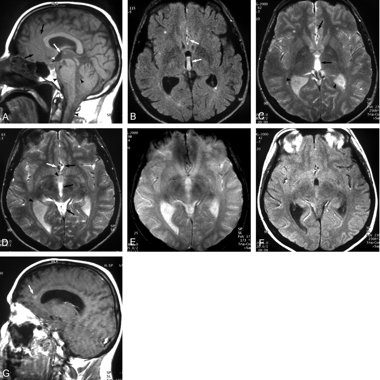
    Images in a 47-year-old woman with SAH.

    A, Sagittal T1-weighted SE image (TR/TE, 584/12) shows increased signal intensity throughout the basilar cisterns, fourth ventricle, and spinal subarachnoid space (black arrowheads). This area of relatively high signal intensity extends through the cerebral aqueduct into the posterior aspect of the third ventricle, with a dependent level just anterior to the massa intermedia (white arrow). The anterior inferior aspect of the interhemispheric fissure also shows slightly increased signal intensity, and the cortical sulci are not visualized in this area (black arrows). The hypointensity that corresponds to the aneurysm (arrowhead), and the A2 segments of the anterior cerebral arteries (ACAs), are seen on the background of SAH.

    B, Axial FLAIR image (TR/TE/TI, 6212/100/1800) shows high signal intensity of the posterior aspect of the third ventricle and in the interhemispheric fissure surrounding the ACAs (arrows). A small, nonspecific area of high signal intensity is incidentally found in the right frontal lobe.

    C, Axial T2-weighted SE image (2008/80) at the same level as in B. The signal intensity in the posterior aspect of the third ventricle and in the interhemispheric fissure (arrows) is higher that that of other CSF-containing spaces (arrowheads).

    D, Axial T2-weighted SE image at the adjacent lower level. The signal intensity of the posterior aspect of the third ventricle, around the ACAs, and in the supravermian cistern (black arrows) is higher than that of the lateral ventricles and the anterior portion of the third ventricle (arrowheads). The larger flow-void area posterior to the ACAs (white arrow) was proved to correspond to the ACoA aneurysm on a subsequent intraarterial angiogram (not shown).

    E, Axial T2*-weighted GRE image (800/35; flip angle, 25°) at approximately the same level as in D. The signal intensities within the third ventricle are reversed compared with those in D. The posterior aspect of the third ventricle is of lower signal intensity. This hypointensity is mild, presumably due to small amount of deoxyhemoglobin, which is partly counterbalanced by extremely high T2 signal intensity.

    F, Axial proton density-weighted SE image (2008/20) at the same level as in D. The signal intensity within the posterior portion of the third ventricle, in the interhemispheric fissure, and in the supravermian cistern, is approximately the same as that in the gray matter.

    G, Sagittal contrast-enhanced T1-weighted SE image (584/12) shows pooling of gadolinium-based contrast agent just anterior to corpus callosum (arrow). Homogeneously low signal intensity of the CSF is present in the lateral ventricle. The image quality is compromised by motion.

Back to top

In this issue

American Journal of Neuroradiology: 24 (3)
American Journal of Neuroradiology
Vol. 24, Issue 3
1 Mar 2003
  • Table of Contents
  • Index by author
Advertisement
Print
Download PDF
Email Article

Thank you for your interest in spreading the word on American Journal of Neuroradiology.

NOTE: We only request your email address so that the person you are recommending the page to knows that you wanted them to see it, and that it is not junk mail. We do not capture any email address.

Enter multiple addresses on separate lines or separate them with commas.
Hyperacute Subarachnoid Hemorrhage on T2-Weighted MR Images
(Your Name) has sent you a message from American Journal of Neuroradiology
(Your Name) thought you would like to see the American Journal of Neuroradiology web site.
CAPTCHA
This question is for testing whether or not you are a human visitor and to prevent automated spam submissions.
Cite this article
Zoran Rumboldt, Miljenko Kalousek, Mauricio Castillo
Hyperacute Subarachnoid Hemorrhage on T2-Weighted MR Images
American Journal of Neuroradiology Mar 2003, 24 (3) 472-475;

Citation Manager Formats

  • BibTeX
  • Bookends
  • EasyBib
  • EndNote (tagged)
  • EndNote 8 (xml)
  • Medlars
  • Mendeley
  • Papers
  • RefWorks Tagged
  • Ref Manager
  • RIS
  • Zotero
0 Responses
Respond to this article
Share
Bookmark this article
Hyperacute Subarachnoid Hemorrhage on T2-Weighted MR Images
Zoran Rumboldt, Miljenko Kalousek, Mauricio Castillo
American Journal of Neuroradiology Mar 2003, 24 (3) 472-475;
del.icio.us logo Twitter logo Facebook logo Mendeley logo
  • Tweet Widget
  • Facebook Like
  • Google Plus One

Jump to section

  • Article
    • Abstract
    • Case Report
    • Discussion
    • Conclusion
    • References
  • Figures & Data
  • Info & Metrics
  • Responses
  • References
  • PDF

Related Articles

  • No related articles found.
  • PubMed
  • Google Scholar

Cited By...

  • Role of Iron in Brain Injury After Intraventricular Hemorrhage
  • Crossref
  • Google Scholar

This article has not yet been cited by articles in journals that are participating in Crossref Cited-by Linking.

More in this TOC Section

  • Progression of Microstructural Damage in Spinocerebellar Ataxia Type 2: A Longitudinal DTI Study
  • Usefulness of Quantitative Susceptibility Mapping for the Diagnosis of Parkinson Disease
  • Evaluating the Effects of White Matter Multiple Sclerosis Lesions on the Volume Estimation of 6 Brain Tissue Segmentation Methods
Show more Brain

Similar Articles

Advertisement

Indexed Content

  • Current Issue
  • Accepted Manuscripts
  • Article Preview
  • Past Issues
  • Editorials
  • Editors Choice
  • Fellow Journal Club
  • Letters to the Editor

Cases

  • Case Collection
  • Archive - Case of the Week
  • Archive - Case of the Month
  • Archive - Classic Case

Special Collections

  • Special Collections

Resources

  • News and Updates
  • Turn around Times
  • Submit a Manuscript
  • Author Policies
  • Manuscript Submission Guidelines
  • Evidence-Based Medicine Level Guide
  • Publishing Checklists
  • Graphical Abstract Preparation
  • Imaging Protocol Submission
  • Submit a Case
  • Become a Reviewer/Academy of Reviewers
  • Get Peer Review Credit from Publons

Multimedia

  • AJNR Podcast
  • AJNR SCANtastic
  • Video Articles

About Us

  • About AJNR
  • Editorial Board
  • Not an AJNR Subscriber? Join Now
  • Alerts
  • Feedback
  • Advertise with us
  • Librarian Resources
  • Permissions
  • Terms and Conditions

American Society of Neuroradiology

  • Not an ASNR Member? Join Now

© 2025 by the American Society of Neuroradiology All rights, including for text and data mining, AI training, and similar technologies, are reserved.
Print ISSN: 0195-6108 Online ISSN: 1936-959X

Powered by HighWire