
Article Figures & Data
Figures
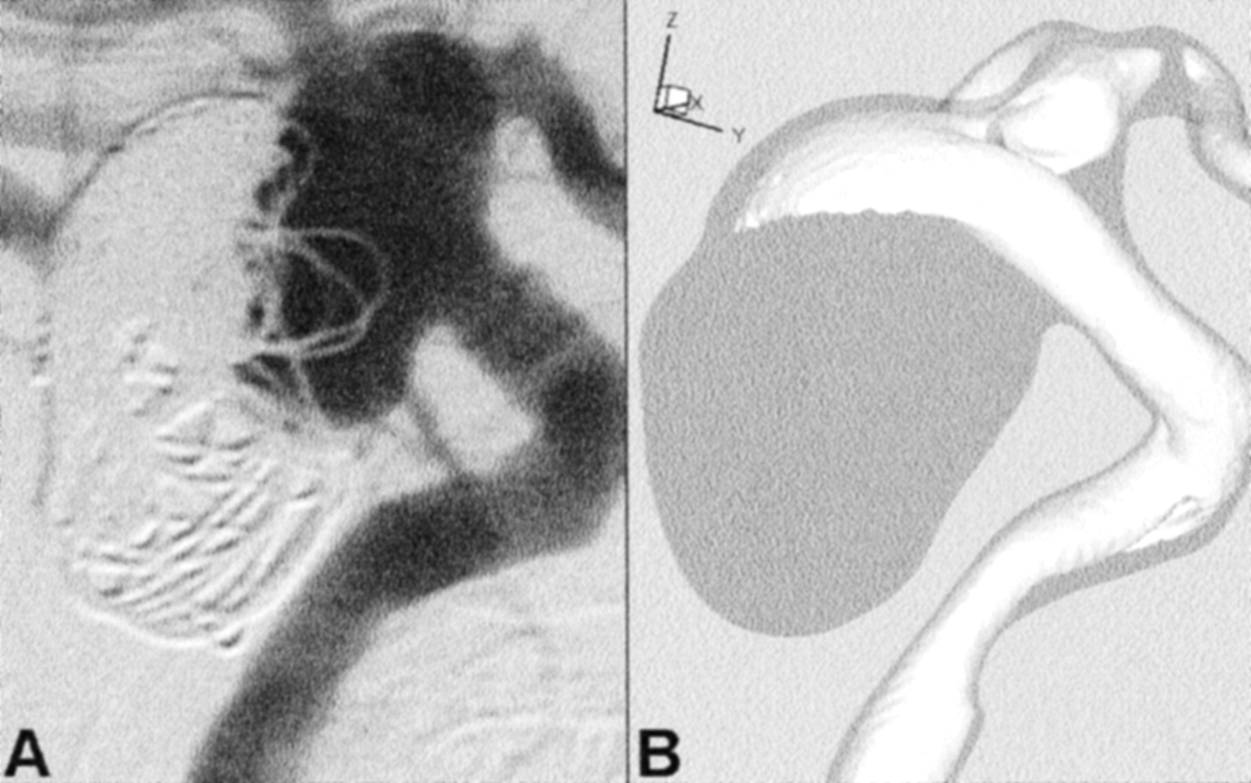
- Fig 1.
Geometry of aneurysm and parent vessel, shown in anteroposterior (left column) and lateral (right column) views.
A and B, Maximum intensity projections obtained through the CT reconstructed data illustrate the geometrical complexity of the aneurysm and parent vessel. Angled arrows identify blebs on the right and posterior sides of the aneurysm sac. Horizontal arrows point to an apparent stenosis of the petrous segment, which may be attributed to compression of the vessel against adjacent bone.
C and D, Corresponding views of the finite element mesh show the geometric faithfulness and spatial resolution of the CFD model; for clarity, only the corner nodes of the quadratic finite elements are shown. Note the model coordinate system: ± x = left/right, ± y = anterior/posterior, ± z = superior/inferior.
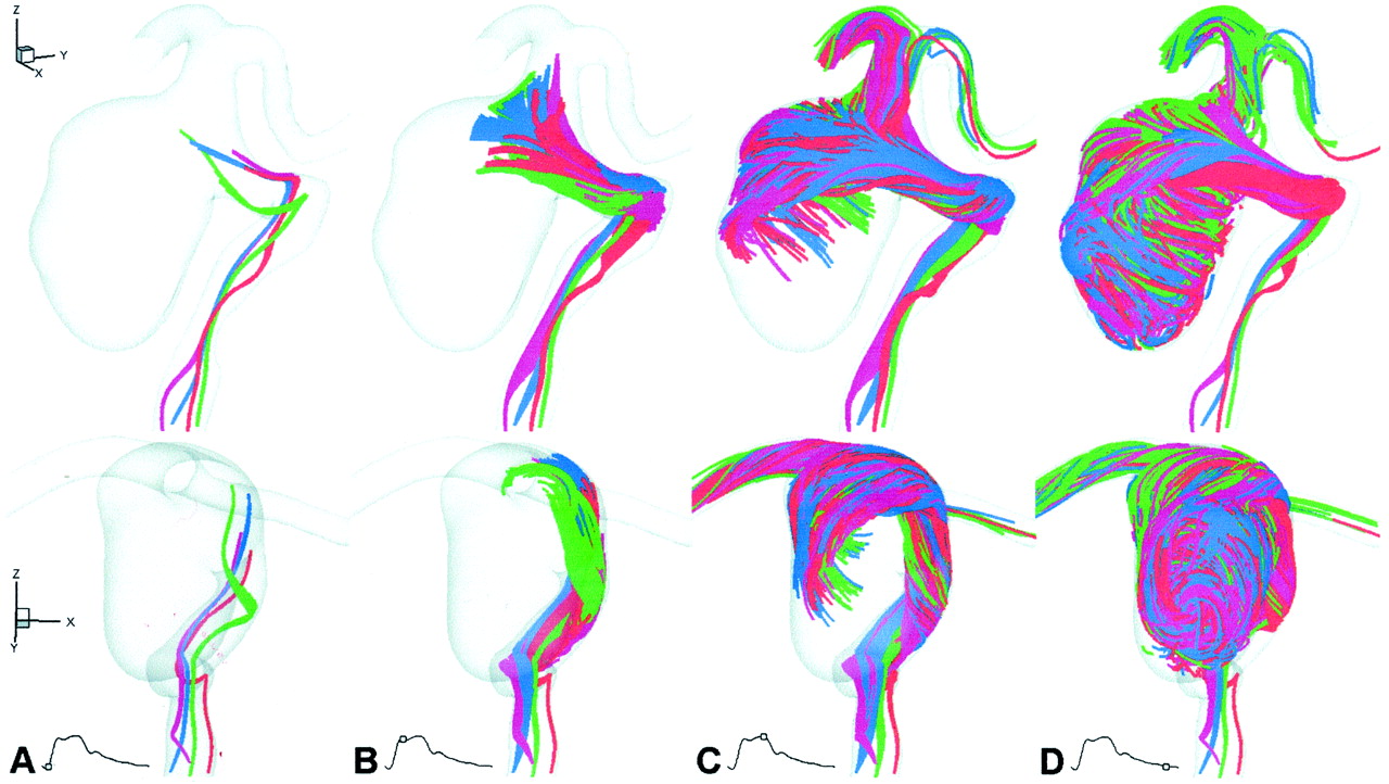
- Fig 2.
Virtual slipstreams, shown at selected times in lateral (top row) and anteroposterior (bottom row) oblique views, provide an overview of the aneurysm hemodynamics.
A, Early systole (t = 50 ms). Slipstreams showed little mixing or spreading as they approached the neck.
B, Peak systole (t = 150 ms). Slipstreams spread and began to mix as they entered both the proximal and distal ends of the neck.
C, Early diastole (t = 300 ms). Slipstreams mixed as they impacted the posterior wall of the aneurysm and swirled in the right and inferior directions.
D, Late diastole (t = 800 ms). Within 0.5 second, the aneurysm was almost entirely opacified by vigorously mixed slipstreams.
- Fig 3.
More detailed views of the complex aneurysm hemodynamics are provided at selected times and for selected planes via conventional field plots of sagittal (top row), coronal (middle row), and axial (bottom row) planes, each from the nominal center of the aneurysm. Contours of velocity magnitude (V, in cm/s) are shown with vectors superimposed to indicate the magnitude and direction of in-plane flow components. White circles identify the approximate center of each vortex. S, superior; I, inferior; A, anterior; P, posterior; R, right; L, left.
A, Peak systole (t = 150 ms).
B, Early diastole (t = 300 ms).
C, Mid-diastole (t = 450 ms).
D, Late diastole (t = 600 ms).
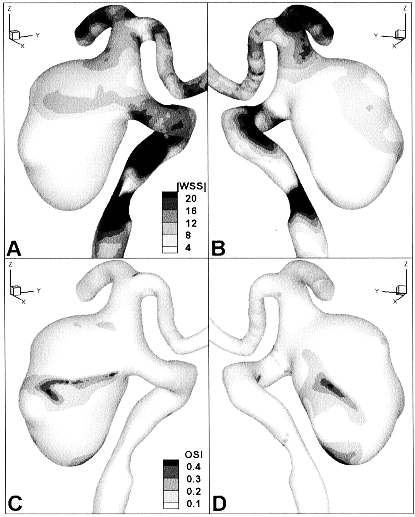
- Fig 4.
Computed wall shear stress patterns are shown in oblique posterior (left panels) and anterior (right panels) views.
A and B, Contours of cycle-averaged wall shear stress magnitude (WSS, dynes/cm2).
C and D, Contours of oscillatory shear index (OSI, dimensionless), a measure of the relative variability of the wall shear stress over the cardiac cycle.
- Fig 5.
Two sequential frames from 2-Hz cine digital subtraction angiograms. These zoomed views correspond to the orientation and extent of the CFD model in the lower panels of Figure 2C and D.
A, Aneurysm filling dynamics, shown approximately 0.5 second after selective injection into the internal carotid artery, show good general agreement with the corresponding virtual slipstreams shown in Figure 2C.
B, One frame (ie, 0.5 second) later, the aneurysm is already largely opacified, which also is consistent with the corresponding virtual slipstreams shown in Figure 2D.
- Fig 6.
Illustration of a possible relationship between coil compaction and computed flow dynamics.
A, Lateral digital subtraction angiogram obtained at 6-month follow-up examination shows compaction of the coil mass away from the neck and toward the posterior wall of the aneurysm.
B, High speed flow entering the aneurysm (shown by using a 15 cm/s isovelocity surface from the cycle-averaged velocity field superimposed on a projection of the aneurysm model approximately corresponding to that in shown in A) is also directed toward the posterior wall of the aneurysm.


In this issue


Jump to section
Related Articles
Cited By...
- Evaluation of predictive models of aneurysm focal growth and bleb development using machine learning techniques
- Youre so vein, you probably think this models about you: opportunities and challenges for computational fluid dynamics in cerebral venous disease
- Evaluation of predictive models of aneurysm focal growth and bleb development using machine learning techniques
- Quantitative and Qualitative Comparison of 4D-DSA with 3D-DSA Using Computational Fluid Dynamics Simulations in Cerebral Aneurysms
- Selective compromise of hypoplastic posterior communicating artery variants with aneurysms treatable by coil embolization: clinical and radiologic outcomes
- Aneurysmal Parent Artery-Specific Inflow Conditions for Complete and Incomplete Circle of Willis Configurations
- Porcine In Vivo Validation of a Virtual Contrast Model: The Influence of Contrast Agent Properties and Vessel Flow Rates
- Simultaneous imaging of blood flow dynamics and vascular remodelling during development
- Quantitative comparison of hemodynamic parameters from steady and transient CFD simulations in cerebral aneurysms with focus on the aneurysm ostium
- CFD: Computational Fluid Dynamics or Confounding Factor Dissemination? The Role of Hemodynamics in Intracranial Aneurysm Rupture Risk Assessment
- Effect of Bifurcation Angle Configuration and Ratio of Daughter Diameters on Hemodynamics of Bifurcation Aneurysms
- Analysis of Intra-Aneurysmal Flow for Cerebral Aneurysms with Cerebral Angiography
- High Shear Stress and Flow Velocity in Partially Occluded Aneurysms Prone to Recanalization
- Hemodynamics and Anatomy of Elastase-Induced Rabbit Aneurysm Models: Similarity to Human Cerebral Aneurysms?
- Association of Hemodynamic Characteristics and Cerebral Aneurysm Rupture
- Quantitative Characterization of the Hemodynamic Environment in Ruptured and Unruptured Brain Aneurysms
- In Vitro Study of Near-Wall Flow in a Cerebral Aneurysm Model with and without Coils
- Computational fluid dynamic simulation to assess flow characteristics of an in vitro aneurysm model
- Quantitative Hemodynamic Analysis of Brain Aneurysms at Different Locations
- An objective approach to digital removal of saccular aneurysms: technique and applications
- Three-dimensional imaging and computational modelling for estimation of wall stresses in arteries
- Magnitude and Role of Wall Shear Stress on Cerebral Aneurysm: Computational Fluid Dynamic Study of 20 Middle Cerebral Artery Aneurysms