
Article Figures & Data
Figures
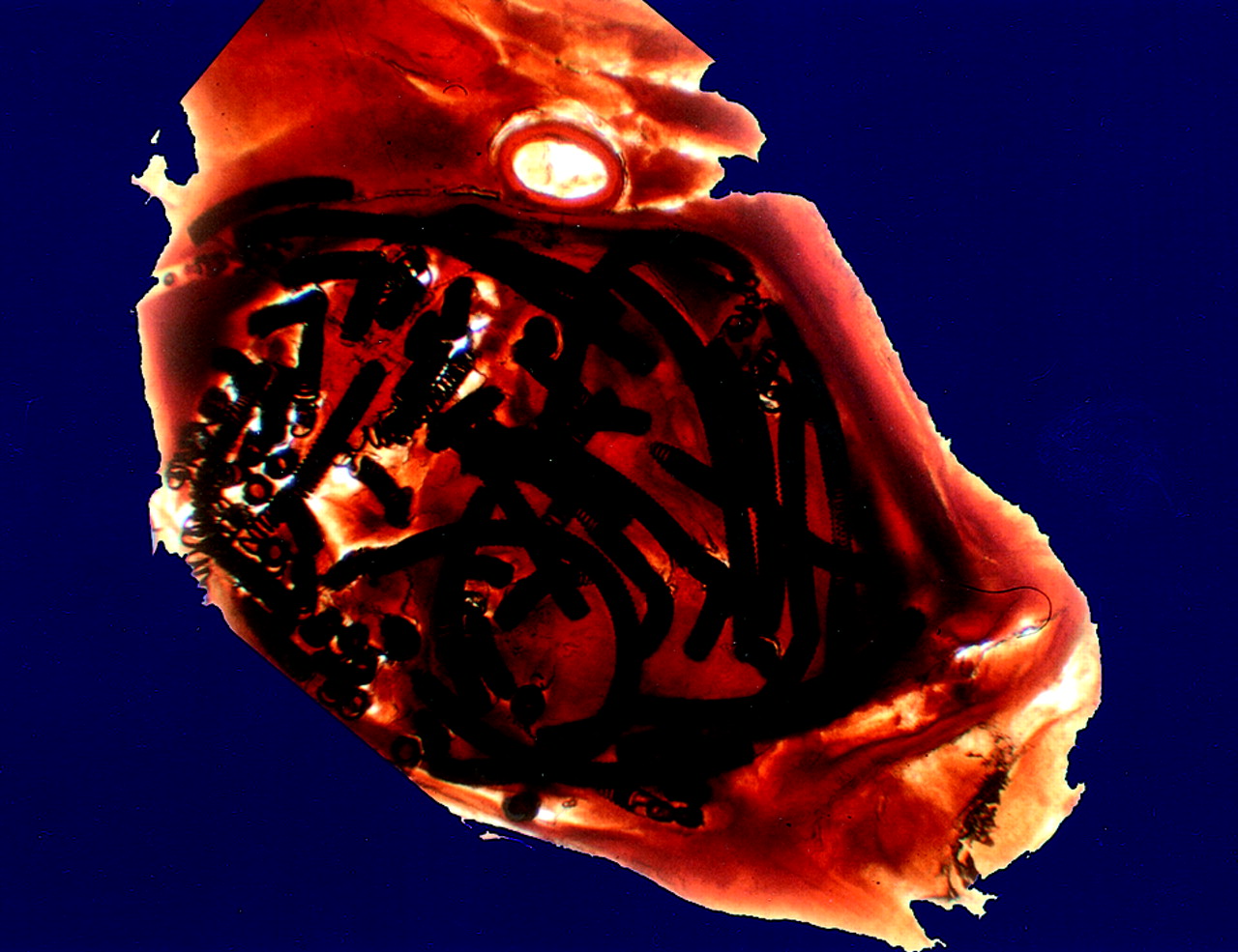
- Fig 1.
Distribution of platinum coils in a plasticized medial cerebral artery aneurysm is shown by this arbitrarily chosen 250-μm-thick section from case 1 (see Table 1). The scale can be extrapolated from the coil diameter of 0.010 in.
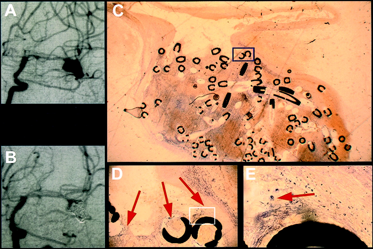
- Fig 2.
Exemplary short-term results from case 1, in which death occurred 5 days after treatment (see Table 1).
A, Angiogram obtained before treatment of a medial cerebral artery aneurysm.
B, Angiogram obtained after GDC treatment of a medial cerebral artery aneurysm.
C, Histologic 5- to 10-μm-thick section of plasticized aneurysm 5 days after treatment depicts a thrombus consisting of fibrin and erythrocytes. The scale can be extrapolated from the coil diameter of 0.01 in.
D, ×10 magnification of the inset shown in C depicts fibrin clotting and erythrocytes (arrows).
E, ×40 magnification of the inset shown in D depicts single macrophages (arrow).
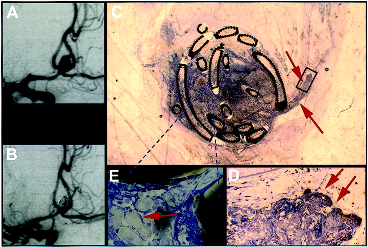
- Fig 3.
Exemplary mid-term results from case 3, in which death occurred 13 days after treatment (see Table 1).
A, Angiogram obtained before treatment of an anterior communicating artery aneurysm.
B, Angiogram obtained after GDC treatment of an anterior communicating artery aneurysm.
C, Histologic 5- to 10-μm-thick section of plasticized aneurysm 13 days after treatment shows thrombus extending into the parent vessel (arrows). The scale can be extrapolated from the coil diameter of 0.010 in.
D, ×20 magnification of the inset in shown in C depicts that the thrombus projecting into the feeding vessel is partially coated with endothelium (arrows).
E, ×100 magnification of the area in C indicated by the dashed lines depicts foamy giant cells between the platinum coils (arrow).
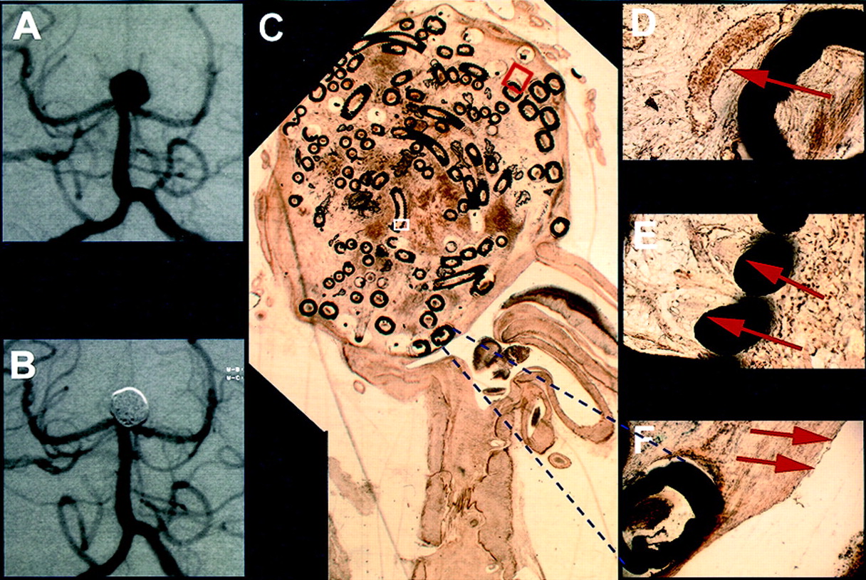
- Fig 4.
Exemplary long-term results from case 6, in which death occurred 272 days after treatment (Table 1).
A, Angiogram obtained before treatment of a basilar tip aneurysm.
B, Angiogram obtained after treatment of a basilar tip aneurysm.
C, Histologic 5- to 10-μm-thick section of plasticized aneurysm depicts scar-like connective tissue in the former aneurysm lumen 272 days after treatment. The scale can be extrapolated from the two coil diameters of 0.010 and 0.015 in.
D, ×20 magnification of the red inset shown in C depicts vascularized connecting tissue surrounding platinum coils (arrow).
E, ×40 magnification of the white inset shown in C depicts foreign body giant cells with multiple nuclei adjacent to coils (arrows).
F, ×10 magnification of the area indicated by the dashed lines depicts scar tissue covered by a layer of long slender cells, resembling endothelium, demarcating the aneurysm orifice against the parent vessel (arrows).


Tables
Patients, aneurysms, and clinical characteristics in six autopsied cases
Case No. Sex Age at Time of Death H&H Aneurysm Size Time Lapse (days) Aneurysm Location Cause of Death Survival (days) 1* M 43 III l 1 Medial cerebral artery Brain swelling 5 2 F 41 III m 1 Superior cerebellar artery Brain swelling 12 3* F 48 IV m 2 Anterior communicating artery Brain swelling 13 4 F 58 III s 1 Anterior communicating artery Vasospasm 20 5 F 43 IV m 1 Basilary tip Brain swelling 26 6* F 72 V m 1 Basilary tip Frontal cerebral bleeding 272 Note.—H&H indicates Hunt and Hess Scale grade at admission;
* , case discussed in present text; m, male; f, female; l, large (>15 mm); m, medium (6–15 mm); s, small (<6 mm). Time lapse is number of days between first bleeding and treatment; survival is number of days survived after treatment.
In this issue


Jump to section
Related Articles
Cited By...
- Histopathological analysis of in vivo specimens of recurrent aneurysms after coil embolization
- Characterizing patterns of endothelialization following coil embolization: a whole-mount, dual immunostaining approach
- Immunohistochemical analysis of a ruptured basilar top aneurysm autopsied 22 years after embolization with Guglielmi detachable coils
- Mechanisms of Healing in Coiled Intracranial Aneurysms: A Review of the Literature
- Immunohistochemical analysis of a ruptured basilar top aneurysm autopsied 22 years after embolization with Guglielmi detachable coils
- Temporal Evolution of Susceptibility Artifacts from Coiled Aneurysms on MR Angiography: An In Vivo Canine Study
- Matrix2 Coils in Embolization of Intracranial Aneurysms: 1-Year Outcome and Comparison with Bare Platinum Coil Group in a Single Institution
- In Vivo Experimental Intracranial Aneurysm Models: A Systematic Review
- Computerized Assessment of Angiographic Occlusion Rate and Coil Density in Embolized Human Cerebral Aneurysms
- Fatal Recurrent Subarachnoid Hemorrhage after Endovascular Aneurysm Occlusion from Overdistention of the Aneurysm Wall