
Article Figures & Data
Figures
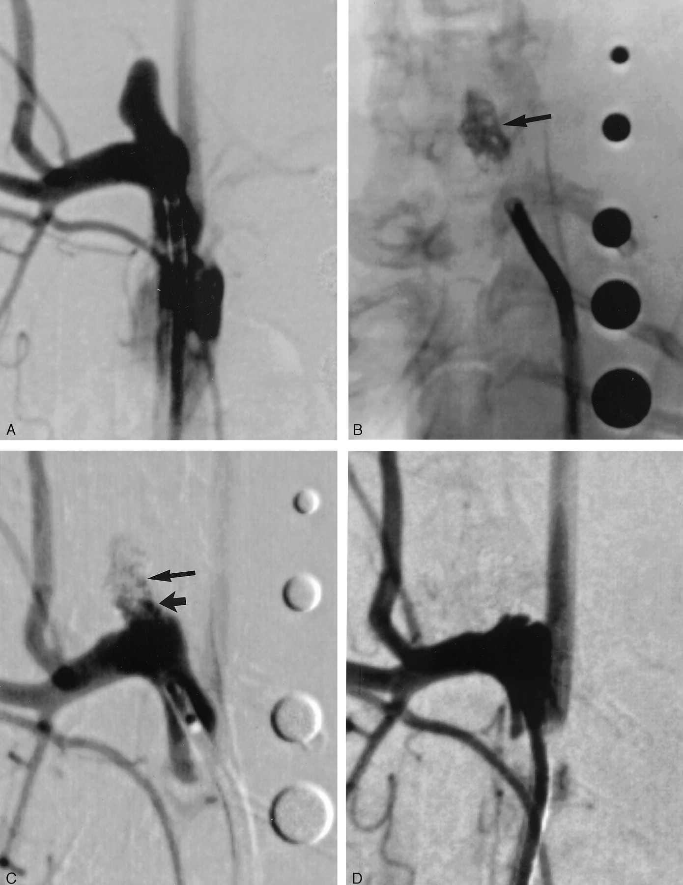
- Fig 1.
Subject 1, aneurysm packed with collagen-based coil (2-week sample).
A, DSA image, showing aneurysm cavity.
B, Radiograph, showing collagen coils after implantation (arrow). Radiopaque sizing markers range from 2 mm to 6 mm in diameter.
C, DSA image, immediately after coil implantation, demonstrating residual flow at the neck (short arrow) and in the body (long arrow) of the aneurysm cavity.
D, DSA image, 2 weeks after implantation, showing interval progressive aneurysm occlusion, with no residual flow in the aneurysm cavity.
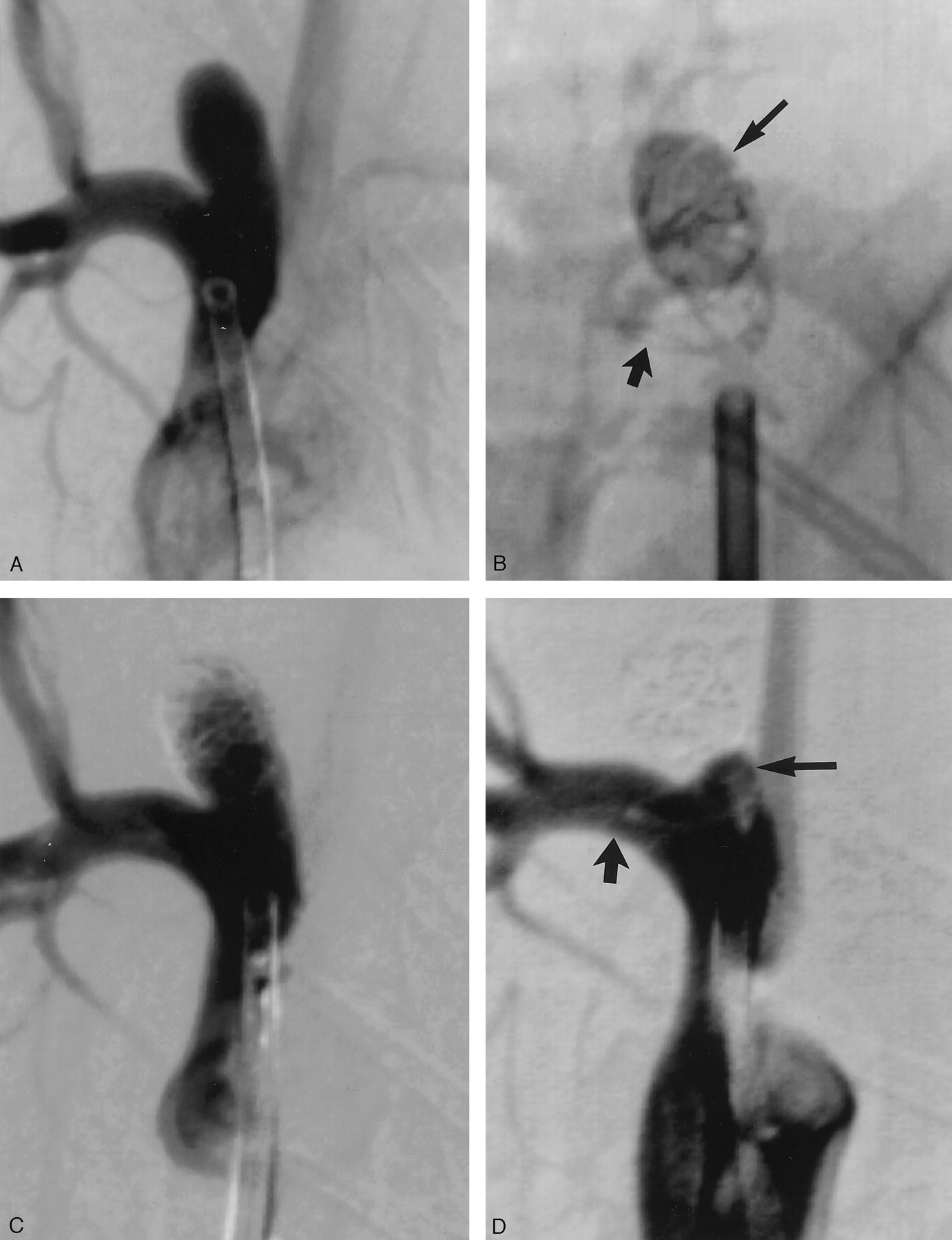
- Fig 2.
Subject 8, aneurysm packed with collagen coil (10-week sample).
A, DSA image, showing aneurysm cavity.
B, Radiograph, showing collagen coils after implantation. Loose packing was present in the aneurysm cavity (long arrow) with a loop of coil present in the parent artery distal to the aneurysm cavity (short arrow).
C, DSA image, immediately after coil implantation, demonstrating large amount of residual flow in the body of the aneurysm cavity. No compromise of flow existed in the parent artery distal to the aneurysm cavity.
D, DSA image, 10 weeks after implantation, showing interval progressive aneurysm occlusion, with no residual flow in the aneurysm cavity and a triangular neck remnant (long arrow). Flow remained unimpeded in the region of the coils in the distal parent artery (short arrow).
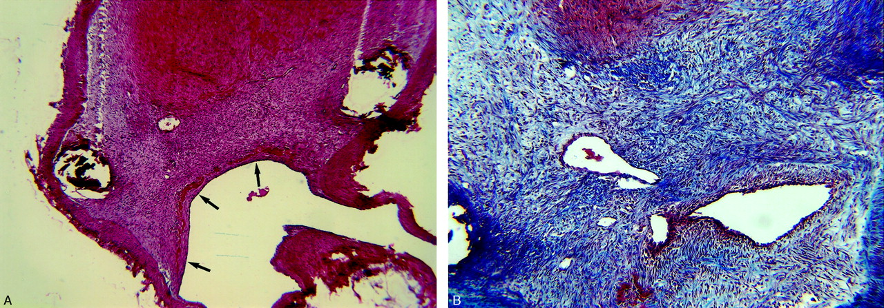
- Fig 3.
Subject 3, aneurysm packed with collagen coil (2-week sample).
A, Histologic specimen showing the neck of the aneurysm covered by a thick layer of tissue consisting of nucleated cells (arrows). (Hematoxylin and eosin stain, original magnification ×40.)
B, Histologic speciment showing dense blue staining indicates collagen deposition within the aneurysm cavity. Vascular channels are also present. (Trichrome stain, original magnification ×100.).
- Fig 4.
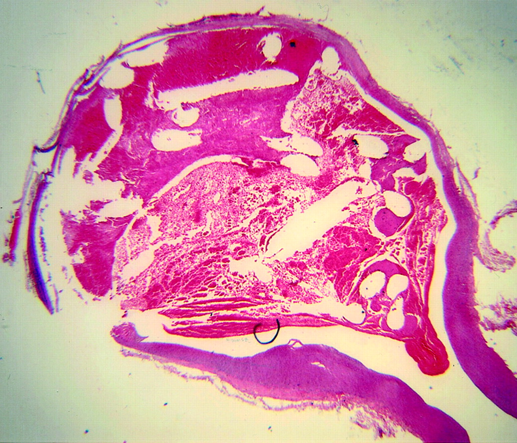
Subject 7, aneurysms packed with collagen-based coil (10-week sample). Histologic specimen showing the aneurysm cavity is filled with dense matrix and spindle cells. (Hematoxylin and eosin stain, original magnification ×20.).
- Fig 5.
Subject 5, aneurysms packed with platinum coil (2-week sample). Histologic specimen showing the aneurysm cavity is filled with unorganized thrombus. The thrombus in the dome is laminated, suggesting ongoing turnover of unorganized thrombus. (Hematoxylin and eosin stain, original magnification ×20.).
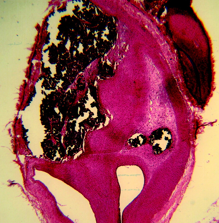
- Fig 6.
Subject 12, aneurysm packed with platinum coil (10-week sample). Histologic specimen showing the aneurysm cavity remains nearly completely filled with unorganized thrombus, with a small amount of loose matrix along the periphery (arrows). (Hematoxylin and eosin stain, original magnification ×20.).


In this issue


Jump to section
Related Articles
Cited By...
- Rabbit Elastase Aneurysm Model Mimics the Recurrence Rate of Human Intracranial Aneurysms following Platinum Coil Embolization
- Rabbit aneurysm models mimic histologic wall types identified in human intracranial aneurysms
- Autologous adipose-derived mesenchymal stem cells improve healing of coiled experimental saccular aneurysms: an angiographic and histopathological study
- Correlation of thrombus formation on 7 T MRI with histology in a rat carotid artery side wall aneurysm model
- Healing of saccular aneurysms following platinum coil embolization: lack of improved efficacy with vitamin C supplementation
- Embolization of intracranial aneurysms with second-generation Matrix-2 detachable coils: mid-term and long-term results
- The Woven EndoBridge: A New Aneurysm Occlusion Device
- Creation of Large Elastase-Induced Aneurysms: Presurgical Arterial Remodeling Using Arteriovenous Fistulas
- Five-Year Follow-Up in Elastase-Induced Aneurysms in Rabbits
- In Vivo Experimental Intracranial Aneurysm Models: A Systematic Review
- Intrinsic Pathway-Mediated Apoptosis in Elastase-Induced Aneurysms in Rabbits
- Morbidity and Mortality Associated with Creation of Elastase-Induced Saccular Aneurysms in a Rabbit Model
- Endovascular Histologic Effects of Ultrathin Gold- or Vitronectin-Coated Platinum Aneurysm Coils in a Rodent Arterial Occlusion Model: A Preliminary Investigation
- Control of Aneurysm Volume by Adjusting the Position of Ligation During Creation of Elastase-Induced Aneurysms: A Prospective Study
- Interventional Neuroradiology