
Article Figures & Data
Figures
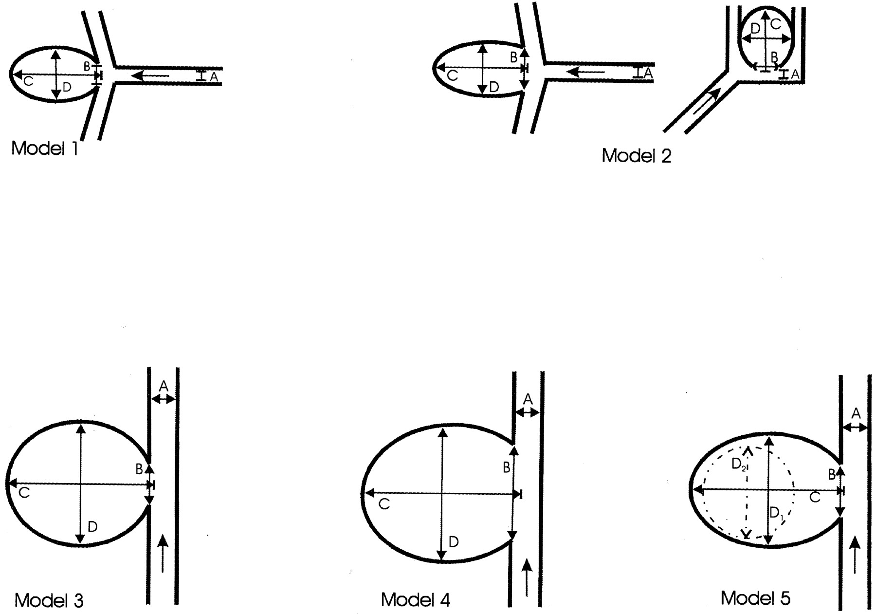
- Fig 1.
Aneurysm models 1–5. Arrows indicate the direction of blood flow. A indicates parent artery; B, ostium width, C, dome height; and D, dome diameter.
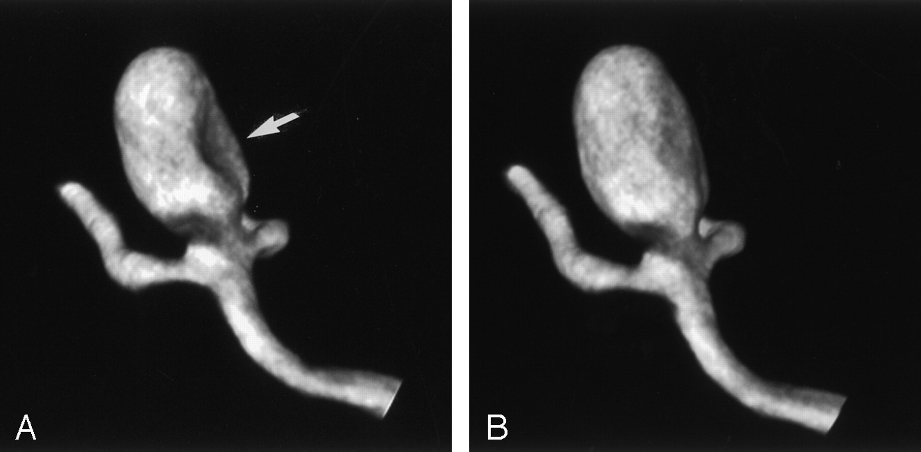
- Fig 2.
Model 2, wide-necked aneurysm in the MCA bifurcation and AcoA. Voxel size, 0.25 mm.
A, The 5-second rotation with continuous flow shows filling defects in both aneurysms (arrows).
B, The 8-second rotation with continuous flow shows a slight defect in the MCA aneurysm (arrow).
C, Accurate visualization of the aneurysm is achieved in the 8-second rotation with the bolus injection.
D, The 14-second rotation with continuous flow has higher plasticity, but it does not provide any additional information about the geometry of the aneurysm.
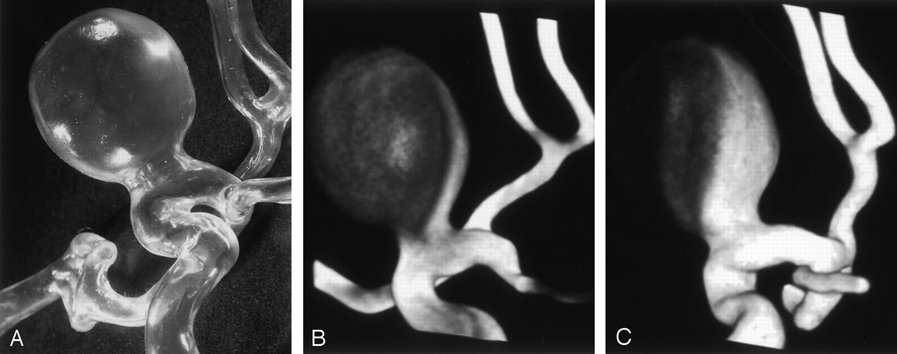
- Fig 3.
Model 1, small-necked aneurysm in the MCA bifurcation. Voxel size, 0.13 mm.
A, The 8-second rotation with continuous flow shows a filling defect at the medial wall of the aneurysm (arrow).
B, The 8-second rotation with bolus injection provides accurate 3D visualization of the shape of the aneurysm.
- Fig 4.
Model 3, small-necked giant lateral aneurysm. Voxel size, 0.16 mm.
A, The true shape of the aneurysm was not visualized on the 3D reconstruction.
B, The 14-second rotation with a bolus injection reveals only the distal aneurysmal shell.
C, In the 14-second rotation, the proximal wall of the aneurysm is not clearly delineated, even after the administration of contrast material at a rate of 5 mL/s.
- Fig 5.
Model 4, large-necked giant lateral aneurysm with large neck. Voxel size, 0.18 mm. The 8-second rotation with a bolus injection enables complete visualization of the shape of the aneurysm.


Tables
Model and Parent Artery* Ostium Width, mm Dome Height, mm Dome Diameter, mm 1, MCA 3 × 3 4.5 16.5 10 2, MCA 3 × 3 8 16.5 10 AcoA 2 × 2 6 11 10 3, ICA 5 × 5.5 8 27 23 4, ICA 5 × 5.5 17 28 25.5 × 24 5, ICA 5 × 5.5 10.5 28 21 Second lobule 12 17 17 * AcoA indicates anterior communicating artery.
Protocol Duration of Rotational Run 5 Seconds 8 Seconds 14 Seconds Continuous flow, mL/s 4.0 2.5 2.5 Initial bolus injection, mL/s First phase 5.0 over 2 s 5.0 over 2 s 4.0 over 4 s Second phase 3.3 over 3 s 1.7 over 6 s 1.9 over 10 s
In this issue


Jump to section
Related Articles
Cited By...
- Influence of observers, threshold values, and measurement methods on volumetric analysis of cerebral aneurysms with three-dimensional rotational angiography
- Artery Length Sensitivity in Patient-Specific Cerebral Aneurysm Simulations
- The utility of cone beam volume CT in the evaluation of thrombosed intracranial aneurysms in subarachnoid hemorrhage
- The utility of cone beam volume CT in the evaluation of thrombosed intracranial aneurysms in subarachnoid hemorrhage