Advertisement

Main menu

  • Home
  • Content
    • Current Issue
    • Accepted Manuscripts
    • Article Preview
    • Past Issue Archive
    • AJNR Case Collection
    • Case of the Week Archive
    • Classic Case Archive
    • Case of the Month Archive
  • Special Collections
    • Spinal CSF Leak Articles (Jan 2020-June 2024)
    • 2024 AJNR Journal Awards
    • Most Impactful AJNR Articles
  • Multimedia
    • AJNR Podcast
    • AJNR Scantastics
    • Video Articles
  • For Authors
    • Submit a Manuscript
    • Author Policies
    • Fast publishing of Accepted Manuscripts
    • Graphical Abstract Preparation
    • Manuscript Submission Guidelines
    • Imaging Protocol Submission
    • Submit a Case for the Case Collection
  • About Us
    • About AJNR
    • Editorial Board
  • More
    • Become a Reviewer/Academy of Reviewers
    • Subscribers
    • Permissions
    • Alerts
    • Feedback
    • Advertisers
    • ASNR Home
  • Other Publications
    • ajnr

User menu

  • Alerts
  • Log in

Search

  • Advanced search
American Journal of Neuroradiology

American Journal of Neuroradiology

ASHNR American Society of Functional Neuroradiology ASHNR American Society of Pediatric Neuroradiology ASSR
  • Alerts
  • Log in

Advanced Search

  • Home
  • Content
    • Current Issue
    • Accepted Manuscripts
    • Article Preview
    • Past Issue Archive
    • AJNR Case Collection
    • Case of the Week Archive
    • Classic Case Archive
    • Case of the Month Archive
  • Special Collections
    • Spinal CSF Leak Articles (Jan 2020-June 2024)
    • 2024 AJNR Journal Awards
    • Most Impactful AJNR Articles
  • Multimedia
    • AJNR Podcast
    • AJNR Scantastics
    • Video Articles
  • For Authors
    • Submit a Manuscript
    • Author Policies
    • Fast publishing of Accepted Manuscripts
    • Graphical Abstract Preparation
    • Manuscript Submission Guidelines
    • Imaging Protocol Submission
    • Submit a Case for the Case Collection
  • About Us
    • About AJNR
    • Editorial Board
  • More
    • Become a Reviewer/Academy of Reviewers
    • Subscribers
    • Permissions
    • Alerts
    • Feedback
    • Advertisers
    • ASNR Home
  • Follow AJNR on Twitter
  • Visit AJNR on Facebook
  • Follow AJNR on Instagram
  • Join AJNR on LinkedIn
  • RSS Feeds

Welcome to the new AJNR, Updated Hall of Fame, and more. Read the full announcements.


AJNR is seeking candidates for the position of Associate Section Editor, AJNR Case Collection. Read the full announcement.


OtherINTERVENTIONAL

Preliminary Study of Tagged MR Image Velocimetry in a Replica of an Intracranial Aneurysm

Haruo Isoda, Shoichi Inagawa, Hiroyasu Takeda, Satoshi Isogai, Yasuo Takehara and Harumi Sakahara
American Journal of Neuroradiology April 2003, 24 (4) 604-607;
Haruo Isoda
  • Find this author on Google Scholar
  • Find this author on PubMed
  • Search for this author on this site
Shoichi Inagawa
  • Find this author on Google Scholar
  • Find this author on PubMed
  • Search for this author on this site
Hiroyasu Takeda
  • Find this author on Google Scholar
  • Find this author on PubMed
  • Search for this author on this site
Satoshi Isogai
  • Find this author on Google Scholar
  • Find this author on PubMed
  • Search for this author on this site
Yasuo Takehara
  • Find this author on Google Scholar
  • Find this author on PubMed
  • Search for this author on this site
Harumi Sakahara
  • Find this author on Google Scholar
  • Find this author on PubMed
  • Search for this author on this site
  • Article
  • Figures & Data
  • Info & Metrics
  • Responses
  • References
  • PDF
Loading

Article Figures & Data

Figures

  • Fig 1.
    • Download figure
    • Open in new tab
    • Download powerpoint
    Fig 1.

    Images of the replica of an anterior communicating artery aneurysm.

    A, On an original axial image, transverse through the center of the aneurysm, an aneurysmal sac (left arrow) shows relatively low signal intensity compared with the A1 segments (arrowheads) of the anterior cerebral artery. This signal intensity is due to the relatively slow flow in the intracranial aneurysm. Stronger signal intensity (right arrow) at the left aspect of the replica is observed; this is due to stronger flow from the right A1 segment into the left aspect of the aneurysm.

    B–D, Anteroposterior (B), inferosuperior (C), and left-to-right (D) projections of MIP MR angiograms show the anterior communicating artery aneurysm (arrow).

  • Fig 2.
    • Download figure
    • Open in new tab
    • Download powerpoint
    Fig 2.

    Schematic diagram of the PIV system. The PIV technique is used to calculate the flow vector on the basis of the positional changes of the particles in the flowing fluid; for this, two sequential digital images are used.
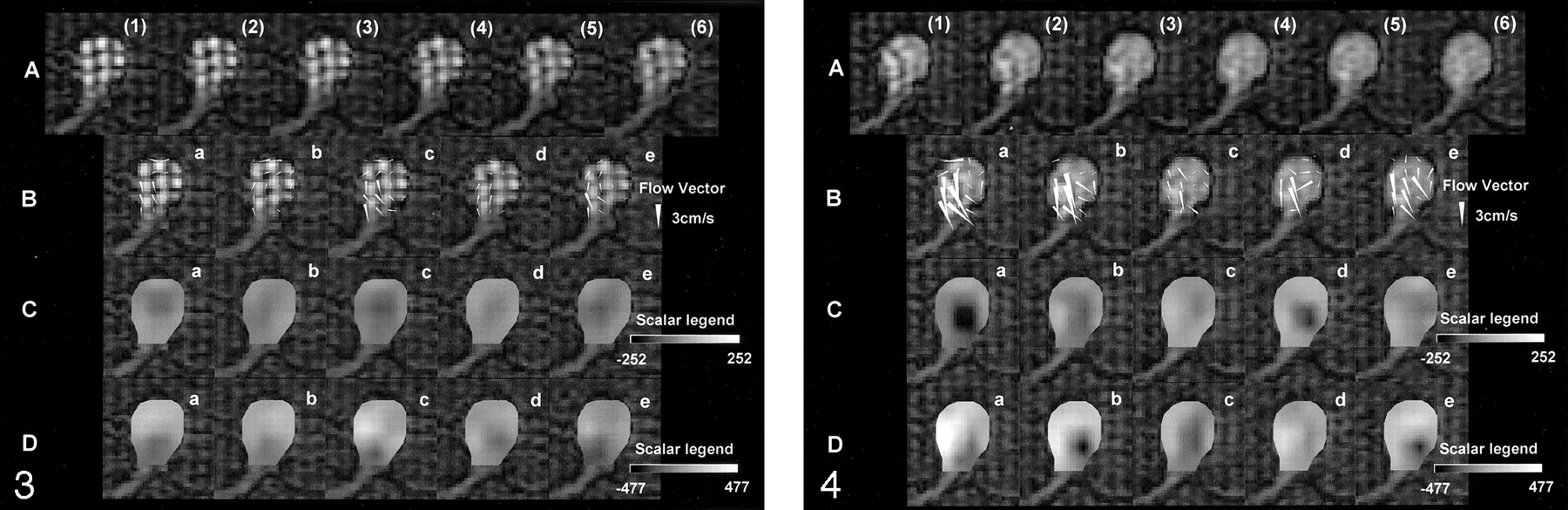
  • Fig 3.
    • Download figure
    • Open in new tab
    • Download powerpoint
    Fig 3.

    Tagged MR images and data processed with PIV software during the diastolic phase. Row A, Six tagged MR images, (1)–(6), were obtained with an interval of 9 milliseconds during the diastolic phase. In the aneurysm replica, counterclockwise rotation of the tag lines is observed. The quantitative velocity is not known; however, we can estimate the hemodynamics in the aneurysm. The tag lines gradually disappear due to the longitudinal relaxation of the spin in the flowing fluid and due to the mixture of the spin. Rows B–D, Flow-vector (row B), vorticity (row C), and shear-strain (row D) images were processed with the PIV software by using the data of tagged MR images. Each image—a, b, c, d, and e—was obtained from the following datasets, respectively: (1) and (2), (2) and (3), (3) and (4), (4) and (5), and (5) and (6). The aneurysm is displayed in the same orientation as in Figure 1A. The flow-vector images reveal the flow vector (arrowheads) of the flowing fluid at each location in the replica every 9 ms. We could measure the quantitative velocity and the direction of flowing fluid. The maximum flow was 2.2 cm/s. Vorticity and shear-strain images are shown in gray scale. In the scalar legends depicted, gray at the midpoint means no vorticity or shear strain. During the diastolic phase, the absolute value of vorticity and shear strain seem to be low, according to the color shown on the images.

  • Fig 4.
    • Download figure
    • Open in new tab
    • Download powerpoint
    Fig 4.

    Tagged MR images and data processed with PIV software during the systolic phase. Row A, Six tagged MR images, (1)–(6), were obtained with an interval of 9 milliseconds during the systolic phase. In the aneurysm replica, counterclockwise rotation of the tag lines is observed. The quantitative velocity is not known; however, we can estimate the hemodynamics in the aneurysm. Tag lines move more quickly in systolic phase than in the diastolic phase (Fig 3A). They also disappear sooner. Rows B–D, Flow vector images (B), vorticity images (C) and shear strain images (D) processed with PIV software from the data of tagged MR images (A) are shown. Each image—a, b, c, d, and e—was obtained from the following datasets, respectively: (1) and (2), (2) and (3), (3) and (4), (4) and (5), and (5) and (6). The aneurysm is displayed in the same orientation as in Figure 1A. Flow-vector images reveal the flow vector (arrowheads) of the flowing fluid at each location in the replica every 9 milliseconds. These velocities obtained in systolic phase (row B) are faster than those in the diastolic phase (Fig 3B). The maximum velocity is 4.5 cm/s, which is slower than expected. This velocity is due to the short neck of the aneurysm. Fewer flow vectors were noted in the left aspect of the aneurysm, where stronger inflow was estimated to be present; the tag lines were not clearly shown because of faster flow. Vorticity and shear-strain images are shown in gray scale. In the scalar legends depicted, gray at the midpoint means no vorticity or shear strain. Some vorticity images show black areas in the center of the aneurysm; these indicate counterclockwise vorticity. Vorticity is stronger during the systolic phase (row C) than during the diastolic phase (Fig 3C). The inflow portion of the left aspect of the replica shows a darker area, which indicates stronger shear strain (row D). On the other hand, the contralateral side of the replica demonstrated a lighter area, which indicates stronger shear strain. On the shear-strain images, differences in color reflect the differences in the direction of shear deformation. The difference in direction is not important because it is not related to the absolute value of the shear strain. Shear strains are stronger during the systolic phase (row D) than during diastolic phase (Fig 3D). Shear strain multiplied by viscosity equals shear stress. Therefore, the aneurysmal wall near the site with strong shear strain is estimated to have strong shear stress.

Back to top

In this issue

American Journal of Neuroradiology: 24 (4)
American Journal of Neuroradiology
Vol. 24, Issue 4
1 Apr 2003
  • Table of Contents
  • Index by author
Advertisement
Print
Download PDF
Email Article

Thank you for your interest in spreading the word on American Journal of Neuroradiology.

NOTE: We only request your email address so that the person you are recommending the page to knows that you wanted them to see it, and that it is not junk mail. We do not capture any email address.

Enter multiple addresses on separate lines or separate them with commas.
Preliminary Study of Tagged MR Image Velocimetry in a Replica of an Intracranial Aneurysm
(Your Name) has sent you a message from American Journal of Neuroradiology
(Your Name) thought you would like to see the American Journal of Neuroradiology web site.
CAPTCHA
This question is for testing whether or not you are a human visitor and to prevent automated spam submissions.
Cite this article
Haruo Isoda, Shoichi Inagawa, Hiroyasu Takeda, Satoshi Isogai, Yasuo Takehara, Harumi Sakahara
Preliminary Study of Tagged MR Image Velocimetry in a Replica of an Intracranial Aneurysm
American Journal of Neuroradiology Apr 2003, 24 (4) 604-607;

Citation Manager Formats

  • BibTeX
  • Bookends
  • EasyBib
  • EndNote (tagged)
  • EndNote 8 (xml)
  • Medlars
  • Mendeley
  • Papers
  • RefWorks Tagged
  • Ref Manager
  • RIS
  • Zotero
0 Responses
Respond to this article
Share
Bookmark this article
Preliminary Study of Tagged MR Image Velocimetry in a Replica of an Intracranial Aneurysm
Haruo Isoda, Shoichi Inagawa, Hiroyasu Takeda, Satoshi Isogai, Yasuo Takehara, Harumi Sakahara
American Journal of Neuroradiology Apr 2003, 24 (4) 604-607;
del.icio.us logo Twitter logo Facebook logo Mendeley logo
  • Tweet Widget
  • Facebook Like
  • Google Plus One

Jump to section

  • Article
    • Abstract
    • Methods
    • Results
    • Discussion
    • Conclusion
    • Footnotes
    • References
  • Figures & Data
  • Info & Metrics
  • Responses
  • References
  • PDF

Related Articles

  • No related articles found.
  • PubMed
  • Google Scholar

Cited By...

  • No citing articles found.
  • Crossref
  • Google Scholar

This article has not yet been cited by articles in journals that are participating in Crossref Cited-by Linking.

More in this TOC Section

  • SAVE vs. Solumbra Techniques for Thrombectomy
  • CT Perfusion&Reperfusion in Acute Ischemic Stroke
  • Delayed Reperfusion Post-Thrombectomy&Thrombolysis
Show more Interventional

Similar Articles

Advertisement

Indexed Content

  • Current Issue
  • Accepted Manuscripts
  • Article Preview
  • Past Issues
  • Editorials
  • Editors Choice
  • Fellow Journal Club
  • Letters to the Editor

Cases

  • Case Collection
  • Archive - Case of the Week
  • Archive - Case of the Month
  • Archive - Classic Case

Special Collections

  • Special Collections

Resources

  • News and Updates
  • Turn around Times
  • Submit a Manuscript
  • Author Policies
  • Manuscript Submission Guidelines
  • Evidence-Based Medicine Level Guide
  • Publishing Checklists
  • Graphical Abstract Preparation
  • Imaging Protocol Submission
  • Submit a Case
  • Become a Reviewer/Academy of Reviewers
  • Get Peer Review Credit from Publons

Multimedia

  • AJNR Podcast
  • AJNR SCANtastic
  • Video Articles

About Us

  • About AJNR
  • Editorial Board
  • Not an AJNR Subscriber? Join Now
  • Alerts
  • Feedback
  • Advertise with us
  • Librarian Resources
  • Permissions
  • Terms and Conditions

American Society of Neuroradiology

  • Not an ASNR Member? Join Now

© 2025 by the American Society of Neuroradiology All rights, including for text and data mining, AI training, and similar technologies, are reserved.
Print ISSN: 0195-6108 Online ISSN: 1936-959X

Powered by HighWire