
Article Figures & Data
Figures
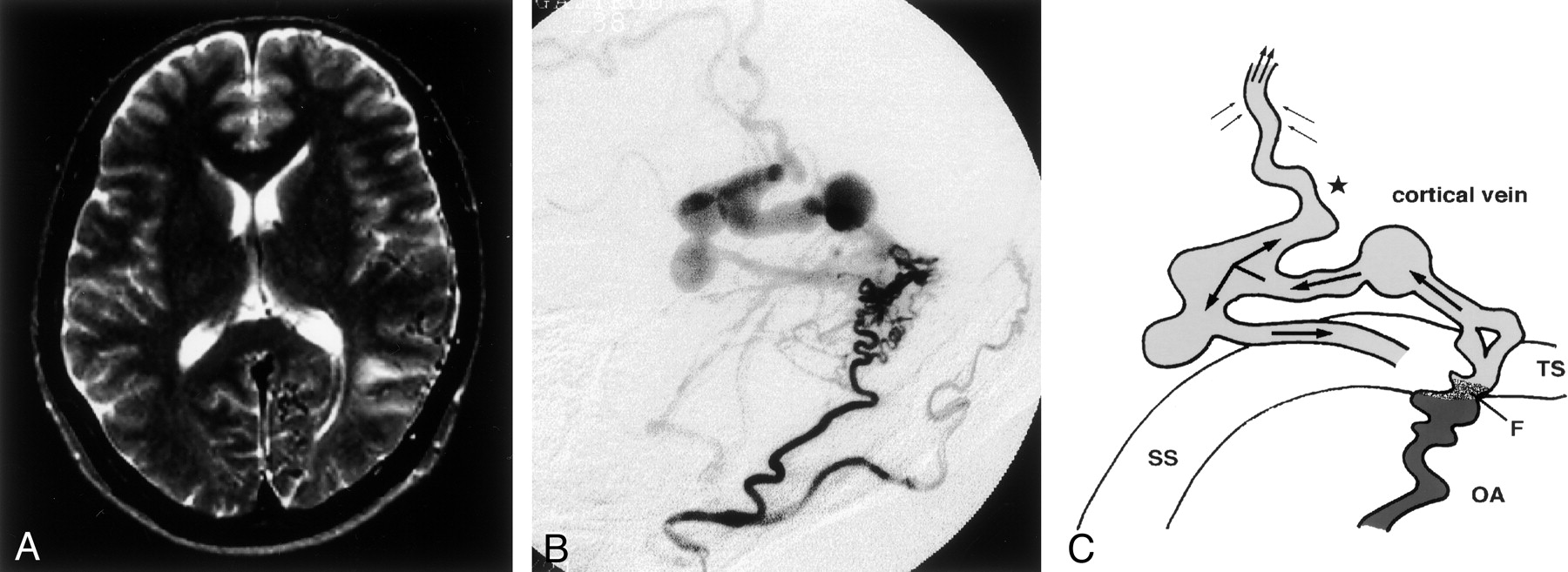
- Fig 1.
Type 1. Case 7 in a 67-year-old man. with cerebral hemorrhage.
A, T2-weighted MR image reveals no hyperintense lesion in the left temporo-parietal lobe.
B, Left external carotid angiogram, lateral projection, shows DAVFs adjacent to the left transverse sinus. Venous drainage is retrograde into the left transverse sinus. An accessory drainage route into the superior sagittal sinus is recognized. Multiple varices are seen in the venous drainage path.
C, Schematic diagram of a DAVF with an accessory route (star) in the retrograde venous drainage (single arrows). The accessory route with retrograde flow (top double arrows) and the surrounding venous flow (left and right double arrows) drain into another sinus through this accessory route. F indicates the fistula point; OA, occipital artery; SS, sigmoid sinus; and TS, transverse sinus.
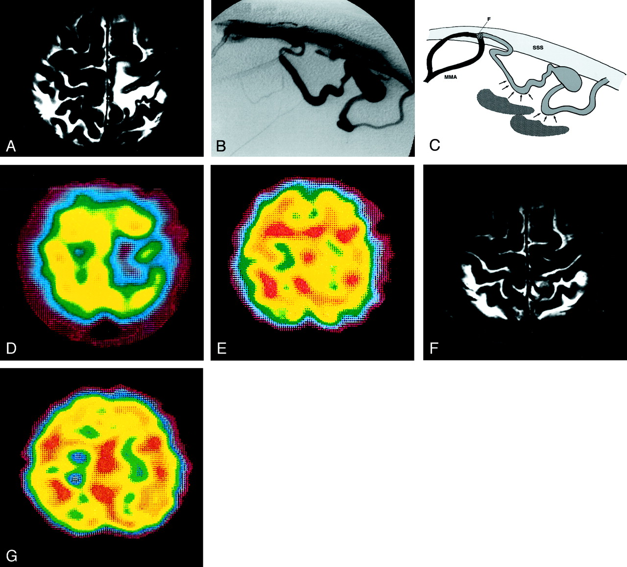
- Fig 2.
Type 2a. Case 13 in a 58-year-old man.
A, T2-weighted MR image reveals a hyperintense lesion in the left parietal lobe.
B, External carotid angiogram, lateral projection, of the left middle meningeal artery, reveals DAVFs at the superior sagittal sinus. The feeder middle meningeal arteries drain directly into the cortical vein. Final venous drainage is into the superior sagittal sinus via a varix. No accessory route is recognized.
C, Schematic drawing of a DAVF without an accessory route in the retrograde venous drainage. The surrounding flow (arrows) cannot drain into another area, resulting in severe venous congestion (shadow). F indicates fistula point; MMA, middle meningeal artery; and SSS, superior sagittal sinus
D, 99mTc-HMPAO SPECT scan shows an area of hypoperfusion at the site of the lesion.
E, After an acetazolamide challenge, the area of hypoperfusion is increased.
F, Post-treatment T2-weighted MR image shows the disappearance of the hyperintense area.
G, Post-treatment SPECT image reveals normal perfusion of the left parietal lobe.
- Fig 3.
Type 2b. Case 21 in a 63-year-old man.
A, T2-weighted MR image reveals a hyperintense lesion in the left temporo-occipital lobe.
B, External carotid angiogram, lateral projection, of the left occipital artery shows DAVFs adjacent to the left transverse sinus. No accessory route is recognized.
C, 99mTc-HMPAO SPECT scan shows a hypoperfused area at the site of the lesion.
D, The hypoperfused area is not increased after the acetazolamide challenge.
E, After treatment, the hyperintense area seen on the T2-weighted MR image persists and expands to the left parietal lobe.
F, SPECT image obtained immediately after treatment reveals hyperperfusion in the left parietal lobe.
G, SPECT image obtained 6 months after treatment demonstrates hypoperfusion in the left parietal lobe.
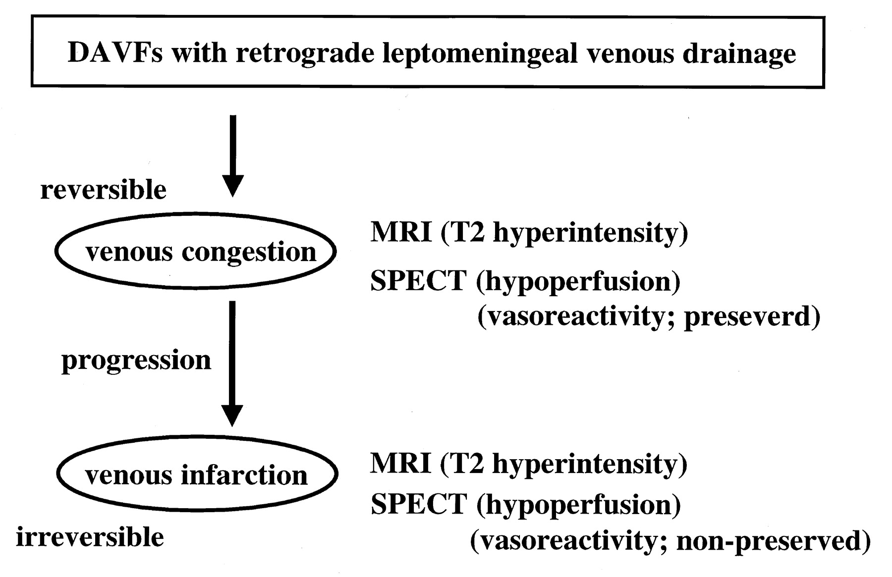
- Fig 4.
Diagram summarizes our MR imaging and SPECT findings in DAVFs with RLVD.


Tables
Patient/Age, y/Sex Clinical Presentation Angiographic Findings Location* Varices Drainage Pattern Accessory Route A: Type 1, Non-hyperintensity on T2-Weighted MR Images 1/66/M Mental disturbance SSS Multiple RLVD only Yes 2/66/F Exophthalmos CS None RLVD only Yes 3/62/F Exophthalmos CS None RLVD only Yes 4/63/M Subarachnoid hemorrhage T-SS Single RLVD only Yes 5/68/F Double vision CS None RLVD only Yes 6/49/F Cerebral hematoma T-SS Single Sinus, RLVD Yes 7/67/M Cerebral hematoma Sig S Multiple Sinus, RLVD Yes 8/59/M Mental disturbance CS Single Sinus, RLVD Yes 9/52/F Mental disturbance CS Single Sinus, RLVD Yes 10/60/F Mental disturbance CS None Sinus, RLVD Yes 11/53/F Double vision CS Single Sinus, RLVD Yes B: Type 2, Hyperintensity on T2-Weighted MR Images 12/51/F Mental disturbance T-SS Single Sinus, RLVD No 13/58/M Lower-limb hypesthesia SSS Single RLVD only No 14/66/F Exophthalmos CS None RLVD only No 15/31/F Exophthalmos CS None Sinus, RLVD No 16/50/M Exophthalmos CS None Sinus, RLVD No 17/71/M Headache T-SS None Sinus, RLVD No 18/63/F Headache CS None Sinus, RLVD No 19/47/M Visual field disturbance T-SS None RLVD only No 20/50/F Mental disturbance T-SS None RLVD only No 21/63/M Mental disturbance T-SS None RLVD only No 22/70/F Exophthalmos CS None RLVD only No * CS indicates cavernous sinus; SSS, superior sagittal sinus; T-SS, transverse-sigmoid sinus; and SigS, sigmoid sinus.
- TABLE 2:
MR Imaging and SPECT findings in nine patients with DAVF hyperintensity on T2-Weighted MR Images
Patient Before Treatmen Findings After Treatment Findings Outcome T2-Weighted MR Imaging SPECT T2-Weighted MR Imaging SPECT At Rest With Acetazolamide At Rest With Acetazolamide Type 2a 12 Hyperintensity Hypoperfusion Preserved T2-normalization at 1 mo Normal Not done Good 13 Hyperintensity Hypoperfusion Preserved T2-normalization at 2 wk Normal Not done Good 14 Hyperintensity Hypoperfusion Preserved T2-normalization at 2 wk Normal Not done Good 15 Hyperintensity Hypoperfusion Preserved T2-normalization at 1 wk Normal Not done Good 16 Hyperintensity Hypoperfusion Preserved T2-normalization at 3 wk Normal Not done Good 17 Hyperintensity Hypoperfusion Preserved T2-normalization at 2 wk Normal Not done Good 18 Hyperintensity Hypoperfusion Preserved T2-normalization at 3 wk Normal Not done Good Type 2b 19 Hyperintensity Hypoperfusion Non-preserved T2-hyperintensity continued, subcortical hemorrhage Hypoperfusion, peripheral hyperperfusion Non-preserved Poor 20 Hyperintensity Hypoperfusion Non-preserved T2-hyperintensity continued Hypoperfusion Non-preserved Poor 21 Hyperintensity Hypoperfusion Non-preserved T2-hyperintensity continued Hypoperfusion Non-preserved Poor 22 Hyperintensity Hypoperfusion Non-preserved T2-hyperintensity continued Hypoperfusion Non-preserved Poor
In this issue


Jump to section
Related Articles
Cited By...
- No citing articles found.