
Article Figures & Data
Figures
- Fig 1.
MR images obtained in a 70-year-old man with metastatic brain tumor.
A, T1-weighted spin-echo image does not clearly show distinction between lesion (arrow) and surrounding edematous tissue.
B, T1-weighted FLAIR image clearly shows enhancing lesion (arrow).
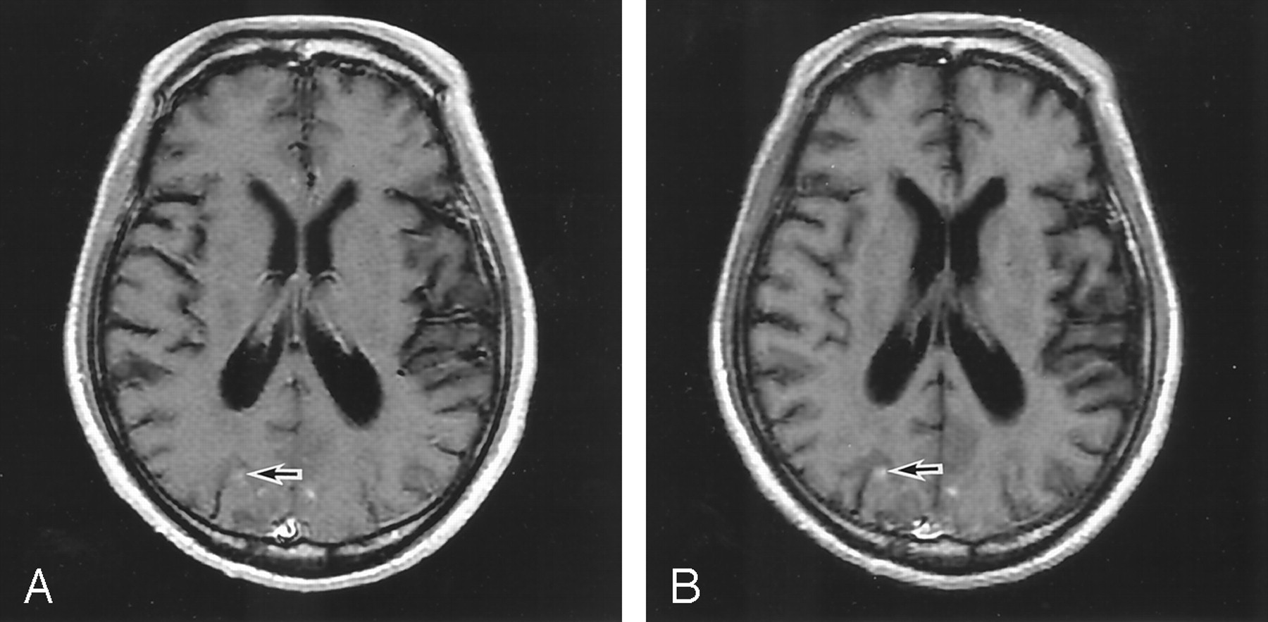
- Fig 2.
MR images obtained in a 60-year-old man with multiple metastatic brain tumors.
A, T1-weighted spin-echo image has poor lesion-WM CNR (arrow).
B, T1-weighted FLAIR image clearly shows enhancing lesion (arrow).


Tables
Lesion-WM Lesion-CSF GM-WM CSF-WM T1-weighted spin-echo CNR 8.07 ± 4.84 30.6 ± 5.71 5.13 ± 2.14 22.4 ± 3.29 T1-weighted FLAIR CNR 14.1 ± 8.78 41.5 ± 9.10 9.10 ± 3.07 27.1 ± 4.26 Note.—WM signifies white matter; CSF, cerebrospinal fluid; GM, gray matter; CNR, contrast-to-noise ratio; and FLAIR, fluid-attenuated inversion recovery.
* Values represent the mean ± SD.
T1-weighted FLAIR Before T1-weighted/ Spin-Echo Imaging T1-weighted FLAIR After T1-weighted/ Spin-Echo Imaging T1-weighted spin-echo imaging 35.5 ± 7.18 37.5 ± 4.05 T1-weighted FLAIR imaging 41.6 ± 5.64 48.7 ± 11.4 Note.—FLAIR signifies fluid-attenuated inversion recovery.
* Values represent the mean ± SD.
Lesion Conspicuity Image Artifact Image Contrast Radiologist 1 3.90 ± 0.91 3.00 ± 0.46 4.25 ± 0.72 Radiologist 2 4.20 ± 0.95 3.75 ± 0.64 3.95 ± 0.89 Radiologist 3 3.55 ± 0.60 3.05 ± 0.39 3.95 ± 0.69 * Values represent mean ± SD.


