
Article Figures & Data
Figures
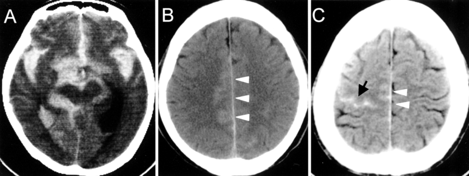
- Fig 1.
Representative CT findings in patients presenting with SAH.
A, Diffuse thick SAH in the most subarachnoid cisterns due to bleeding from arterial dissection at A1.
B, Thin localized SAH in the interhemispheric fissure (arrowheads) due to bleeding from arterial dissection at A3.
C, Very thin SAH in the interhemispheric cistern (arrowheads) and in the sulcus of the convexity of the cerebral hemisphere (arrow) due to bleeding from arterial dissection at A2 in the combined group.
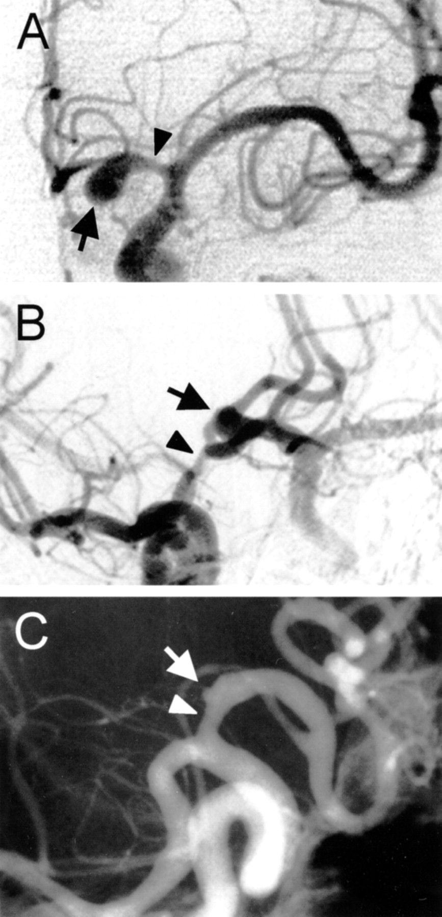
- Fig 2.
Representative findings of cerebral angiography in the ischemic cases.
A, Lateral view angiogram of the right carotid artery in case 9. Stenosis (arrowheads) with dilation (arrow) at the A2 portion of the ACA.
B, Oblique view angiogram of the left carotid artery in case 13. Stenosis (closed arrowhead) with dilation (open arrowheads) at the A2 portion of the ACA accompanied by an intimal flap (arrow).
C, Oblique view angiogram of the left carotid artery in case 8. Stenosis (closed arrowheads) accompanied by the double lumen sign (open arrowheads).
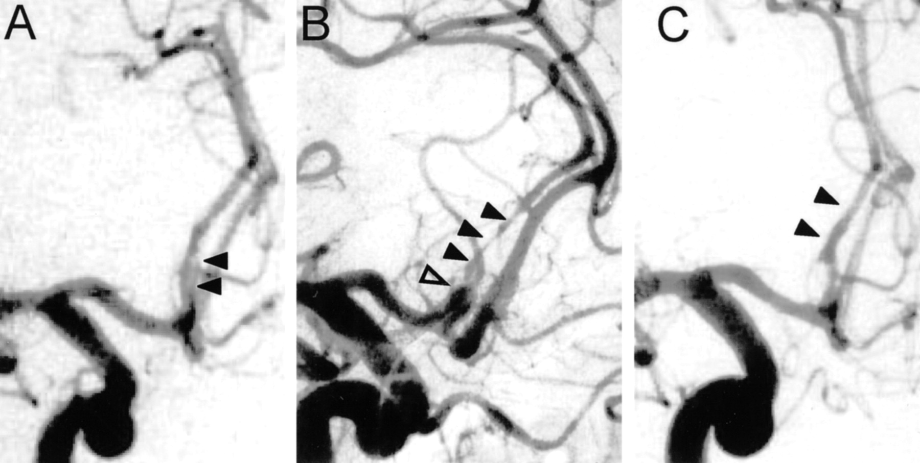
- Fig 3.
Cerebral angiograms of the cases presenting with bleeding from arterial dissection at A1. Slight stenosis (arrowhead) and aneurysmal dilation (arrow) are seen in all cases.
A, Anteroposterior view angiogram of the left carotid artery in case 1.
B, Oblique view angiogram of the right carotid artery in case 3.
C, Oblique view angiogram of the right carotid artery in case 2.
- Fig 4.
Representative findings of serial changes of the lesion on cerebral angiograms.
A, Oblique view angiogram of the right carotid artery, obtained at admission, shows mild stenosis accompanied by double lumen (arrowheads) at A2.
B, Oblique view angiogram of the right carotid artery shows progression to severe stenosis (closed arrowheads) with aneurysmal dilation (open arrowhead) 2 weeks after onset.
C, Oblique view angiogram of the right carotid artery shows resolution 5 months after onset (arrowheads).
- Fig 5.
Bar graph shows serial changes of the stenotic portion as seen on cerebral angiograms. The changes were analyzed by using the 15 follow-up angiography studies.
- Fig 6.
Images from case 12.
A, T1-weighted MR image shows hyperintensity around the signal intensity void.
B, Lateral view angiogram of the left carotid artery, obtained on the same day as the image presented in A, shows stenosis (closed arrowheads) with dilation (open arrowheads).
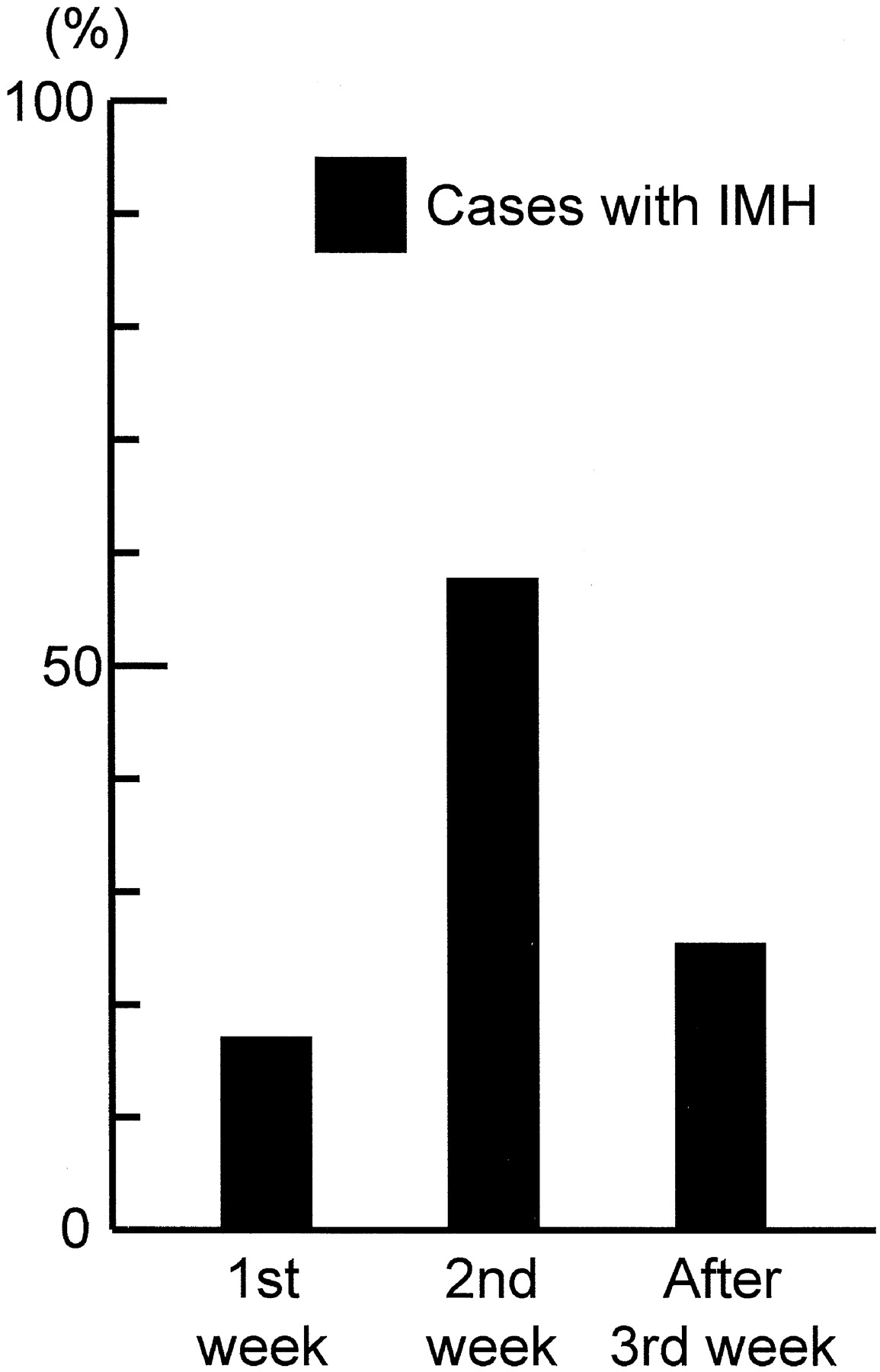
- Fig 7.
Bar graph shows hyperintensity around the signal intensity void, as seen on T1-weighted MR images, considered to be due to intramural hematoma (IMH) based on the timing of the examination.
- Fig 8.
Example comparisons of MR angiograms and cerebral angiograms.
A, MR angiogram from case 7 shows stenosis (arrow) in the left ACA.
B, Lateral view angiogram of the left carotid artery shows compatible findings.
C, MR angiogram from case 9 shows stenosis (arrowheads) and dilation (arrow) in the right ACA.
D, Anteroposterior view angiogram of the right carotid artery shows compatible findings.
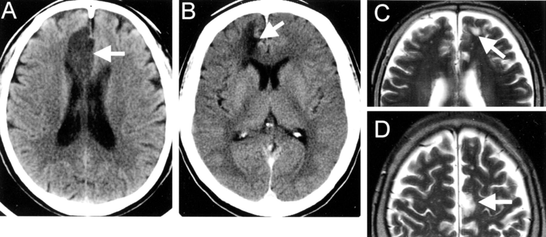
- Fig 9.
CT scans and MR images show infarction in patients presenting with cerebral ischemia.
A, CT scan shows infarction along the interhemispheric fissure (arrow).
B, CT scan shows infarction at the watershed area (arrow).
C, MR image shows multiple patchy infarctions in the ACA territory (arrow).
D, MR image shows patchy infarctions in the ACA territory (arrow).


Tables
- TABLE 1:
Clinical characteristics and neuroradiologic findings in 18 cases of anterior cerebral artery dissection
Case No. Age (yr)/Sex Presentation at Onset Site of Lesion Initial Angiographic Findings Confirmation of Dissection Outcome 1 39/F Bleeding A1 S + D Ope VS 2 66/F Bleeding A1 S + D Ope VS 3 66/F Bleeding A1 S + D Ope MD 4 49/F Bleeding A2 S DL† GR 5 53/M Bleeding A3 S DL GR 6 43/F Ischemia A1–2 S IMH GR 7 50/F Ischemia A2 S DL† GR 8 65/M Ischemia A2 S DL GR 9 56/M Ischemia A2 S + D DL† GR 10 67/F Ischemia A1–2 S + D DL† GR 11 52/F Ischemia A2 S + D IMH GR 12 36/M Ischemia A2 S IMH MD 13 56/F Ischemia A2 S + D DL GR 14 50/F Ischemia A2 S IMH, DL GR 15 40/M Combined* A2 S IMH, DL GR 16 64/F Combined* A2 S + D IMH, DL GR 17 49/F Combined* A2 S + D Ope GR 18 49/M Combined* A2–3 S DL GR Note.—F indicates female; M, male; S + D, stenosis with dilation; S, stenosis without dilation; Ope, discoloration of affected artery around aneurysmal dilation due to intramural hematoma seen during operation; DL, double lumen sign seen on cerebral angiograms; IMH, hyperintensity around signal void due to intramural hematoma seen on T1-weighted MR images; VS, vegetative state; MD, moderate disability; GR, good recovery.
* Four cases showed ischemic symptoms and CT evidence of slight subarachnoid hemorrhage on initial CT scans.
† In 4 cases, the double lumen sign was seen on follow-up angiography.
- TABLE 2:
Clinical parameters and angiographic findings in cases of anterior cerebral artery dissection according to clinical presentations
Total Bleeding Group Ischemic Group Combined Group No. of cases 18 5 9 4 Age (yr) (mean ± SD) 52.8 ± 9.8 54.6 ± 11.6 52.8 ± 9.8 50.5 ± 9.9 Sex ratio (male:female) 6:12 1:3 3:6 2:2 Main site of lesion (n) A1 3 3 0 0 A2 14 1 9 4 A3 1 1 0 0 Initial angiographic findings (n) Stenosis without dilation 8 2 4 2 Stenosis with dilation 10 3 5 2 Double lumen or intimal flap 7* 1* 3* 3* Dilation only 0 0 0 0 Prognosis at 1 year after onset (n) Good recovery 14 2 8 4 Moderate disability 2 1 1 0 Severe disability 0 0 0 0 Vegetative state 2 2 0 0 Death 0 0 0 0 * Double lumen or intimal flap was seen accompanied by the other angiographic findings.
- TABLE 3:
Clinical parameters and angiographic findings in reported cases of anterior cerebral artery dissection
Total Bleeding Group Ischemic Group No. of cases 15 7 8 Age (yr) (mean ± SD) 49.2 ± 10.4 52.6 ± 13.9 46.3 ± 5.3 Sex ratio (male:female) 12:3 5:2 7:1 Site of lesion (n) A1 3 2 1 A2 9 2 7 A3, 4 3 3 0 Angiographic findings (n) Stenosis without dilation 0 0 0 Stenosis with dilation 8 2 6 Double lumen or intimal flap 5 1 4* Dilation only 4 4 0 Occlusion 2 0 2 Outcome (n) Good recovery 12 5 7 Moderate disability 1 0 1 Severe disability 1 1 0 Vegetative state 0 0 0 Death 1 1 0 * Double lumen or intimal flap was seen in four cases accompanied by the other angiographic findings.
In this issue


Jump to section
Related Articles
Cited By...
- No citing articles found.