
Article Figures & Data
Figures
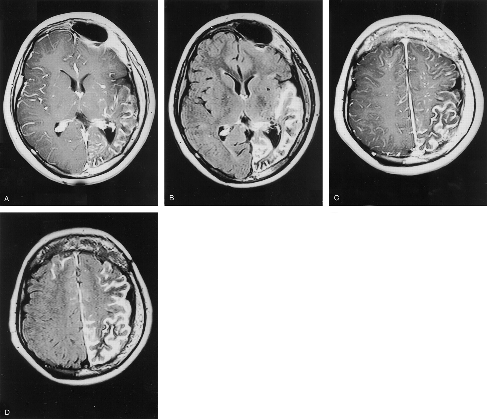
- Fig 1.
Images from the case of a 9-year-old child with suspected left-sided Sturge-Weber syndrome.
A, Axial view contrast-enhanced T1-weighted image obtained at the level of the Monro foramina. Note the prominent but normal enhancement of vascular structures in the right hemisphere.
B, Axial view contrast-enhanced FLAIR image obtained at the level of the Monro foramina shows the improved visualization of leptomeningeal disease in the left temporal lobe. Note that enhancement of normal vascular structures in the right hemisphere is suppressed.
C, Axial view contrast-enhanced T1-weighted image obtained toward the vertex.
D, Axial view contrast-enhanced FLAIR image obtained toward the vertex shows considerably more enhancement in the left frontal lobe as compared with the T1-weighted image. More importantly, abnormal leptomeningeal enhancement is shown over the right frontal lobe, indicating bilateral disease. The presence of bilateral disease precluded surgical intervention.
- Fig 2.
Images from the case of a 3-year-old patient who presented with early morning headache and vomiting headache. A mass in the fourth ventricle was shown to be a medulloblastoma.
A, Axial view contrast-enhanced T1-weighted image obtained at presentation showed extensive leptomeningeal spread.
B, Significant response to treatment was achieved by 6 months, with no residual disease evident on the T1-weighted image.
C, Contrast-enhanced FLAIR image did show leptomeningeal enhancement in the right frontal lobe (arrows) that could be seen in retrospect on the T1-weighted image.
D, Contrast-enhanced T1-weighted image obtained at 12 months showed progressive disease in the right frontal lobe.
E, Contrast-enhanced T1-weighted image obtained at 18 months also showed progressive disease in the right frontal lobe.
- Fig 3.
Images from the case of a 2-year-old patient who presented with symptoms and signs suggestive of a posterior fossa mass.
A, Coronal view contrast-enhanced T1-weighted image shows a large mass centered in the right cerebellar hemisphere that was confirmed to be medulloblastoma. No evidence of intracranial metastases was observed at presentation.
B, Contrast-enhanced FLAIR image obtained at 6 months shows definite abnormality in the meninges overlying the left parietal lobe (arrow).
C, In retrospect, this region is viewed as abnormal on the contrast-enhanced T1-weighted image, although it was reported as normal.
D, Contrast-enhanced T1-weighted image obtained at 12 months shows definite progression.
E, Contrast-enhanced T1-weighted image obtained at 18 months also shows definite progression.


In this issue


Jump to section
Related Articles
Cited By...
- Gadolinium-Enhanced T2 FLAIR Is an Imaging Biomarker of Radiation Necrosis and Tumor Progression in Patients with Brain Metastases
- Leptomeningeal Enhancement in Multiple Sclerosis and Other Neurological Diseases: A Systematic Review and Meta-Analysis
- Leptomeningeal gadolinium enhancement across the spectrum of chronic neuroinflammatory diseases
- Gadolinium-based MRI characterization of leptomeningeal inflammation in multiple sclerosis
- Elevated Cerebral Blood Volume Contributes to Increased FLAIR Signal in the Cerebral Sulci of Propofol-Sedated Children
- Comparison of the Added Value of Contrast-Enhanced 3D Fluid-Attenuated Inversion Recovery and Magnetization-Prepared Rapid Acquisition of Gradient Echo Sequences in Relation to Conventional Postcontrast T1-Weighted Images for the Evaluation of Leptomeningeal Diseases at 3T
- A Spectrum of Unusual Neuroimaging Findings in Patients with Suspected Sturge-Weber Syndrome