
Article Figures & Data
Figures
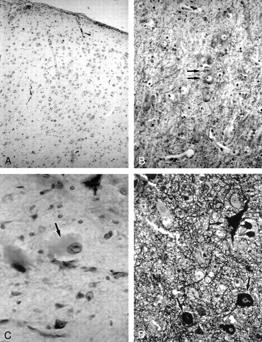
- Fig 1.
Photomicrographs show the histologic characteristics of the FCD subtypes.
A, Architectural dysplasia characterized by moderate derangement of cortical lamination, with neurons of the same shape and size scattered throughout the cortex (Kluver-Barrera stain; original magnification, ×250).
B, Cytoarchitectural dysplasia. Note the cluster of cytomegalic neurons with satellitosis (arrows) (Kluver-Barrera stain; original magnification, ×250).
C, Taylor’s FCD with balloon cells. Note large balloon cell characterized by homogeneous eosinophilic cytoplasm and peripheral nucleus with prominent nucleolus (arrow) (hematoxylin-eosin stain; original magnification, ×250).
D, Taylor’s FCD with balloon cells. Note large dysmorphic neurons containing abundant cytoplasmic neurofilaments (arrows) (Bielchowsky stain; original magnification, ×250).
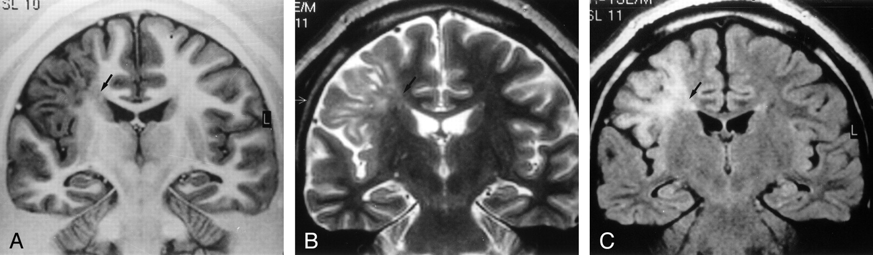
- Fig 2.
MR images of Taylor’s FCD with balloon cells.
A, Coronal turbo SE IR T1-weighted image (3000/20/400/2) demonstrates thickening of the right frontal cortex with loss of demarcation between gray and white matter and decreased white matter signal intensity (arrow) tapering toward the ventricle.
B, Coronal turbo SE T2-weighted image (2300/100/4) and C, coronal turbo SE FLAIR T2-weighted image (6000/100/2000/3), obtained at the same level as A, show increased signal intensity (arrow) of the subcortical white matter extending to the ventricle as a radial band. No mass effect is present.
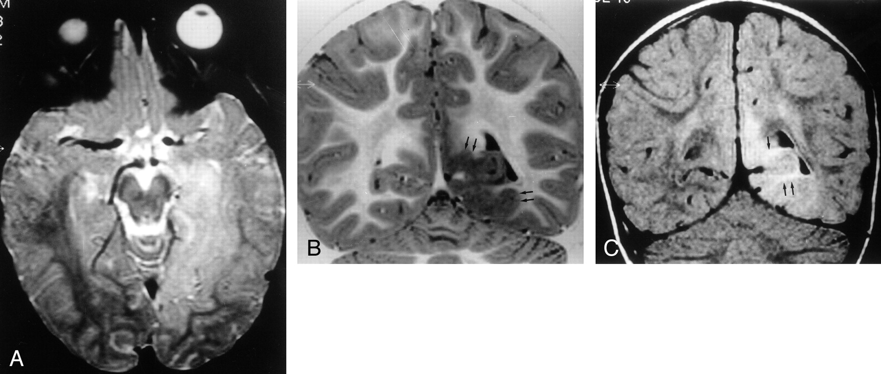
- Fig 3.
MR images of Taylor’s FCD without balloon cells.
A, Transverse SE T2-weighted image (2300/90/1) shows extensive hyperintense lesion in the left temporo-occipitobasal region, with no mass effect on adjacent structures.
B, Coronal turbo SE IR T1-weighted image (3000/20/400/2) better demonstrates thickening of the cortex (arrows) with blurring of the gray-white matter junction and subcortical white matter hypointensity.
C, Coronal turbo SE FLAIR T2-weighted image (6000/100/2000/3) reveals that the hyperintensity of the lesion mainly involves the subcortical white matter (arrows). The ventricular trigone is enlarged on the left.
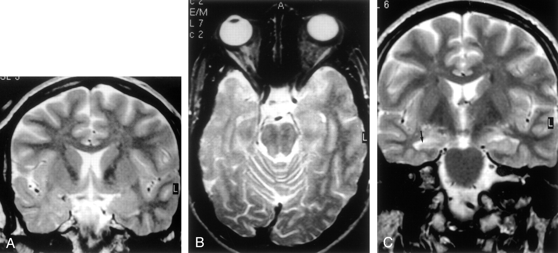
- Fig 4.
MR images of cytoarchitectural dysplasia.
A, Coronal turbo SE IR T1-weighted image (3000/20/400/2), B, coronal turbo SE T2-weighted image (2300/100/4), and C, sagittal turbo SE T2-weighted image of a 3D volume acquisition (2100/130/1). Note reduced demarcation of the gray-white matter boundary in the right temporal lobe, with marked signal intensity alterations of subcortical white matter, which is hypointense on the T1-weighted image in A and hyperintense on the T2-weighted images in B and C. These alterations induced us to diagnose Taylor’s FCD. Note, however, that the thickening of the cortex (arrows in A and B), is only mild. This is a case of histologic cytoarchitectural dysplasia resembling Taylor’s FCD.
- Fig 5.
MR images of architectural dysplasia and ipsilateral hippocampal sclerosis (dual abnormality).
A, Coronal turbo SE T2-weighted image (2300/100/4) reveals hypoplasia of right temporal pole with white matter hyperintensity.
B, Transverse SE T2-weighted image (2300/90/1) confirms reduced volume of right temporal pole compared with the contralateral side, with enlargement of the overlying subarachnoid space.
C, Coronal turbo SE T2-weighted image (2300/100/4) shows right hippocampal head (arrow), characterized by atrophy and signal hyperintensity, suggesting hippocampal sclerosis.


Tables
Classification Characteristics Architectural dysplasia Heterotopic neurons in white matter Derangement of cortical lamination Cytoarchitectural dysplasia Heterotopic neurons in white matter Derangement of cortical lamination Giant neurons Taylor’s FCD Without balloon cells Heterotopic neurons in white matter Derangement of cortical lamination Giant neurons Dysmorphic neurons With balloon cells Heterotopic neurons in white matter Derangement of cortical lamination Giant neurons Dysmorphic neurons Balloon cells Sex (F/M) Age at Surgery (y)* Age at Clinical Onset (y)* Seizure Duration (y)* No. of Seizures per month* Surgical Outcome Engel Class Ia† Whole series (n = 49) 27/22 2–42 (24 ± 11) 0–26 (6 ± 7) 1–42 (17 ± 10) 1–600 (69 ± 117) 25 (51) Histologic TFCD (n = 15) 7/8 2–35 (19 ± 11) 0–22 (6 ± 7) 2–27 (13 ± 8) 1–400 (97 ± 103) 11 (73) Histologic CD (n = 6) 3/3 4–42 (20 ± 16) 0–26 (6 ± 10) 1–42 (14 ± 16) 1–300 (152 ± 139) 2 (33) Histologic AD (n = 28) 17/11 2–41 (27 ± 9) 0–24 (7 ± 6) 2–34 (20 ± 8) 1–600 (39 ± 112) 12 (43) * Data are ranges. Numbers in parentheses are the mean ± SD.
† Data are number (%) of patients seizure free at ≥1 year.
Note.—TFCD indicates Taylor’s FCD; CD = cytoarchitectural dysplasia; AD = architectural dysplasia.
- TABLE 3:
MR Findings in 49 Patients with Histologically Diagnosed FCD Who Were Operated On for Drug-Resistant Partial Epilepsy
MR Findings No. (%) of Patients MR Diagnosis TFCD Non-TFCD Unrevealing 15 (31) HS 3 (6) – – FCD 17 (35) 12 5 Dual abnormality (FCD + HS) 14 (28) 1 13 Note.—TFCD indicates Taylor’s FCD; HS = hippocampal sclerosis.
Histopathologic Diagnosis (n = 49) MR Negative for FCD (n = 19 [39]) MR Positive for FCD (n = 30 [61])* Detailed MR Diagnosis TFCD 13 non-TFCD 17* TFCD (n = 15) 5 (33) 10 (67) 9 1 CD (n = 6) 3 (50) 3 (50) 2 1 AD (n = 28) 11 (39) 17 (61) 2 15 Note.—TFCD indicates Taylor’s FCD; CD = cytoarchitectural dysplasia; AD = architectural dysplasia.
Data are number of patients. Numbers in parentheses are percentages.
* One MR-diagnosed non-TFCD case is not included because it could not be verified histologically (MR abnormalities in temporal lobe, but ipsilateral frontal region resected).
MR Diagnosis Cortical Thickening GM-WM Blurring WM Hyperintensity on T2WI WM Hypo-intensity on T1WI IR Tapering to Ventricle GM Hyper-intensity on T2WI Focal Hypoplasia WM Core Atrophy Histopathologic Diagnosis Severe Moderate TFCD (n = 13) 10 13 13 – 10 3 6 3 – 7 8 8 – 6 3 2 2 – TFCD with BC (n = 8) 1 1 1 – 1 – 1 – – TFCD without BC (n = 1) 1 2 2 – 2 – 2 – – CD (n = 2) 1 2 2 – 1 – 1 1 – AD (n = 2) Non-TFCD (n = 17) 2 5 – 14 6 – 2 17 16 – – – 1 – – – 1 1 TFCD with BC (n = 1) – – – 1 – – – 1 – CD (n = 1) 2 5 – 12 6 – 2 15 15 AD (n = 15) Note.—GM indicates gray matter; WM = white matter; WI = weighted images; TFCD = Taylor’s FCD; BC = balloon cells.
Data are number of patients.
In this issue


Jump to section
Related Articles
Cited By...
- Striking MRI Changes of Focal Cortical Dysplasia Over Time: A Case Series and Literature Review
- Optimizing the Detection of Subtle Insular Lesions on MRI When Insular Epilepsy Is Suspected
- The surgically remediable syndrome of epilepsy associated with bottom-of-sulcus dysplasia
- Optimizing MR Imaging Detection of Type 2 Focal Cortical Dysplasia: Best Criteria for Clinical Practice
- Diffusion Tensor Imaging Assessment of the Epileptogenic Zone in Children with Localization-Related Epilepsy
- Individual Differences in Verbal Abilities Associated with Regional Blurring of the Left Gray and White Matter Boundary
- FDG-PET improves surgical outcome in negative MRI Taylor-type focal cortical dysplasias
- Evaluation of Focal Cortical Dysplasia and Mixed Neuronal and Glial Tumors in Pediatric Epilepsy Patients Using 18F-FDG and 11C-Methionine PET
- Disorders of Cortical Formation: MR Imaging Features
- Focal cortical dysplasia: long term seizure outcome after surgical treatment
- Terminology and classification of the cortical dysplasias