Advertisement

Main menu

  • Home
  • Content
    • Current Issue
    • Accepted Manuscripts
    • Article Preview
    • Past Issue Archive
    • AJNR Case Collection
    • Case of the Week Archive
    • Classic Case Archive
    • Case of the Month Archive
  • Special Collections
    • Spinal CSF Leak Articles (Jan 2020-June 2024)
    • 2024 AJNR Journal Awards
    • Most Impactful AJNR Articles
  • Multimedia
    • AJNR Podcast
    • AJNR Scantastics
    • Video Articles
  • For Authors
    • Submit a Manuscript
    • Author Policies
    • Fast publishing of Accepted Manuscripts
    • Graphical Abstract Preparation
    • Manuscript Submission Guidelines
    • Imaging Protocol Submission
    • Submit a Case for the Case Collection
  • About Us
    • About AJNR
    • Editorial Board
  • More
    • Become a Reviewer/Academy of Reviewers
    • Subscribers
    • Permissions
    • Alerts
    • Feedback
    • Advertisers
    • ASNR Home
  • Other Publications
    • ajnr

User menu

  • Alerts
  • Log in

Search

  • Advanced search
American Journal of Neuroradiology

American Journal of Neuroradiology

ASHNR American Society of Functional Neuroradiology ASHNR American Society of Pediatric Neuroradiology ASSR
  • Alerts
  • Log in

Advanced Search

  • Home
  • Content
    • Current Issue
    • Accepted Manuscripts
    • Article Preview
    • Past Issue Archive
    • AJNR Case Collection
    • Case of the Week Archive
    • Classic Case Archive
    • Case of the Month Archive
  • Special Collections
    • Spinal CSF Leak Articles (Jan 2020-June 2024)
    • 2024 AJNR Journal Awards
    • Most Impactful AJNR Articles
  • Multimedia
    • AJNR Podcast
    • AJNR Scantastics
    • Video Articles
  • For Authors
    • Submit a Manuscript
    • Author Policies
    • Fast publishing of Accepted Manuscripts
    • Graphical Abstract Preparation
    • Manuscript Submission Guidelines
    • Imaging Protocol Submission
    • Submit a Case for the Case Collection
  • About Us
    • About AJNR
    • Editorial Board
  • More
    • Become a Reviewer/Academy of Reviewers
    • Subscribers
    • Permissions
    • Alerts
    • Feedback
    • Advertisers
    • ASNR Home
  • Follow AJNR on Twitter
  • Visit AJNR on Facebook
  • Follow AJNR on Instagram
  • Join AJNR on LinkedIn
  • RSS Feeds

Welcome to the new AJNR, Updated Hall of Fame, and more. Read the full announcements.


AJNR is seeking candidates for the position of Associate Section Editor, AJNR Case Collection. Read the full announcement.


Research ArticlePEDIATRICS

Brain Imaging in the Unsedated Pediatric Patient: Comparison of Periodically Rotated Overlapping Parallel Lines with Enhanced Reconstruction and Single-Shot Fast Spin-Echo Sequences

Kirsten P. Forbes, James G. Pipe, John P. Karis, Victoria Farthing and Joseph E. Heiserman
American Journal of Neuroradiology May 2003, 24 (5) 794-798;
Kirsten P. Forbes
  • Find this author on Google Scholar
  • Find this author on PubMed
  • Search for this author on this site
James G. Pipe
  • Find this author on Google Scholar
  • Find this author on PubMed
  • Search for this author on this site
John P. Karis
  • Find this author on Google Scholar
  • Find this author on PubMed
  • Search for this author on this site
Victoria Farthing
  • Find this author on Google Scholar
  • Find this author on PubMed
  • Search for this author on this site
Joseph E. Heiserman
  • Find this author on Google Scholar
  • Find this author on PubMed
  • Search for this author on this site
  • Article
  • Figures & Data
  • Info & Metrics
  • Responses
  • References
  • PDF
Loading

Article Figures & Data

Figures

  • Fig 1.
    • Download figure
    • Open in new tab
    • Download powerpoint
    Fig 1.

    Illustration of PROP k-space data acquisition. Data are acquired in a series of rotating blades, each of which collects data from the central area of k-space. Each blade contains several phase-encode lines.

  • Fig 2.
    • Download figure
    • Open in new tab
    • Download powerpoint
    Fig 2.

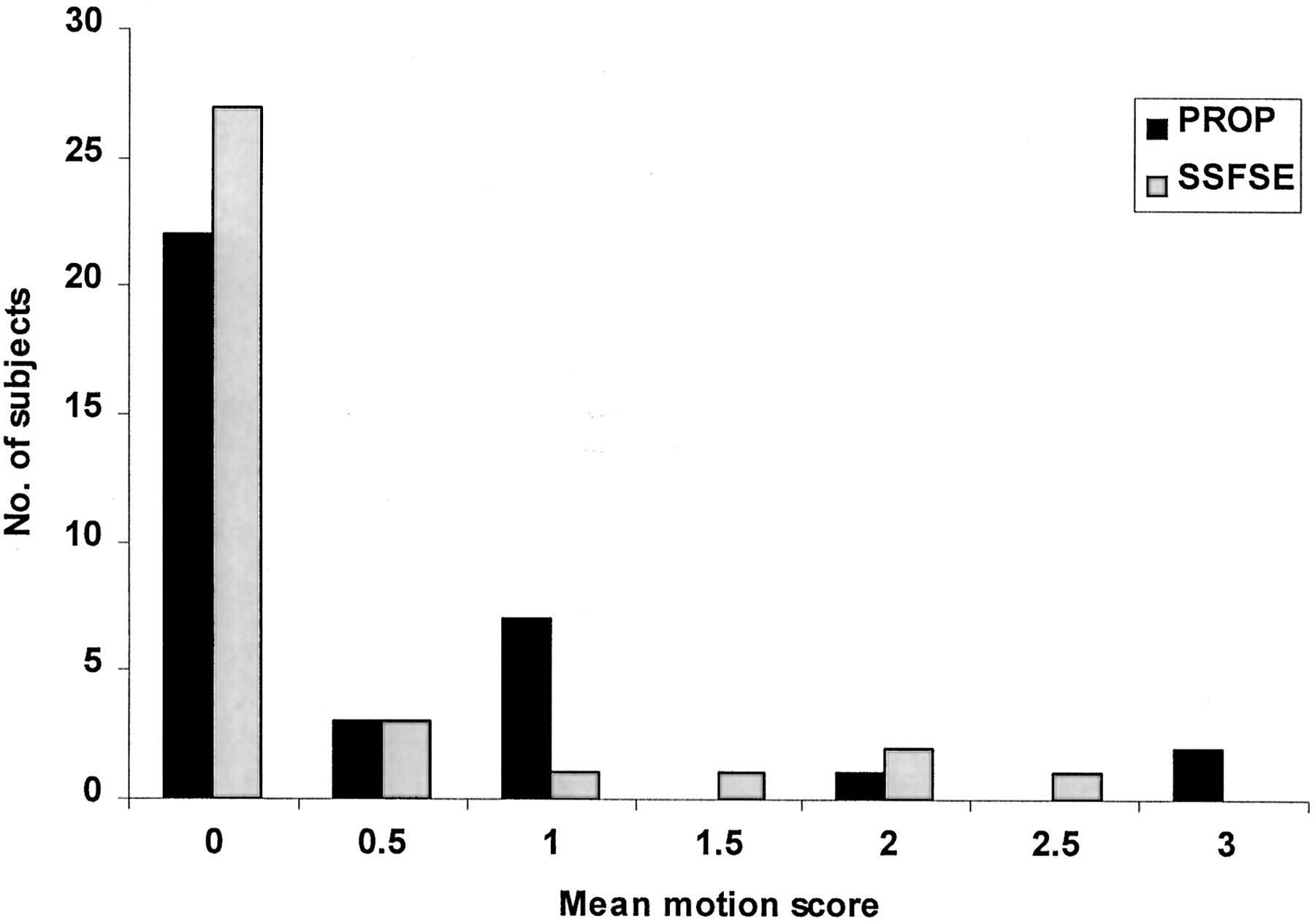
    Bar graph shows the mean score of the two observers in grading both PROP and SS-FSE images for visible effects of motion. Scores were as follows: 0, no motion; 1, mild motion; 2, moderate motion; and 3, severe motion. On most images, no motion effects were seen, and PROP and SS-FSE performed equally in terms of motion correction.

  • Fig 3.
    • Download figure
    • Open in new tab
    • Download powerpoint
    Fig 3.

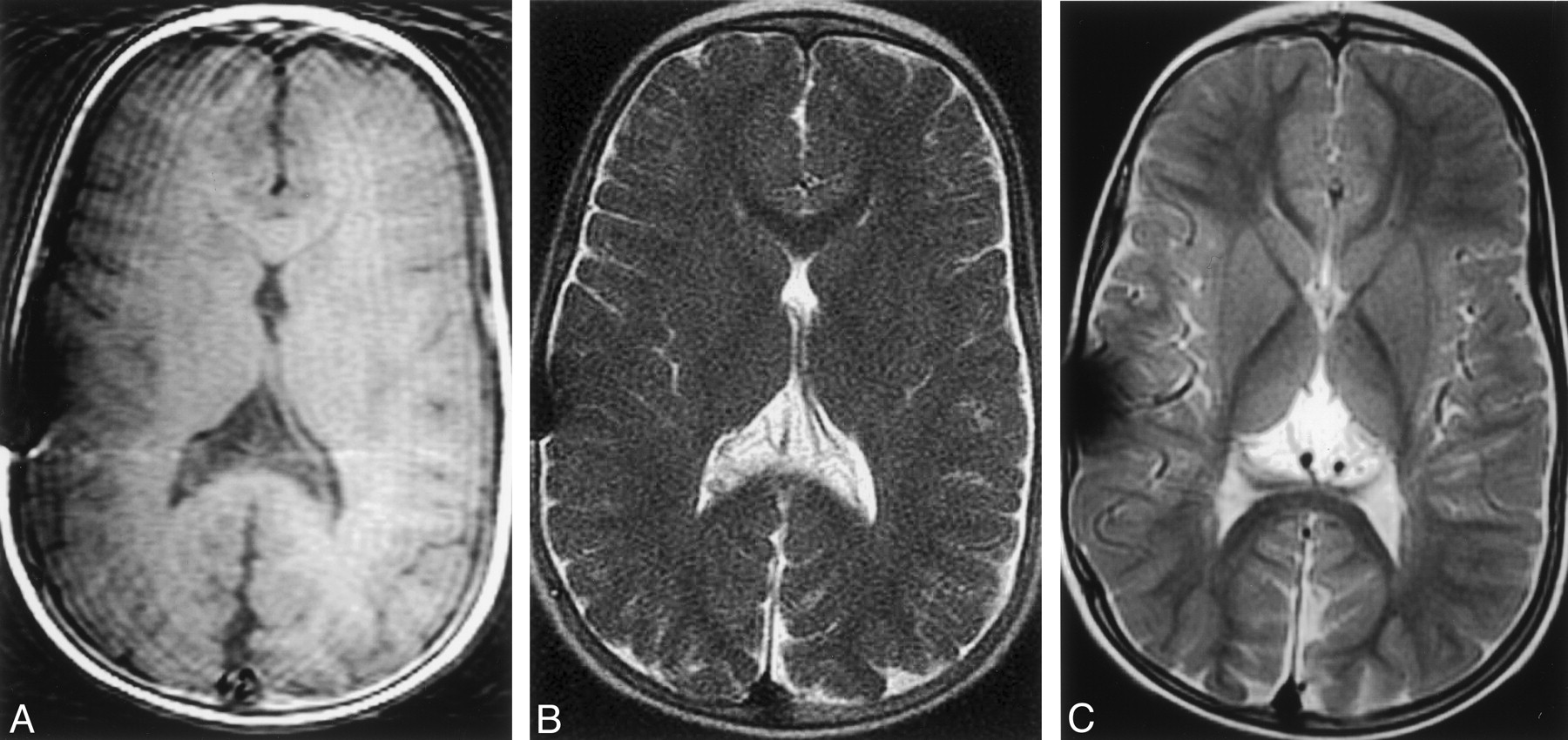
    Compensation of moderate head motion with SS-FSE and PROP FSE imaging.

    A, T1-weighted image shows the effects of head motion. Motion artifact and variable signal intensity are demonstrated across the image.

    B, SS-FSE image shows no evidence of motion artifact and enables a good assessment of ventricular size.

    C, PROP FSE image offers improved gray matter-white matter differentiation. Note that artifact overlying the right side of the skull. This was caused by a ventricular catheter and was worse on this study than on others because of a higher receiver bandwidth.

  • Fig 4.
    • Download figure
    • Open in new tab
    • Download powerpoint
    Fig 4.

    Compensation of severe head motion with SS-FSE and PROP FSE imaging.

    A, T1-weighted image shows the effects of severe head motion, with notable motion artifact and image blurring.

    B, SS-FSE image shows a marked reduction in motion artifact, although the artifact remains even when a parent is holding the child’s head still.

    C, PROP FSE image also offers a substantial reduction in motion artifact, although the image still demonstrates some blurring.

  • Fig 5.
    • Download figure
    • Open in new tab
    • Download powerpoint
    Fig 5.

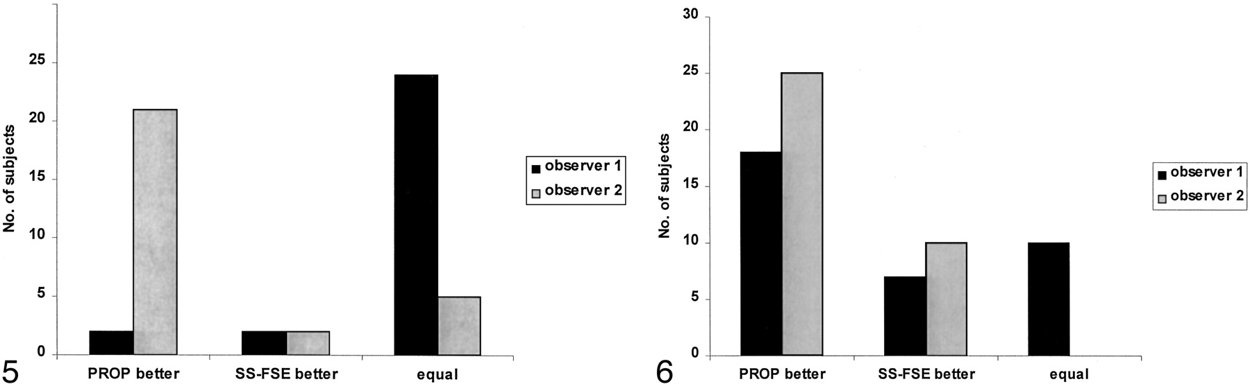
    Visibility of pathology on SS-FSE and PROP images. Bar graph shows the impressions of the two observers regarding the images that depicted pathology most clearly. While observer 1 judged most of the images to be equal, observer 2 thought that PROP images offered an advantage.

  • Fig 6.
    • Download figure
    • Open in new tab
    • Download powerpoint
    Fig 6.

    Bar graph reveals the sequences that the observers preferred. Both observers clearly preferred PROP images because of improvements in the contrast of the brain parenchyma.
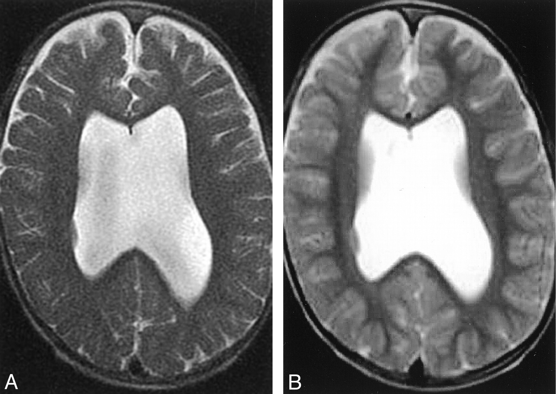
  • Fig 7.
    • Download figure
    • Open in new tab
    • Download powerpoint
    Fig 7.

    Improved image contrast with PROP MR imaging.

    A, SS-FSE image of the brain allows assessment of ventricular size and subarachnoid spaces, but assessment of the brain parenchyma is limited.

    B, PROP FSE image offers improved contrast, which allows a more-detailed assessment of both gray matter and white matter. This improvement allows the identification of a small focus of gray matter heterotopia in the lateral wall of the right lateral ventricle.

Back to top

In this issue

American Journal of Neuroradiology: 24 (5)
American Journal of Neuroradiology
Vol. 24, Issue 5
1 May 2003
  • Table of Contents
  • Index by author
Advertisement
Print
Download PDF
Email Article

Thank you for your interest in spreading the word on American Journal of Neuroradiology.

NOTE: We only request your email address so that the person you are recommending the page to knows that you wanted them to see it, and that it is not junk mail. We do not capture any email address.

Enter multiple addresses on separate lines or separate them with commas.
Brain Imaging in the Unsedated Pediatric Patient: Comparison of Periodically Rotated Overlapping Parallel Lines with Enhanced Reconstruction and Single-Shot Fast Spin-Echo Sequences
(Your Name) has sent you a message from American Journal of Neuroradiology
(Your Name) thought you would like to see the American Journal of Neuroradiology web site.
CAPTCHA
This question is for testing whether or not you are a human visitor and to prevent automated spam submissions.
Cite this article
Kirsten P. Forbes, James G. Pipe, John P. Karis, Victoria Farthing, Joseph E. Heiserman
Brain Imaging in the Unsedated Pediatric Patient: Comparison of Periodically Rotated Overlapping Parallel Lines with Enhanced Reconstruction and Single-Shot Fast Spin-Echo Sequences
American Journal of Neuroradiology May 2003, 24 (5) 794-798;

Citation Manager Formats

  • BibTeX
  • Bookends
  • EasyBib
  • EndNote (tagged)
  • EndNote 8 (xml)
  • Medlars
  • Mendeley
  • Papers
  • RefWorks Tagged
  • Ref Manager
  • RIS
  • Zotero
0 Responses
Respond to this article
Share
Bookmark this article
Brain Imaging in the Unsedated Pediatric Patient: Comparison of Periodically Rotated Overlapping Parallel Lines with Enhanced Reconstruction and Single-Shot Fast Spin-Echo Sequences
Kirsten P. Forbes, James G. Pipe, John P. Karis, Victoria Farthing, Joseph E. Heiserman
American Journal of Neuroradiology May 2003, 24 (5) 794-798;
del.icio.us logo Twitter logo Facebook logo Mendeley logo
  • Tweet Widget
  • Facebook Like
  • Google Plus One

Jump to section

  • Article
    • Abstract
    • Methods
    • Results
    • Discussion
    • Conclusion
    • References
  • Figures & Data
  • Info & Metrics
  • Responses
  • References
  • PDF

Related Articles

  • No related articles found.
  • PubMed
  • Google Scholar

Cited By...

  • Ultrafast Brain MRI Can Be Used for Indications beyond Shunted Hydrocephalus in Pediatric Patients
  • Diagnostic Performance of Ultrafast Brain MRI for Evaluation of Abusive Head Trauma
  • Retrospective Review of Rapid Pediatric Brain MR Imaging at an Academic Institution Including Practice Trends and Factors Affecting Scan Times
  • Motion-Compensation Techniques in Neonatal and Fetal MR Imaging
  • Radiation Risk Due to Shunted Hydrocephalus and the Role of MR Imaging-Safe Programmable Valves
  • Comparison of Brain MR Images at 1.5T Using BLADE and Rectilinear Techniques for Patients Who Move during Data Acquisition
  • BLADE in Sagittal T2-Weighted MR Imaging of the Cervical Spine
  • Improved Delineation of Ventricular Shunt Catheters Using Fast Steady-State Gradient Recalled-Echo Sequences in a Rapid Brain MR Imaging Protocol in Nonsedated Pediatric Patients
  • Crossref
  • Google Scholar

This article has not yet been cited by articles in journals that are participating in Crossref Cited-by Linking.

More in this TOC Section

  • Comparison of Image Quality and Radiation Dose in Pediatric Temporal Bone CT Using Photon-Counting Detector CT and Energy-Integrating Detector CT
  • SyMRI & MR Fingerprinting in Brainstem Myelination
  • Pons&Vermis Localization on Fetal MRI Using U-Net
Show more Pediatrics

Similar Articles

Advertisement

Indexed Content

  • Current Issue
  • Accepted Manuscripts
  • Article Preview
  • Past Issues
  • Editorials
  • Editors Choice
  • Fellow Journal Club
  • Letters to the Editor

Cases

  • Case Collection
  • Archive - Case of the Week
  • Archive - Case of the Month
  • Archive - Classic Case

Special Collections

  • Special Collections

Resources

  • News and Updates
  • Turn around Times
  • Submit a Manuscript
  • Author Policies
  • Manuscript Submission Guidelines
  • Evidence-Based Medicine Level Guide
  • Publishing Checklists
  • Graphical Abstract Preparation
  • Imaging Protocol Submission
  • Submit a Case
  • Become a Reviewer/Academy of Reviewers
  • Get Peer Review Credit from Publons

Multimedia

  • AJNR Podcast
  • AJNR SCANtastic
  • Video Articles

About Us

  • About AJNR
  • Editorial Board
  • Not an AJNR Subscriber? Join Now
  • Alerts
  • Feedback
  • Advertise with us
  • Librarian Resources
  • Permissions
  • Terms and Conditions

American Society of Neuroradiology

  • Not an ASNR Member? Join Now

© 2025 by the American Society of Neuroradiology All rights, including for text and data mining, AI training, and similar technologies, are reserved.
Print ISSN: 0195-6108 Online ISSN: 1936-959X

Powered by HighWire