Advertisement

Main menu

  • Home
  • Content
    • Current Issue
    • Accepted Manuscripts
    • Article Preview
    • Past Issue Archive
    • AJNR Case Collection
    • Case of the Week Archive
    • Classic Case Archive
    • Case of the Month Archive
  • Special Collections
    • Spinal CSF Leak Articles (Jan 2020-June 2024)
    • 2024 AJNR Journal Awards
    • Most Impactful AJNR Articles
  • Multimedia
    • AJNR Podcast
    • AJNR Scantastics
    • Video Articles
  • For Authors
    • Submit a Manuscript
    • Author Policies
    • Fast publishing of Accepted Manuscripts
    • Graphical Abstract Preparation
    • Manuscript Submission Guidelines
    • Imaging Protocol Submission
    • Submit a Case for the Case Collection
  • About Us
    • About AJNR
    • Editorial Board
  • More
    • Become a Reviewer/Academy of Reviewers
    • Subscribers
    • Permissions
    • Alerts
    • Feedback
    • Advertisers
    • ASNR Home
  • Other Publications
    • ajnr

User menu

  • Alerts
  • Log in

Search

  • Advanced search
American Journal of Neuroradiology

American Journal of Neuroradiology

ASHNR American Society of Functional Neuroradiology ASHNR American Society of Pediatric Neuroradiology ASSR
  • Alerts
  • Log in

Advanced Search

  • Home
  • Content
    • Current Issue
    • Accepted Manuscripts
    • Article Preview
    • Past Issue Archive
    • AJNR Case Collection
    • Case of the Week Archive
    • Classic Case Archive
    • Case of the Month Archive
  • Special Collections
    • Spinal CSF Leak Articles (Jan 2020-June 2024)
    • 2024 AJNR Journal Awards
    • Most Impactful AJNR Articles
  • Multimedia
    • AJNR Podcast
    • AJNR Scantastics
    • Video Articles
  • For Authors
    • Submit a Manuscript
    • Author Policies
    • Fast publishing of Accepted Manuscripts
    • Graphical Abstract Preparation
    • Manuscript Submission Guidelines
    • Imaging Protocol Submission
    • Submit a Case for the Case Collection
  • About Us
    • About AJNR
    • Editorial Board
  • More
    • Become a Reviewer/Academy of Reviewers
    • Subscribers
    • Permissions
    • Alerts
    • Feedback
    • Advertisers
    • ASNR Home
  • Follow AJNR on Twitter
  • Visit AJNR on Facebook
  • Follow AJNR on Instagram
  • Join AJNR on LinkedIn
  • RSS Feeds

Welcome to the new AJNR, Updated Hall of Fame, and more. Read the full announcements.


AJNR is seeking candidates for the position of Associate Section Editor, AJNR Case Collection. Read the full announcement.


Research ArticlePEDIATRICS

The Usefulness of MR Imaging in the Diagnosis of Dysembryoplastic Neuroepithelial Tumor in Children: A Study of 14 Cases

Carla Fernandez, Nadine Girard, Armando Paz Paredes, Corinne Bouvier-Labit, Gabriel Lena and Dominique Figarella-Branger
American Journal of Neuroradiology May 2003, 24 (5) 829-834;
Carla Fernandez
  • Find this author on Google Scholar
  • Find this author on PubMed
  • Search for this author on this site
Nadine Girard
  • Find this author on Google Scholar
  • Find this author on PubMed
  • Search for this author on this site
Armando Paz Paredes
  • Find this author on Google Scholar
  • Find this author on PubMed
  • Search for this author on this site
Corinne Bouvier-Labit
  • Find this author on Google Scholar
  • Find this author on PubMed
  • Search for this author on this site
Gabriel Lena
  • Find this author on Google Scholar
  • Find this author on PubMed
  • Search for this author on this site
Dominique Figarella-Branger
  • Find this author on Google Scholar
  • Find this author on PubMed
  • Search for this author on this site
  • Article
  • Figures & Data
  • Info & Metrics
  • Responses
  • References
  • PDF
Loading

Article Figures & Data

Figures

  • Tables
  • Fig 1.
    • Download figure
    • Open in new tab
    • Download powerpoint
    Fig 1.

    Typical DNT findings.

    A, Sagittal T1-weighted MR image shows a large lesion of low signal intensity involving the temporal lobe, without edema or mass effect and corresponding to a complex form of DNT. The lesion is divided by septations leading to an alveolar aspect.

    B, The lesion is of high signal intensity on this T2-weighted MR image. The septations appear to be of low signal intensity.

    C, Sagittal T1-weighted MR image shows a frontoparietal DNT with sharp boundaries and a rectangular pattern of distribution.

    D, Coronal T2-weighted MR image illustrates the triangular pattern of distribution typical of DNT, with a tumor width that is maximal at the cortical level and decreases toward brain ventricles.

    E, Low-magnification view showing the cortical location and the nodular architecture typical of DNT (hematoxylin phloxin-saffron, magnification ×10).

    F, The glio-neuronal specific element is composed of oligodendrocyte-like cells surrounding areas of mucoid substance containing “floating neurons” (hematoxylin phloxin-saffron, magnification ×300).
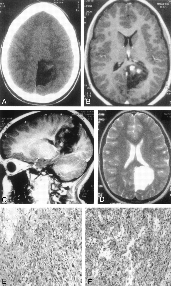
  • Fig 2.
    • Download figure
    • Open in new tab
    • Download powerpoint
    Fig 2.

    DNT involving the occipital lobe and presenting hemorrhagic changes (case 7).

    A, On CT scan, the lesion appears of low attenuation and shows a nodular enhancement after contrast injection.

    B and C, On transverse (B) and sagittal (C) T1-weighted MR images, the DNT is of low signal intensity, displays septations, but shows three areas of hyperintensity after gadolinium injection, mimicking a glioma.

    D, Transverse T2-weighted MR image shows the absence of edema and mass effect on median structure.

    E, Marked nuclear atypias can be observed in the glial areas of DNT (hematoxylin phloxin-saffron, magnification ×200).

    F, An oligodendroglioma-like area showing major hemorrhagic changes characterized by numerous hemosiderin-laden histiocytes (hematoxylin phloxin-saffron, magnification ×200).

Tables

  • Figures
    • View popup
    TABLE 1:

    Clinical findings

    Patient (no.)Age (y)/SexSymptom DurationSeizure TypeExtent of ResectionType of ResectionSeizure Frequency at Last Medical Examination/Anticonvulsive TreatmentTumor RecurrenceFollow-up (Months)
    111/M7 monthsPartial*TotalTumorectomy0/NoNo91
    215/M4 yearsPartial*TotalTumorectomy0/NoNo148
    37/M9 monthsPartial*TotalTumorectomy0/NoNo165
    414/M6 monthsPartialTotalTumorectomy0/NoNo29
    59/F8 monthsPartialTotalTumorectomy0/YesNo32
    69/M2 yearsPartial*TotalLobectomy0/NoNo63
    712/F9 monthsPartialTotalTumorectomy0/NoNo25
    84/F3 yearsPartial*TotalTumorectomy0/NoNo99
    911/F7 yearsPartial*TotalTumorectomy0/NoNo87
    108/M4 monthsPartialTotalTumorectomyUnchanged/yesNo36
    113/M5 monthsPartial*TotalTumorectomy0/YesNo36
    1218/F14 yearsPartial*TotalTumorectomy0/NoNo167
    136/M2 yearsPartial*TotalTumorectomy0 then recurrence/yesYes125
    1414/M2 weeksPartialTotalTumorectomy0/NoNo117
    • * Drug-resistant seizures.

    • View popup
    TABLE 2:

    Neuroradiologic findings

    Patient (no.)LocationCTMRDistributionSeptationsSkull Erosion
    AttenuationContrastT1/T2Contrast
    1FrontalHypoattenuatedNA↓/↑−Rectangular++
    2TemporalNANA↓/↑−Triangular+−
    3TemporalHypoattenuated−↓/↑NATriangular+−
    4FrontalHypoattenuated−↓/↑−Rectangular++
    5TemporalNANA↓/↑NodularRound++
    6TemporalHypoattenuated−↓/↑NATriangular++
    7OccipitalHeterogeneousNodularH/↑NodularTriangular+−
    8FrontoparietalNANA↓/↑NodularRectangular+−
    9TemporalNANA↓/↑−Triangular−−
    10FrontalHypoattenuated−↓/↑NATriangular++
    11TemporalNANA↓/↑−Triangular−−
    12FrontalNANA↓/↑−Triangular+−
    13FrontalHypoattenuated−↓/↑NARound++
    14FrontalHypoattenuated−↓/↑−Round+−
    • Note.—NA, not available; MRI, T1/T2 = T1-weighted image/T2-weighted image; ↓, hypointense lesion; ↑, hyperintense lesion; H, heterogeneous lesion; −, negative; +, positive.

    • View popup
    TABLE 3:

    Pathologic findings

    PatientCortical DysplasiaGlial Nodule Cell TypeCalcificationsAtypiasEndothelial ProliferationPerivascular InflammationMeningeal Involvement
    1C.U.+O−−−−+
    2M.I.NAO + P+++−−+
    3R.E.+O+−−−−
    4S.C.+O−−−−+
    5D.E.NAO+−−−−
    6B.E.+O + A−−−+−
    7B.O.−O + P−+−++
    8L.A.+O−−−++
    9Z.E.+O+++−+
    10A.N.NAO−−−−−
    11C.H.+O−−−−−
    12A.Y.+None−−−−−
    13P.I.NANone−−−−−
    14C.E.NANone−−−−−
    • Note.—O, oligodendrocytes; P, piloid cells; A, fibrillary astrocytes; −, negative; +, positive; NA, not available.

Back to top

In this issue

American Journal of Neuroradiology: 24 (5)
American Journal of Neuroradiology
Vol. 24, Issue 5
1 May 2003
  • Table of Contents
  • Index by author
Advertisement
Print
Download PDF
Email Article

Thank you for your interest in spreading the word on American Journal of Neuroradiology.

NOTE: We only request your email address so that the person you are recommending the page to knows that you wanted them to see it, and that it is not junk mail. We do not capture any email address.

Enter multiple addresses on separate lines or separate them with commas.
The Usefulness of MR Imaging in the Diagnosis of Dysembryoplastic Neuroepithelial Tumor in Children: A Study of 14 Cases
(Your Name) has sent you a message from American Journal of Neuroradiology
(Your Name) thought you would like to see the American Journal of Neuroradiology web site.
CAPTCHA
This question is for testing whether or not you are a human visitor and to prevent automated spam submissions.
Cite this article
Carla Fernandez, Nadine Girard, Armando Paz Paredes, Corinne Bouvier-Labit, Gabriel Lena, Dominique Figarella-Branger
The Usefulness of MR Imaging in the Diagnosis of Dysembryoplastic Neuroepithelial Tumor in Children: A Study of 14 Cases
American Journal of Neuroradiology May 2003, 24 (5) 829-834;

Citation Manager Formats

  • BibTeX
  • Bookends
  • EasyBib
  • EndNote (tagged)
  • EndNote 8 (xml)
  • Medlars
  • Mendeley
  • Papers
  • RefWorks Tagged
  • Ref Manager
  • RIS
  • Zotero
0 Responses
Respond to this article
Share
Bookmark this article
The Usefulness of MR Imaging in the Diagnosis of Dysembryoplastic Neuroepithelial Tumor in Children: A Study of 14 Cases
Carla Fernandez, Nadine Girard, Armando Paz Paredes, Corinne Bouvier-Labit, Gabriel Lena, Dominique Figarella-Branger
American Journal of Neuroradiology May 2003, 24 (5) 829-834;
del.icio.us logo Twitter logo Facebook logo Mendeley logo
  • Tweet Widget
  • Facebook Like
  • Google Plus One

Jump to section

  • Article
    • Abstract
    • Methods
    • Results
    • Discussion
    • Conclusion
    • Acknowledgments
    • Footnotes
    • References
  • Figures & Data
  • Info & Metrics
  • Responses
  • References
  • PDF

Related Articles

  • No related articles found.
  • PubMed
  • Google Scholar

Cited By...

  • Dysembryoplastic neuroepithelial tumors in childhood: Long-term outcome and prognostic features
  • Crossref
  • Google Scholar

This article has not yet been cited by articles in journals that are participating in Crossref Cited-by Linking.

More in this TOC Section

  • Comparison of Image Quality and Radiation Dose in Pediatric Temporal Bone CT Using Photon-Counting Detector CT and Energy-Integrating Detector CT
  • SyMRI & MR Fingerprinting in Brainstem Myelination
  • Pons&Vermis Localization on Fetal MRI Using U-Net
Show more Pediatrics

Similar Articles

Advertisement

Indexed Content

  • Current Issue
  • Accepted Manuscripts
  • Article Preview
  • Past Issues
  • Editorials
  • Editors Choice
  • Fellow Journal Club
  • Letters to the Editor

Cases

  • Case Collection
  • Archive - Case of the Week
  • Archive - Case of the Month
  • Archive - Classic Case

Special Collections

  • Special Collections

Resources

  • News and Updates
  • Turn around Times
  • Submit a Manuscript
  • Author Policies
  • Manuscript Submission Guidelines
  • Evidence-Based Medicine Level Guide
  • Publishing Checklists
  • Graphical Abstract Preparation
  • Imaging Protocol Submission
  • Submit a Case
  • Become a Reviewer/Academy of Reviewers
  • Get Peer Review Credit from Publons

Multimedia

  • AJNR Podcast
  • AJNR SCANtastic
  • Video Articles

About Us

  • About AJNR
  • Editorial Board
  • Not an AJNR Subscriber? Join Now
  • Alerts
  • Feedback
  • Advertise with us
  • Librarian Resources
  • Permissions
  • Terms and Conditions

American Society of Neuroradiology

  • Not an ASNR Member? Join Now

© 2025 by the American Society of Neuroradiology All rights, including for text and data mining, AI training, and similar technologies, are reserved.
Print ISSN: 0195-6108 Online ISSN: 1936-959X

Powered by HighWire