Advertisement

Main menu

  • Home
  • Content
    • Current Issue
    • Accepted Manuscripts
    • Article Preview
    • Past Issue Archive
    • AJNR Case Collection
    • Case of the Week Archive
    • Classic Case Archive
    • Case of the Month Archive
  • Special Collections
    • Spinal CSF Leak Articles (Jan 2020-June 2024)
    • 2024 AJNR Journal Awards
    • Most Impactful AJNR Articles
  • Multimedia
    • AJNR Podcast
    • AJNR Scantastics
    • Video Articles
  • For Authors
    • Submit a Manuscript
    • Author Policies
    • Fast publishing of Accepted Manuscripts
    • Graphical Abstract Preparation
    • Manuscript Submission Guidelines
    • Imaging Protocol Submission
    • Submit a Case for the Case Collection
  • About Us
    • About AJNR
    • Editorial Board
  • More
    • Become a Reviewer/Academy of Reviewers
    • Subscribers
    • Permissions
    • Alerts
    • Feedback
    • Advertisers
    • ASNR Home
  • Other Publications
    • ajnr

User menu

  • Alerts
  • Log in

Search

  • Advanced search
American Journal of Neuroradiology

American Journal of Neuroradiology

ASHNR American Society of Functional Neuroradiology ASHNR American Society of Pediatric Neuroradiology ASSR
  • Alerts
  • Log in

Advanced Search

  • Home
  • Content
    • Current Issue
    • Accepted Manuscripts
    • Article Preview
    • Past Issue Archive
    • AJNR Case Collection
    • Case of the Week Archive
    • Classic Case Archive
    • Case of the Month Archive
  • Special Collections
    • Spinal CSF Leak Articles (Jan 2020-June 2024)
    • 2024 AJNR Journal Awards
    • Most Impactful AJNR Articles
  • Multimedia
    • AJNR Podcast
    • AJNR Scantastics
    • Video Articles
  • For Authors
    • Submit a Manuscript
    • Author Policies
    • Fast publishing of Accepted Manuscripts
    • Graphical Abstract Preparation
    • Manuscript Submission Guidelines
    • Imaging Protocol Submission
    • Submit a Case for the Case Collection
  • About Us
    • About AJNR
    • Editorial Board
  • More
    • Become a Reviewer/Academy of Reviewers
    • Subscribers
    • Permissions
    • Alerts
    • Feedback
    • Advertisers
    • ASNR Home
  • Follow AJNR on Twitter
  • Visit AJNR on Facebook
  • Follow AJNR on Instagram
  • Join AJNR on LinkedIn
  • RSS Feeds

Welcome to the new AJNR, Updated Hall of Fame, and more. Read the full announcements.


AJNR is seeking candidates for the position of Associate Section Editor, AJNR Case Collection. Read the full announcement.


Research ArticleBRAIN

Parent Vessel Occlusion for Vertebrobasilar Fusiform and Dissecting Aneurysms

R. Leibowitz, H.M. Do, M.L. Marcellus, S.D. Chang, G.K. Steinberg and M.P. Marks
American Journal of Neuroradiology May 2003, 24 (5) 902-907;
R. Leibowitz
  • Find this author on Google Scholar
  • Find this author on PubMed
  • Search for this author on this site
H.M. Do
  • Find this author on Google Scholar
  • Find this author on PubMed
  • Search for this author on this site
M.L. Marcellus
  • Find this author on Google Scholar
  • Find this author on PubMed
  • Search for this author on this site
S.D. Chang
  • Find this author on Google Scholar
  • Find this author on PubMed
  • Search for this author on this site
G.K. Steinberg
  • Find this author on Google Scholar
  • Find this author on PubMed
  • Search for this author on this site
M.P. Marks
  • Find this author on Google Scholar
  • Find this author on PubMed
  • Search for this author on this site
  • Article
  • Figures & Data
  • Info & Metrics
  • Responses
  • References
  • PDF
Loading

Article Figures & Data

Figures

  • Tables
  • Fig 1.
    • Download figure
    • Open in new tab
    • Download powerpoint
    Fig 1.

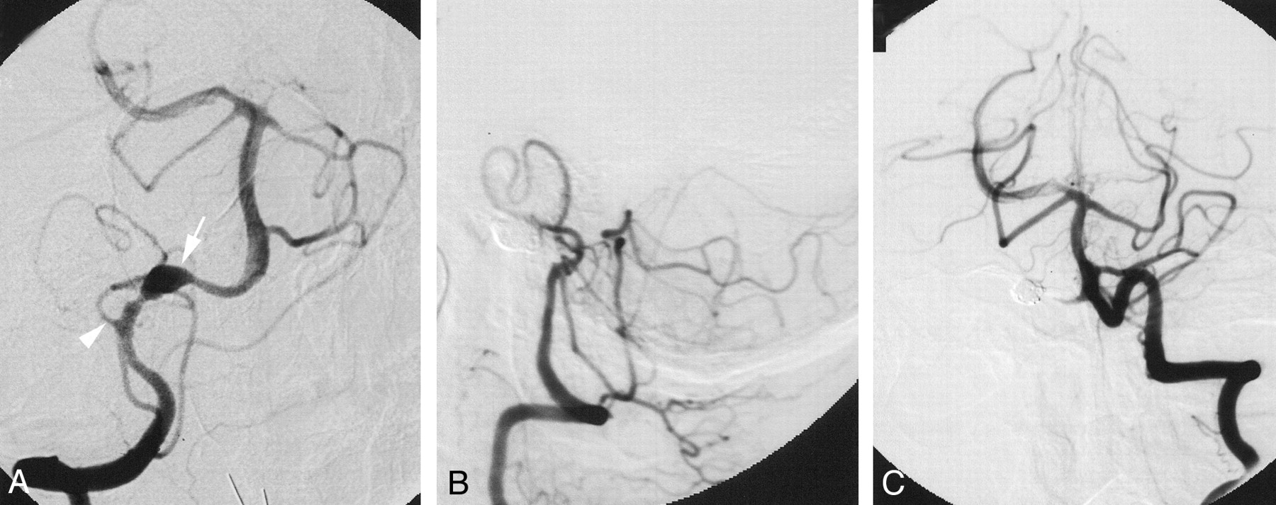
    Images from the case of a 46-year-old man (patient 3 in group I) who suffered SAH from a fusiform aneurysm of the distal intracranial right vertebral artery.

    A, Anteroposterior projection angiogram of the right vertebral artery disclosed a fusiform aneurysm of the distal intracranial portion (arrow) that is proximal to the vertebrobasilar junction and distal to the right posterior-inferior cerebellar artery (PICA, arrowhead).

    B, Aneurysm and distal vertebral artery were embolized with coils. Lateral projection control angiogram of the right vertebral artery, obtained after embolization, shows preservation of flow to the right posterior-inferior cerebellar and basilar arteries.

    C, Anteroposterior projection angiogram of the left vertebral artery shows preservation of flow to the right posterior-inferior cerebellar and basilar arteries. The patient achieved complete recovery and remained neurologically normal.

  • Fig 2.
    • Download figure
    • Open in new tab
    • Download powerpoint
    Fig 2.

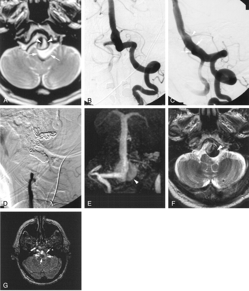
    Images from the case of a 71-year-old man (patient 4 in group II) with a fusiform vertebrobasilar aneurysm with mass effect on the left medulla, causing referable symptoms.

    A, Initial T2-weighted MR image of the posterior fossa shows mass effect and compression of the left medulla (arrows) from the aneurysmally dilated distal left vertebral artery (arrowheads). Note the high signal intensity within the aneurysm, signifying slow disturbed flow.

    B, Anteroposterior projection angiogram of the left vertebral artery.

    C, Oblique projection angiogram of the left vertebral artery shows a fusiform aneurysm extending from the distal intracranial left vertebral artery into the proximal basilar artery (arrows). Contrast material refluxed into the right vertebral artery. Note that the origin of the left posterior-inferior cerebellar artery is from the proximal basilar artery (arrowhead). The patient tolerated a 30-min temporary balloon occlusion of the left vertebral artery just proximal to the vertebrobasilar junction.

    D, Lateral projection angiogram of the left vertebral artery, obtained after embolization, confirms coil occlusion of the artery.

    E, Follow-up MR angiogram, obtained at 24 hr, shows preservation of blood flow to the posterior fossa via the right vertebral artery with high signal intensity slow flow and/or thrombus within the distal left vertebral aneurysm (arrowhead).

    F, Patient’s symptoms improved after embolization. Axial view T2-weighted MR image obtained 18 months after embolization shows thrombosis of the proximal aneurysmal sac, as evidenced by low signal intensity (arrowhead).

    G, MR angiogram obtained 18 months after embolization shows thrombosis of the proximal aneurysmal sac, as evidenced by lack of flow-related enhancement (arrows). Continued flow-related enhancement can be seen in the basilar artery (arrowhead).

Tables

  • Figures
    • View popup
    TABLE 1:

    Clinical outcomes for patients undergoing parent vessel occlusion of the vertebral artery

    Age (y)/SexGroupAneurysm Location and TypeMode of PresentationMethod of EmbolizationMonths of Follow-upClinical Presentation (Modified Rankin Score)Clinical Outcome (Modified Rankin Score)
    148/FIR VA disSAHCoil242
    256/MIL VA fusSAHCoil1454
    346/MIR VA fusSAHCoil1010
    452/MIL VA disSAHBalloon2351
    567/FIR VA fusSAHCoil7253
    676/MIL VA fusSAHCoil7653
    Mean5825.3
    752/MIIL VB fusSAHBalloon1434
    845/MIIB VA dis L VA paSAHCoil1254
    978/MIIL VB fusMass effectCoil3036
    1061/MIIBA fus L VA disSAHBalloon + coil1*56
    1171/MIIL VB fusMass effectCoil1911
    1266/MIIL VB fusMass effectCoil946
    1378/MIIR VB fusMass effectBalloon4**36
    Mean6412.7
    Overall mean6122.0
    • Note.—I indicates parent vessel occlusion for angiographic cure; II, parent vessel occlusion for angiographic palliation; R, right; L, left; VA, vertebral artery; dis, acute dissecting aneurysm; fus, chronic fusiform aneurysm; VB, vertebrobasilar; pa, pseudoaneurysm; SAH, subarachnoid hemorrhage.

    • * Deceased at 1 month after procedure.

    • ** Deceased at 4 months after procedure; died in sleep.

Back to top

In this issue

American Journal of Neuroradiology: 24 (5)
American Journal of Neuroradiology
Vol. 24, Issue 5
1 May 2003
  • Table of Contents
  • Index by author
Advertisement
Print
Download PDF
Email Article

Thank you for your interest in spreading the word on American Journal of Neuroradiology.

NOTE: We only request your email address so that the person you are recommending the page to knows that you wanted them to see it, and that it is not junk mail. We do not capture any email address.

Enter multiple addresses on separate lines or separate them with commas.
Parent Vessel Occlusion for Vertebrobasilar Fusiform and Dissecting Aneurysms
(Your Name) has sent you a message from American Journal of Neuroradiology
(Your Name) thought you would like to see the American Journal of Neuroradiology web site.
CAPTCHA
This question is for testing whether or not you are a human visitor and to prevent automated spam submissions.
Cite this article
R. Leibowitz, H.M. Do, M.L. Marcellus, S.D. Chang, G.K. Steinberg, M.P. Marks
Parent Vessel Occlusion for Vertebrobasilar Fusiform and Dissecting Aneurysms
American Journal of Neuroradiology May 2003, 24 (5) 902-907;

Citation Manager Formats

  • BibTeX
  • Bookends
  • EasyBib
  • EndNote (tagged)
  • EndNote 8 (xml)
  • Medlars
  • Mendeley
  • Papers
  • RefWorks Tagged
  • Ref Manager
  • RIS
  • Zotero
0 Responses
Respond to this article
Share
Bookmark this article
Parent Vessel Occlusion for Vertebrobasilar Fusiform and Dissecting Aneurysms
R. Leibowitz, H.M. Do, M.L. Marcellus, S.D. Chang, G.K. Steinberg, M.P. Marks
American Journal of Neuroradiology May 2003, 24 (5) 902-907;
del.icio.us logo Twitter logo Facebook logo Mendeley logo
  • Tweet Widget
  • Facebook Like
  • Google Plus One

Jump to section

  • Article
    • Abstract
    • Methods
    • Results
    • Discussion
    • References
  • Figures & Data
  • Info & Metrics
  • Responses
  • References
  • PDF

Related Articles

  • No related articles found.
  • PubMed
  • Google Scholar

Cited By...

  • The Fate of Unruptured Intracranial Vertebrobasilar Dissecting Aneurysm with Brain Stem Compression According to Different Treatment Modalities
  • Treatment of posterior circulation non-saccular aneurysms with flow diverters: a single-center experience and review of 56 patients
  • Safety of Unilateral Endovascular Occlusion of the Cervical Segment of the Vertebral Artery without Antecedent Balloon Test Occlusion
  • Incidence and Risk Factors of Recurrence After Endovascular Treatment of Intracranial Vertebrobasilar Dissecting Aneurysms
  • Treatment of Brain Aneurysms
  • Mechanically-induced proximal arterial occlusion and stent-within-a-stent technique for the treatment of bilateral vertebral artery dissecting aneurysms.
  • Crossref
  • Google Scholar

This article has not yet been cited by articles in journals that are participating in Crossref Cited-by Linking.

More in this TOC Section

  • Optimal MRI Sequence for Identifying Occlusion Location in Acute Stroke: Which Value of Time-Resolved Contrast-Enhanced MRA?
  • SWI or T2*: Which MRI Sequence to Use in the Detection of Cerebral Microbleeds? The Karolinska Imaging Dementia Study
  • Progression of Microstructural Damage in Spinocerebellar Ataxia Type 2: A Longitudinal DTI Study
Show more Brain

Similar Articles

Advertisement

Indexed Content

  • Current Issue
  • Accepted Manuscripts
  • Article Preview
  • Past Issues
  • Editorials
  • Editors Choice
  • Fellow Journal Club
  • Letters to the Editor

Cases

  • Case Collection
  • Archive - Case of the Week
  • Archive - Case of the Month
  • Archive - Classic Case

Special Collections

  • Special Collections

Resources

  • News and Updates
  • Turn around Times
  • Submit a Manuscript
  • Author Policies
  • Manuscript Submission Guidelines
  • Evidence-Based Medicine Level Guide
  • Publishing Checklists
  • Graphical Abstract Preparation
  • Imaging Protocol Submission
  • Submit a Case
  • Become a Reviewer/Academy of Reviewers
  • Get Peer Review Credit from Publons

Multimedia

  • AJNR Podcast
  • AJNR SCANtastic
  • Video Articles

About Us

  • About AJNR
  • Editorial Board
  • Not an AJNR Subscriber? Join Now
  • Alerts
  • Feedback
  • Advertise with us
  • Librarian Resources
  • Permissions
  • Terms and Conditions

American Society of Neuroradiology

  • Not an ASNR Member? Join Now

© 2025 by the American Society of Neuroradiology All rights, including for text and data mining, AI training, and similar technologies, are reserved.
Print ISSN: 0195-6108 Online ISSN: 1936-959X

Powered by HighWire