
Article Figures & Data
Figures
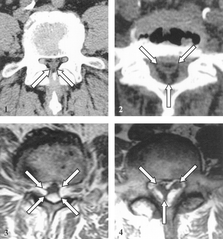
- Fig 1.
Axial CT scan at the L5 level in a 60-year-old patient with epidural lipomatosis shows low-attenuation fat surrounding the dural sac in the spinal canal; the sac shows polygonal deformation with spiculated contours (arrows).
- Fig 2.
Axial CT scan at the L5-Sl level in a 66-year-old patient with epidural lipomatosis shows low-attenuation fat surrounding the dural sac in the spinal canal; the sac shows Y-shaped or inverted triangular deformation (arrows). Disk bulge (anteriorly) and ligamenta flava (posterolaterally) lie peripheral to the fat.
- Fig 3.
MR imaging appearance of epidural lipomatosis in a 70-year-old patient. Axial T1-weighted MR image at the L4 level shows high-signal-intensity fat surrounding the dural sac in the spinal canal; the sac shows polygonal (stellar) deformation with spiculated contours (arrows).
- Fig 4.
MR imaging appearance of epidural lipomatosis in another 66-year-old patient. Axial T1-weighted MR image at the L5-S1 level shows high-signal-intensity fat surrounding the dural sac in the spinal canal; the sac shows Y-shaped or triangular deformation (arrows).
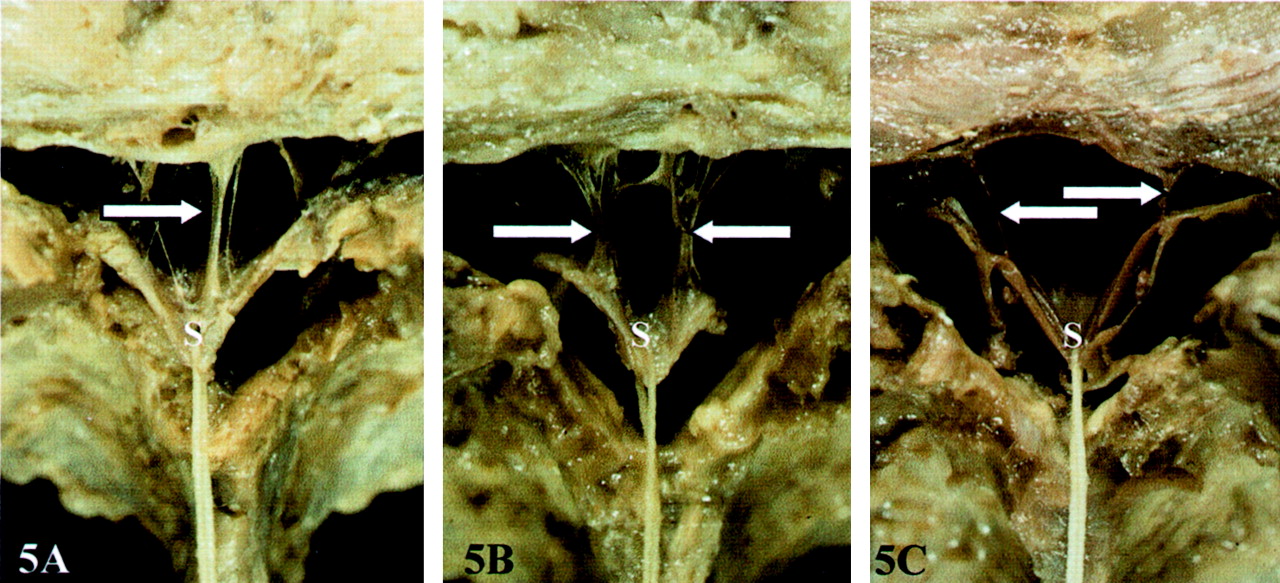
- Fig 5.
Macroscopic study in an adult spine.
A-C, Transverse sections of the lumbar spinal canal show median (arrow in A), bilateral paramedian (arrows in B), and lateral (arrows in C) locations of the meningovertebral ligaments in the anterior epidural space. The thecal sac (S) is collapsed by drawing the dura mater posteriorly with a thread.
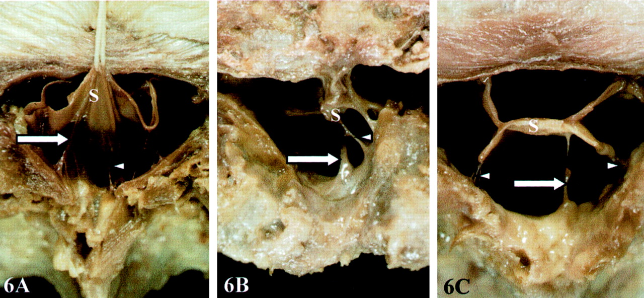
- Fig 6.
Macroscopic study in an adult spine. Transverse sections of the lumbar spinal canal show different locations of the meningovertebral ligaments in the posterior epidural space.
A, Paramedian (arrow) and median (arrowhead) locations of meningovertebral ligaments. The thecal sac (S) is collapsed by drawing the dura mater forward with a thread.
B and C, Paramedian (arrow) and lateral (arrowheads) locations of meningovertebral ligaments. The thecal sac (S) is collapsed centrally.
- Fig 7.
Macroscopic study in an adult spine shows lateral location (arrow) of meningovertebral ligaments in the lateral epidural space (the dura mater is drawn laterally).
- Fig 8.
Distribution of the meningovertebral ligaments in the transverse plane in the 70 adult dissected lumbar vertebrae, with corresponding frequencies (numbers are percentages).
- Fig 9.
Microscopic study in a 27-week fetal spine shows relation of meningovertebral ligaments to dura mater and to spinal canal walls.
A, Photomicrograph (Goldner trichrome stain; original magnification, ×12) of a fetal spinal canal at the L1 level. Meningovertebral ligaments extend from the outer surface of the dura mater to the osteofibrous walls of the spinal canal in the lateral areas, representing perivascular bands (arrowheads), and in the median area of the posterior epidural space (arrow). The large box outlines the region of the epidural space that is magnified in B. The small box outlines the region that is magnified in C.
B, Magnified view (immunohistochemical stain for type I collagen) of the large box in A. The meningovertebral ligament anchors the dura mater (curved arrow) to the spinal canal (arrow) and surrounds small vascular structures (arrowhead).
C, Magnified view (immunohistochemical stain for type I collagen) of the small box in A. The meningovertebral ligament (arrowhead), mainly composed of type I collagen, is connected to the outer surface of the dura mater (arrow).
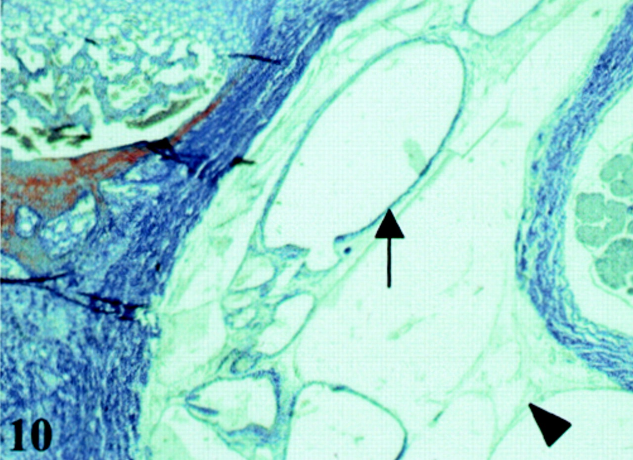
- Fig 10.
Microscopic study of meningovertebral ligament content in fetal spine. Photomicrograph (Goldner trichrome stain; original magnification, ×20) at the L1 level of a 27-week fetal spinal canal. The meningovertebral ligament (arrowhead) contains small vessels (arrow) (same observation as in adult spine in Fig 11).
- Fig 11.
Microscopic study at the L3 level of an adult spinal canal. Photomicrograph (Goldner trichrome stain; original magnification, ×20) shows that the meningovertebral ligament (arrowhead) is connected to the dura mater (long arrow) and contains epidural vessels (short arrows); loose fat is present in the lateral area of the posterior epidural space (asterisk).


Tables
- TABLE 1:
Variations of dural sac shape as seen on axial CT or MR images of the lumbar spine in 26 patients with epidural lipomatosis
Lumbar Level Normal Shape Polygonal (hexagonal, pentagonal, or square) Shape Triangular or Y Shape L3 20 80 0 L4 4 92 4 L5 23 19 58 Note.—Data are percentages.
- TABLE 2:
Variations of dural sac shape as seen on macroscopic photographs of 70 dissected adult lumbar spine segments
Lumbar Level Polygonal (hexagonal, pentagonal, or square) Shape Triangular or Y Shape L1 79 21 L2 79 21 L3 57 43 L4 57 43 L5 64 36 Note.—Data are percentages.
In this issue


Jump to section
Related Articles
Cited By...
- Differentiation between Tuberculous and Pyogenic Spondylodiscitis: The Role of the Anterior Meningovertebral Ligament in Patients with Anterior Epidural Abscess
- Immediate Pain Response to Interlaminar Lumbar Epidural Steroid Administration: Response Characteristics and Effects of Anesthetic Concentration
- Acute spinal cord compression