Advertisement

Main menu

  • Home
  • Content
    • Current Issue
    • Accepted Manuscripts
    • Article Preview
    • Past Issue Archive
    • AJNR Case Collection
    • Case of the Week Archive
    • Classic Case Archive
    • Case of the Month Archive
  • Special Collections
    • Spinal CSF Leak Articles (Jan 2020-June 2024)
    • 2024 AJNR Journal Awards
    • Most Impactful AJNR Articles
  • Multimedia
    • AJNR Podcast
    • AJNR Scantastics
    • Video Articles
  • For Authors
    • Submit a Manuscript
    • Author Policies
    • Fast publishing of Accepted Manuscripts
    • Graphical Abstract Preparation
    • Manuscript Submission Guidelines
    • Imaging Protocol Submission
    • Submit a Case for the Case Collection
  • About Us
    • About AJNR
    • Editorial Board
  • More
    • Become a Reviewer/Academy of Reviewers
    • Subscribers
    • Permissions
    • Alerts
    • Feedback
    • Advertisers
    • ASNR Home
  • Other Publications
    • ajnr

User menu

  • Alerts
  • Log in

Search

  • Advanced search
American Journal of Neuroradiology

American Journal of Neuroradiology

ASHNR American Society of Functional Neuroradiology ASHNR American Society of Pediatric Neuroradiology ASSR
  • Alerts
  • Log in

Advanced Search

  • Home
  • Content
    • Current Issue
    • Accepted Manuscripts
    • Article Preview
    • Past Issue Archive
    • AJNR Case Collection
    • Case of the Week Archive
    • Classic Case Archive
    • Case of the Month Archive
  • Special Collections
    • Spinal CSF Leak Articles (Jan 2020-June 2024)
    • 2024 AJNR Journal Awards
    • Most Impactful AJNR Articles
  • Multimedia
    • AJNR Podcast
    • AJNR Scantastics
    • Video Articles
  • For Authors
    • Submit a Manuscript
    • Author Policies
    • Fast publishing of Accepted Manuscripts
    • Graphical Abstract Preparation
    • Manuscript Submission Guidelines
    • Imaging Protocol Submission
    • Submit a Case for the Case Collection
  • About Us
    • About AJNR
    • Editorial Board
  • More
    • Become a Reviewer/Academy of Reviewers
    • Subscribers
    • Permissions
    • Alerts
    • Feedback
    • Advertisers
    • ASNR Home
  • Follow AJNR on Twitter
  • Visit AJNR on Facebook
  • Follow AJNR on Instagram
  • Join AJNR on LinkedIn
  • RSS Feeds

Welcome to the new AJNR, Updated Hall of Fame, and more. Read the full announcements.


AJNR is seeking candidates for the position of Associate Section Editor, AJNR Case Collection. Read the full announcement.


OtherSPINE

Three-Dimensional Rotational Myelography

Markus Kufeld, Benjamin Claus, Adriana Campi, Wolfgang R. Lanksch and Goetz Benndorf
American Journal of Neuroradiology August 2003, 24 (7) 1290-1293;
Markus Kufeld
aDepartment of Neurosurgery, Humboldt University Charité, Berlin, Germany
  • Find this author on Google Scholar
  • Find this author on PubMed
  • Search for this author on this site
Benjamin Claus
bDepartment of Radiology, Humboldt University Charité, Berlin, Germany
  • Find this author on Google Scholar
  • Find this author on PubMed
  • Search for this author on this site
Adriana Campi
cDepartment of Neuroradiology, University Hospital Zurich, Zurich, Switzerland
  • Find this author on Google Scholar
  • Find this author on PubMed
  • Search for this author on this site
Wolfgang R. Lanksch
aDepartment of Neurosurgery, Humboldt University Charité, Berlin, Germany
  • Find this author on Google Scholar
  • Find this author on PubMed
  • Search for this author on this site
Goetz Benndorf
bDepartment of Radiology, Humboldt University Charité, Berlin, Germany
  • Find this author on Google Scholar
  • Find this author on PubMed
  • Search for this author on this site
  • Article
  • Figures & Data
  • Info & Metrics
  • Responses
  • References
  • PDF
Loading

Article Figures & Data

Figures

  • Fig 1.
    • Download figure
    • Open in new tab
    • Download powerpoint
    Fig 1.

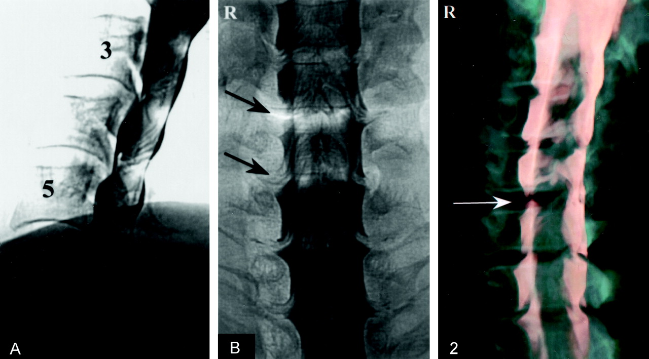
    Images from the case of a 60-year-old man who presented with severe cervicobrachialgia and dysesthesia on the right side.

    A, Lateral myelogram shows multiple anterior defects that indicate general narrowing of the spinal canal with ventral bony spurs and a steep vertebral column. At disk level C5–C6, an additional disk protrusion can only be assumed.

    B, Anteroposterior myelogram shows right-sided local compression of root sleeves C6 and C7, with a major defect at C6 (arrows).

  • Fig 2.
    • Download figure
    • Open in new tab
    • Download powerpoint
    Fig 2.

    Real-time 3D reconstruction of cervical spine, with differentiation of the contrast column from bone, shows relationship between thecal sac and bony structures. This oblique projection, similar to a conventional myelogram, shows the filling defect in the neural foramen of C5–C6 (arrow), indicating major deficit at this level.
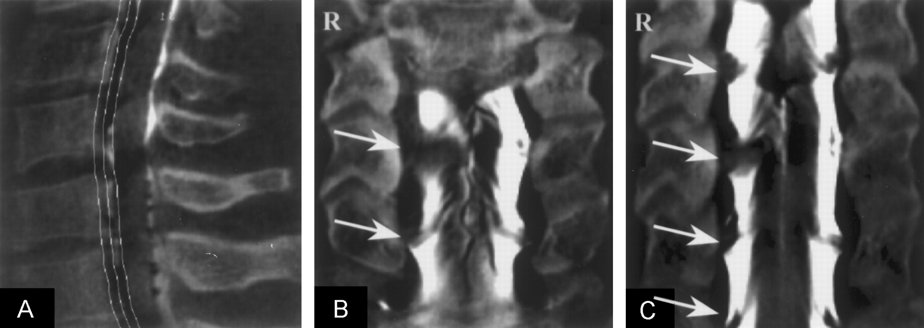
  • Fig 3.
    • Download figure
    • Open in new tab
    • Download powerpoint
    Fig 3.

    Coronal multiplanar reformatting.

    A, Lateral reconstruction localizer in the multiplanar reformatting mode indicates coronal curved planes of reconstruction (see C), following the physiological lordosis of the spine.

    B, Image shows multiplanar reformatting with coronal plane of reconstruction. This plane is straight and limits the visualization and comparison of the root sleeves. Because of the physiological lordosis of the cervical spine, the nerve root sheaths are mostly out of the cutting plane and thus are difficult to compare for evaluation of maximum nerve root compression (arrows).

    C, Image shows reformatting with curved coronal planes of reconstruction, following the physiological lordosis of the spine. The visualization of several nerve root sheaths is possible in the same plane (arrows), allowing simultaneous assessment of multilevel abnormalities. By comparison, straight coronal plane multiplanar reformatting shows only two nerve roots sheaths (see B, arrows).

  • Fig 4.
    • Download figure
    • Open in new tab
    • Download powerpoint
    Fig 4.

    Parasagittal multiplanar reformatting.

    A, Right-sided parasagittal reconstruction of the spine in multiplanar reformatting modus is shown. The defect of the contrast column at the C5–C6 level indicates nerve root sheath compression and also allows distinction between the compressing disk material and the vertebra (arrow). Inset, multiplanar reformatting with axial orientation of the reconstruction plane at the C5–C6 level shows a narrowing of the neural foramen on the right side. The contrast medium is asymmetrically distributed within the spinal canal, indicating a right paramedian soft disk protrusion with compression of the thecal sac and the spinal cord (arrow). White line marks reconstruction plane in relation to spinal canal.

    B, Multiplanar reformatting in oblique parasagittal projection obtained perpendicular to the course of the nerve root canal allows assessment of narrowing of the foramen (arrows). As the inset in A shows (white line shows reconstruction plane), an evaluation of the neural foramen along its course is possible, allowing determination of maximum stenosis.

PreviousNext
Back to top

In this issue

American Journal of Neuroradiology: 24 (7)
American Journal of Neuroradiology
Vol. 24, Issue 7
1 Aug 2003
  • Table of Contents
  • Index by author
Advertisement
Print
Download PDF
Email Article

Thank you for your interest in spreading the word on American Journal of Neuroradiology.

NOTE: We only request your email address so that the person you are recommending the page to knows that you wanted them to see it, and that it is not junk mail. We do not capture any email address.

Enter multiple addresses on separate lines or separate them with commas.
Three-Dimensional Rotational Myelography
(Your Name) has sent you a message from American Journal of Neuroradiology
(Your Name) thought you would like to see the American Journal of Neuroradiology web site.
CAPTCHA
This question is for testing whether or not you are a human visitor and to prevent automated spam submissions.
Cite this article
Markus Kufeld, Benjamin Claus, Adriana Campi, Wolfgang R. Lanksch, Goetz Benndorf
Three-Dimensional Rotational Myelography
American Journal of Neuroradiology Aug 2003, 24 (7) 1290-1293;

Citation Manager Formats

  • BibTeX
  • Bookends
  • EasyBib
  • EndNote (tagged)
  • EndNote 8 (xml)
  • Medlars
  • Mendeley
  • Papers
  • RefWorks Tagged
  • Ref Manager
  • RIS
  • Zotero
0 Responses
Respond to this article
Share
Bookmark this article
Three-Dimensional Rotational Myelography
Markus Kufeld, Benjamin Claus, Adriana Campi, Wolfgang R. Lanksch, Goetz Benndorf
American Journal of Neuroradiology Aug 2003, 24 (7) 1290-1293;
del.icio.us logo Twitter logo Facebook logo Mendeley logo
  • Tweet Widget
  • Facebook Like
  • Google Plus One

Jump to section

  • Article
    • Abstract
    • Case Report
    • Discussion
    • Conclusion
    • References
  • Figures & Data
  • Info & Metrics
  • Responses
  • References
  • PDF

Related Articles

  • No related articles found.
  • PubMed
  • Google Scholar

Cited By...

  • No citing articles found.
  • Crossref
  • Google Scholar

This article has not yet been cited by articles in journals that are participating in Crossref Cited-by Linking.

More in this TOC Section

  • Bern Score Validity for SIH
  • MP2RAGE 7T in MS Lesions of the Cervical Spine
  • Deep Learning for STIR Spine MRI Quality
Show more Spine

Similar Articles

Advertisement

Indexed Content

  • Current Issue
  • Accepted Manuscripts
  • Article Preview
  • Past Issues
  • Editorials
  • Editors Choice
  • Fellow Journal Club
  • Letters to the Editor

Cases

  • Case Collection
  • Archive - Case of the Week
  • Archive - Case of the Month
  • Archive - Classic Case

Special Collections

  • Special Collections

Resources

  • News and Updates
  • Turn around Times
  • Submit a Manuscript
  • Author Policies
  • Manuscript Submission Guidelines
  • Evidence-Based Medicine Level Guide
  • Publishing Checklists
  • Graphical Abstract Preparation
  • Imaging Protocol Submission
  • Submit a Case
  • Become a Reviewer/Academy of Reviewers
  • Get Peer Review Credit from Publons

Multimedia

  • AJNR Podcast
  • AJNR SCANtastic
  • Video Articles

About Us

  • About AJNR
  • Editorial Board
  • Not an AJNR Subscriber? Join Now
  • Alerts
  • Feedback
  • Advertise with us
  • Librarian Resources
  • Permissions
  • Terms and Conditions

American Society of Neuroradiology

  • Not an ASNR Member? Join Now

© 2025 by the American Society of Neuroradiology All rights, including for text and data mining, AI training, and similar technologies, are reserved.
Print ISSN: 0195-6108 Online ISSN: 1936-959X

Powered by HighWire