
Article Figures & Data
Figures
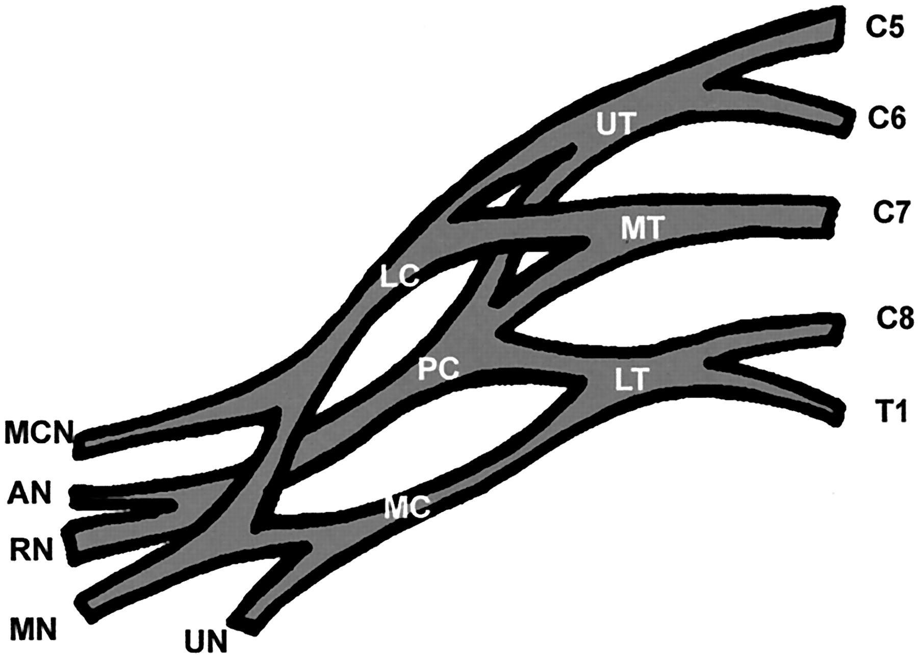
- Fig 1.
Schematic illustration of the different components of the right brachial plexus. C5 indicates fifth nerve root; C6, sixth nerve root; C7, seventh nerve root; C8, eighth nerve root; T1, first thoracic nerve root; UT, upper trunk; MT, middle trunk; LT, lower trunk; LC, lateral nerve cord; PC, posterior nerve cord; MC, medial nerve cord; MCN, musculocutaneous nerve; AN, axillary nerve; RN, radial nerve; MN, median nerve; UN, ulnar nerve.
- Fig 2.
Cervical nerves roots at their extraforaminal part.
A, Position of the sonographic probe to explore cervical nerve roots at their extraforaminal part.
B, Sonogram (90° left rotated) in a 28-year-old female volunteer shows C5 (1), C6 (2), and C7 (3) roots at their extraforaminal part. They appear as oval hypoechoic structures. Note the shadowing of the cervical transverse processes (arrows). S indicates skin.
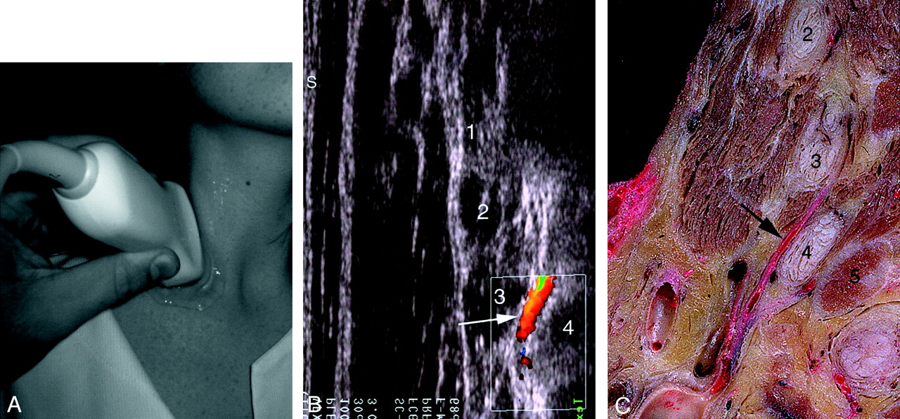
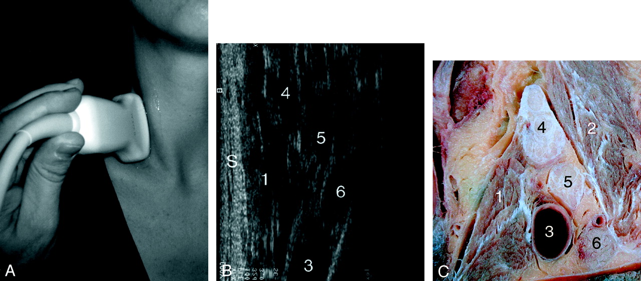
- Fig 3.
Relationship between the deep cervical artery and the C7 and C8 nerve roots.
A, Position of the probe to demonstrate the deep cervical artery.
B, Color Doppler sonogram (90° left rotated) in a 26-year-old female volunteer shows the deep cervical artery (arrow) separating the eighth cervical nerve (4) from the seventh (3).
C, Sagittal gross anatomic section shows the relationship between the deep cervical artery (arrow) and the C7 (3) and C8 (4) nerves roots.
In B and/or C, 1 indicates C5 nerve; 2, C6 nerve; 5, first rib; S, skin.
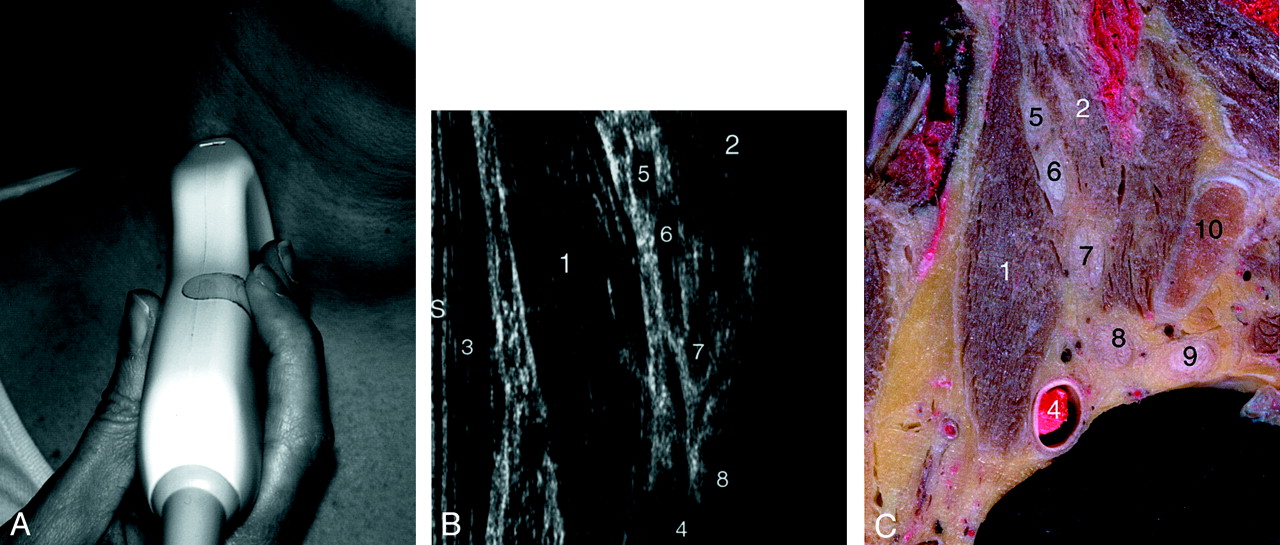
- Fig 4.
Interscalene triangle (nerves roots).
A, Position of the probe to explore the nerve roots at the interscalene triangle.
B, Sonogram (90° left rotated) in a 26-year-old female volunteer shows the C5 (5), C6 (6), and C7 (7) nerve roots at the interscalene triangle.
C, Sagittal gross anatomic section at the interscalene triangle.
In B and/or C, 1 indicates anterior scalene muscle; 2, middle and posterior scalene muscles; 3, sternocleidomastoid muscle; 4, subclavian artery; 5–8, C5 through C8 nerve roots; 9, T1 nerve root; 10, first rib; S, skin.
- Fig 5.
Interscalene triangle (trunks).
A, Position of the probe to explore the trunks at the interscalene triangle.
B, Sonogram (90° left rotated) in a 28-year-old male volunteer shows the upper, middle, and lower trunks at the interscalene triangle.
C, Sagittal gross anatomic section at the interscalene triangle (lateral part).
In B and/or C, 1 indicates anterior scalene muscle; 2, middle and posterior scalene muscles; 3, subclavian artery; 4, upper trunk; 5, middle trunk; 6, lower trunk; S, skin.
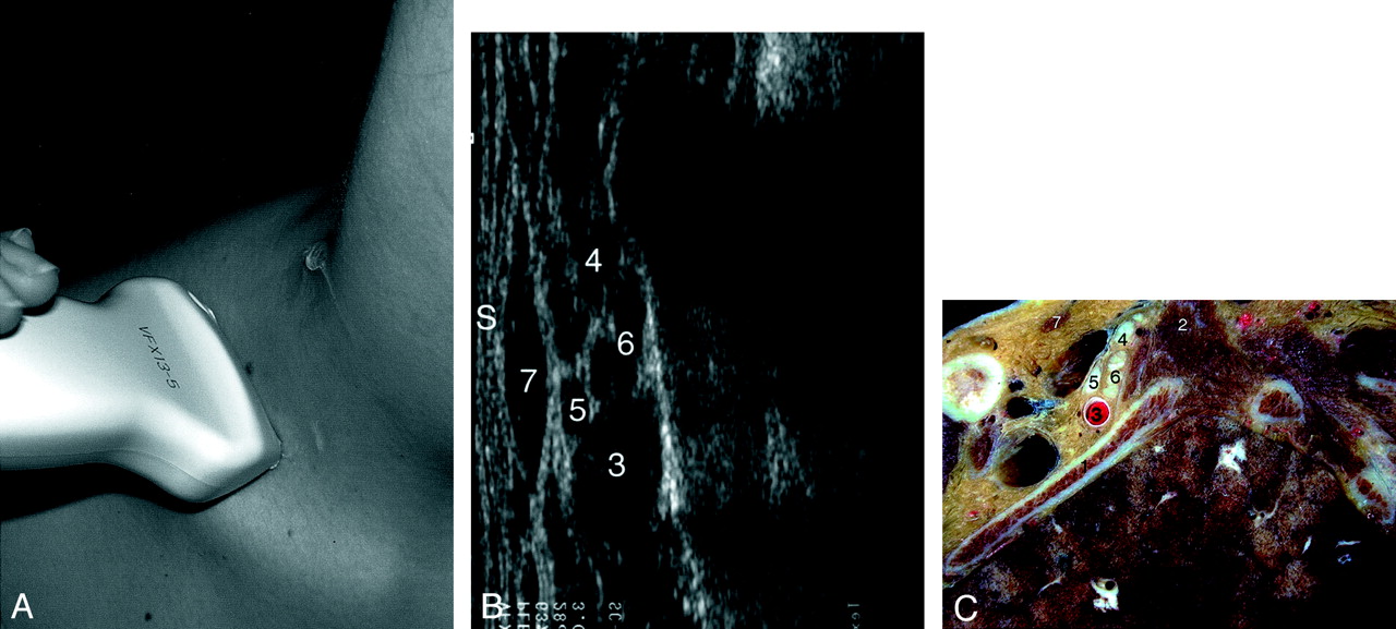
- Fig 6.
Junction between the interscalene triangle and the costoclavicular space (cords).
A, Position of the probe to explore the brachial plexus at the junction between the interscalene triangle and the costoclavicular space.
B, Sonogram (90° left rotated) in a 34-year-old male volunteer shows the nerve cords at the junction between the interscalene triangle and costoclavicular space.
C, Sagittal gross anatomic section at the junction between the interscalene triangle and costoclavicular space.
In B and/or C, 1 indicates first rib; 2, middle and posterior scalene muscles; 3, subclavian artery; 4, posterior nerve cord; 5, lateral nerve cord; 6, medial nerve cord; 7, omohyoid muscle; S, skin.
- Fig 7.
Costoclavicular space (cords).
A, Position of the probe to explore the brachial plexus at the costoclavicular space by an infraclavicular approach.
B, Sonogram (90° left rotated) in a 31-year-old female volunteer shows clusters of round hypoechoic neural fascicles of the different nerve cords at the costoclavicular space.
C, Sagittal gross oblique anatomic section of the costoclavicular space.
In B and/or C, 1 indicates subclavius muscle; 2, pectoralis major muscle; 3, axillary artery; 4, axillary vein; 5, posterior nerve cord; 6, lateral nerve cord; 7, medial nerve cord; S, skin.
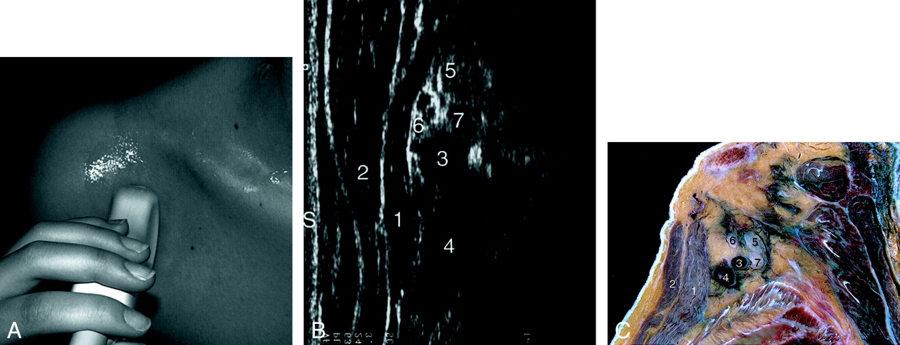
- Fig 8.
Retropectoralis minor space (cords).
A, Position of the probe to explore the brachial plexus at the retropectoralis minor space.
B, Sonogram (90° left rotated) in a 31-year-old female volunteer shows the nerve cords of the brachial plexus at the retropectoralis minor space.
C, Sagittal gross oblique anatomic section of the retropectoralis minor space.
In B and/or C, 1 indicates pectoralis minor muscle; 2, pectoralis major muscle; 3, axillary artery; 4, axillary vein; 5, posterior nerve cord; 6, lateral nerve cord; 7, medial nerve cord; S, skin.
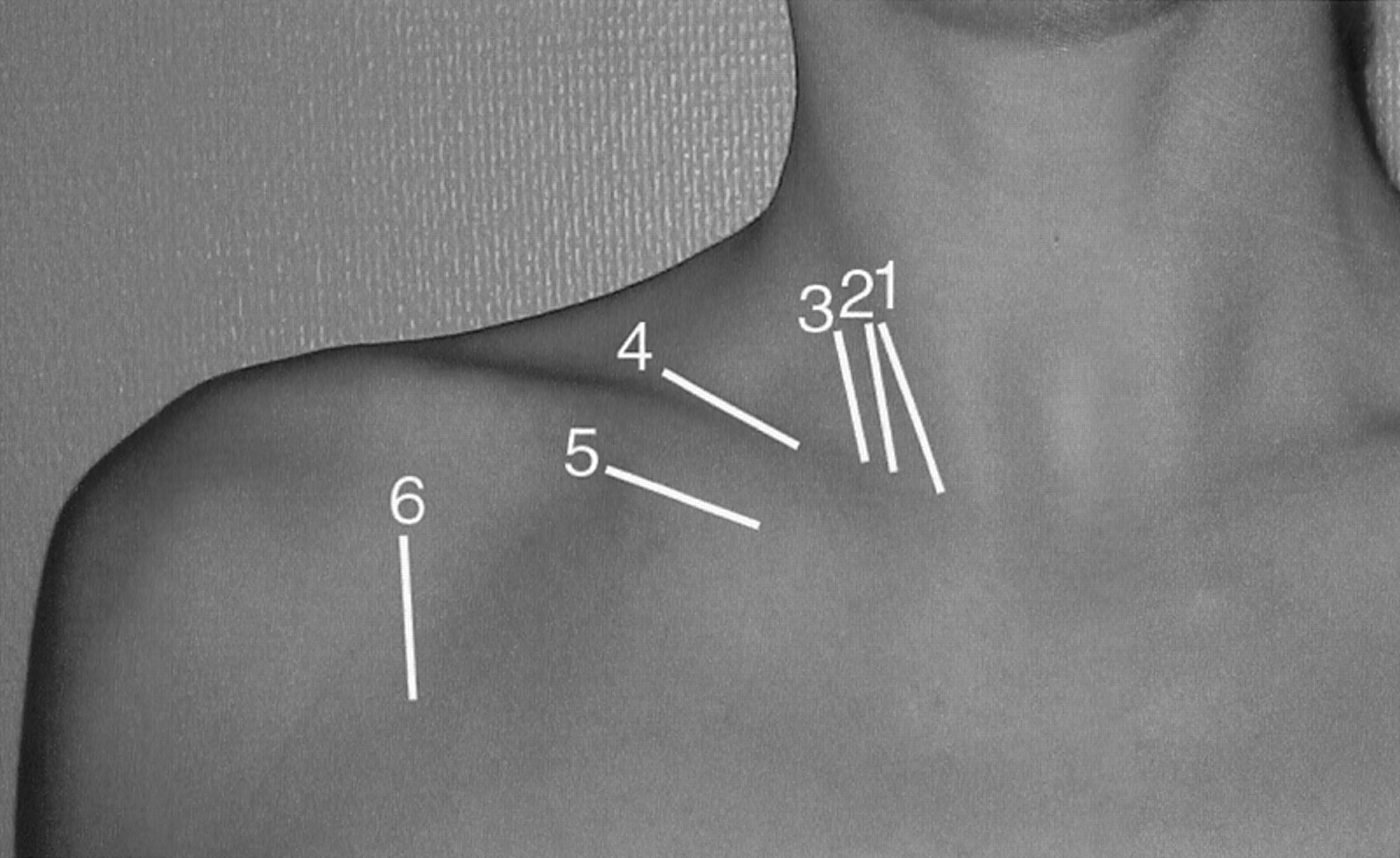
- Fig 9.
Schematic illustration indicates positioning of the probe to analyze 1, the extraforaminal part of nerve roots and the deep cervical artery; 2, the nerve roots at the interscalene triangle; 3, the trunks of the brachial plexus; 4, the cords of the brachial plexus by a supraclavicular approach; 5, the cords of the brachial plexus by an infraclavicular approach; and 6, the cords of the brachial plexus at the retropectoralis minor space.


In this issue


Jump to section
Related Articles
Cited By...
- Evaluating the spread of costoclavicular brachial plexus block: an anatomical study
- Brachial Plexus Ultrasound and MRI in Children with Brachial Plexus Birth Injury
- Ultrasound evaluation of focal neuropathies in athletes: a clinically-focused review
- Spatial mapping of the brachial plexus using three-dimensional ultrasound