Advertisement

Main menu

  • Home
  • Content
    • Current Issue
    • Accepted Manuscripts
    • Article Preview
    • Past Issue Archive
    • AJNR Case Collection
    • Case of the Week Archive
    • Classic Case Archive
    • Case of the Month Archive
  • Special Collections
    • Spinal CSF Leak Articles (Jan 2020-June 2024)
    • 2024 AJNR Journal Awards
    • Most Impactful AJNR Articles
  • Multimedia
    • AJNR Podcast
    • AJNR Scantastics
    • Video Articles
  • For Authors
    • Submit a Manuscript
    • Author Policies
    • Fast publishing of Accepted Manuscripts
    • Graphical Abstract Preparation
    • Manuscript Submission Guidelines
    • Imaging Protocol Submission
    • Submit a Case for the Case Collection
  • About Us
    • About AJNR
    • Editorial Board
  • More
    • Become a Reviewer/Academy of Reviewers
    • Subscribers
    • Permissions
    • Alerts
    • Feedback
    • Advertisers
    • ASNR Home
  • Other Publications
    • ajnr

User menu

  • Alerts
  • Log in

Search

  • Advanced search
American Journal of Neuroradiology

American Journal of Neuroradiology

ASHNR American Society of Functional Neuroradiology ASHNR American Society of Pediatric Neuroradiology ASSR
  • Alerts
  • Log in

Advanced Search

  • Home
  • Content
    • Current Issue
    • Accepted Manuscripts
    • Article Preview
    • Past Issue Archive
    • AJNR Case Collection
    • Case of the Week Archive
    • Classic Case Archive
    • Case of the Month Archive
  • Special Collections
    • Spinal CSF Leak Articles (Jan 2020-June 2024)
    • 2024 AJNR Journal Awards
    • Most Impactful AJNR Articles
  • Multimedia
    • AJNR Podcast
    • AJNR Scantastics
    • Video Articles
  • For Authors
    • Submit a Manuscript
    • Author Policies
    • Fast publishing of Accepted Manuscripts
    • Graphical Abstract Preparation
    • Manuscript Submission Guidelines
    • Imaging Protocol Submission
    • Submit a Case for the Case Collection
  • About Us
    • About AJNR
    • Editorial Board
  • More
    • Become a Reviewer/Academy of Reviewers
    • Subscribers
    • Permissions
    • Alerts
    • Feedback
    • Advertisers
    • ASNR Home
  • Follow AJNR on Twitter
  • Visit AJNR on Facebook
  • Follow AJNR on Instagram
  • Join AJNR on LinkedIn
  • RSS Feeds

Welcome to the new AJNR, Updated Hall of Fame, and more. Read the full announcements.


AJNR is seeking candidates for the position of Associate Section Editor, AJNR Case Collection. Read the full announcement.


Research ArticleHEAD AND NECK

MR Imaging of the Trigeminal Ganglion, Nerve, and the Perineural Vascular Plexus: Normal Appearance and Variants with Correlation to Cadaver Specimens

Lorna Sohn Williams, Ilona M. Schmalfuss, Christopher L. Sistrom, Takuya Inoue, Ryusui Tanaka, Eduardo R. Seoane and Anthony A. Mancuso
American Journal of Neuroradiology August 2003, 24 (7) 1317-1323;
Lorna Sohn Williams
aDepartment of Radiology, JFK Medical Center, Atlantis, FL
  • Find this author on Google Scholar
  • Find this author on PubMed
  • Search for this author on this site
Ilona M. Schmalfuss
bDepartment of Radiology, University of Florida College of Medicine, Gainesville, FL
  • Find this author on Google Scholar
  • Find this author on PubMed
  • Search for this author on this site
Christopher L. Sistrom
bDepartment of Radiology, University of Florida College of Medicine, Gainesville, FL
  • Find this author on Google Scholar
  • Find this author on PubMed
  • Search for this author on this site
Takuya Inoue
cDepartment of Neurosurgery, University of Florida College of Medicine, Gainesville, FL
  • Find this author on Google Scholar
  • Find this author on PubMed
  • Search for this author on this site
Ryusui Tanaka
  • Find this author on Google Scholar
  • Find this author on PubMed
  • Search for this author on this site
Eduardo R. Seoane
cDepartment of Neurosurgery, University of Florida College of Medicine, Gainesville, FL
  • Find this author on Google Scholar
  • Find this author on PubMed
  • Search for this author on this site
Anthony A. Mancuso
bDepartment of Radiology, University of Florida College of Medicine, Gainesville, FL
  • Find this author on Google Scholar
  • Find this author on PubMed
  • Search for this author on this site
  • Article
  • Figures & Data
  • Info & Metrics
  • Responses
  • References
  • PDF
Loading

Article Figures & Data

Figures

  • Tables
  • Fig 1.
    • Download figure
    • Open in new tab
    • Download powerpoint
    Fig 1.

    Trigeminal ganglion in trigeminal cistern in cadaver.

    A, Trigeminal ganglion (TG) is crescentic in shape and proximal divisions of trigeminal nerve (V1, V2, V3) emerge from its anterolateral border. The trigeminal rootlets (*) enter the crescent-shaped TG. V1 exits the TG as the most superior branch. It lies immediately inferior to cranial nerve four (CN4). V2 leaves TG as the middle branch and V3 as the most inferior division. Venous structures are blue- and arterial red-colored silicon. An extensive pericavernous venous plexus surrounds the ganglion and each of the trigeminal nerve branches (white arrows).

    B, Following dissection of the pericavernous venous plexus, the avascular nature of the trigeminal ganglion and its proximal divisions (V1, V2, V3) is seen. The extent of the trigeminal ganglion (between arrowheads) and the trigeminal nerve rootlets (*) are now better delineated. The cranial nerve six (white arrow) is now visible inferior medial to V1. CN indicates cranial nerve
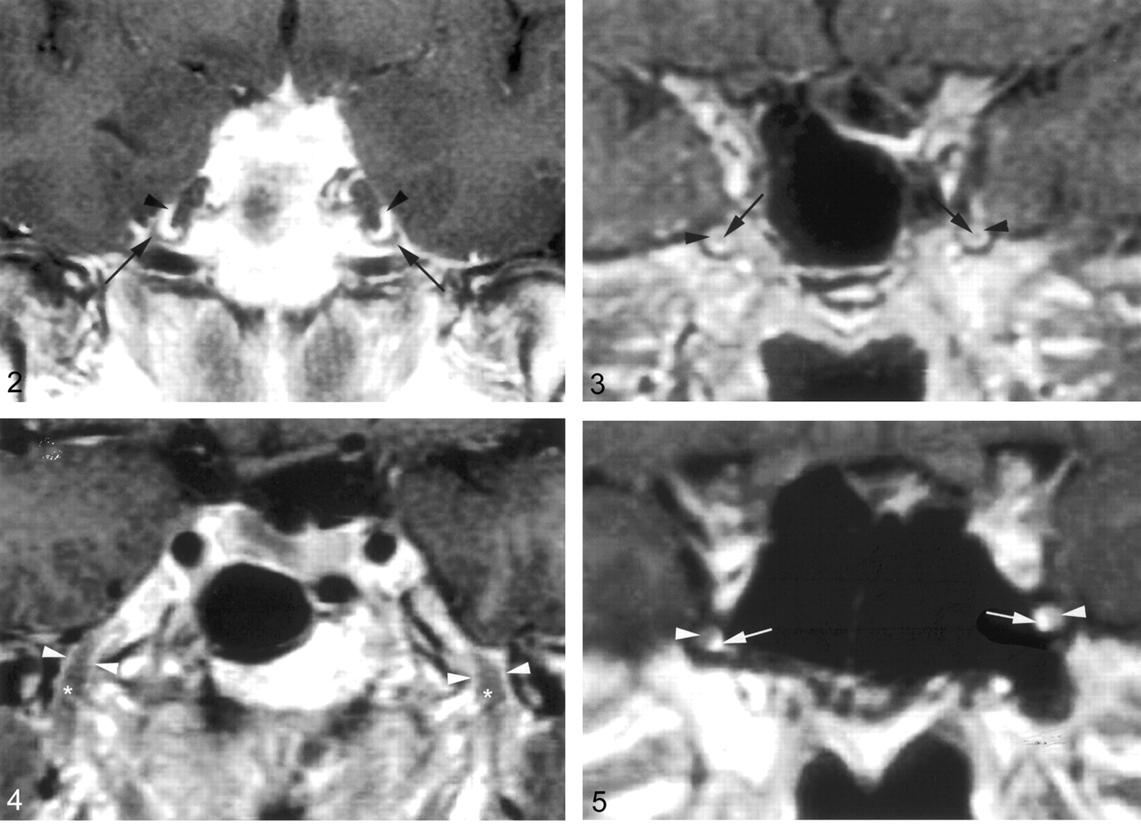
  • Fig 2.
    • Download figure
    • Open in new tab
    • Download powerpoint
    Fig 2.

    Coronal gadolinium-enhanced T1-weighted image (TR/TE/NEX, 400/15/2) depicts the nonenhancing crescent-shaped trigeminal ganglion (arrows) and the prominent perineural venous plexus (arrowheads) superior to it. The venous plexus as well as the ganglion are symmetric in appearance.

  • Fig 3.
    • Download figure
    • Open in new tab
    • Download powerpoint
    Fig 3.

    Coronal gadolinium-enhanced T1-weighted image (TR/TE/NEX, 500/15/2) illustrates the common appearance of V2 (arrows) within the foramen rotundum as central nonenhancing nerve completely surrounded by the perineural venous plexus (arrowheads).

  • Fig 4.
    • Download figure
    • Open in new tab
    • Download powerpoint
    Fig 4.

    Coronal gadolinium-enhanced T1-weighted image (TR/TE/NEX, 500/15/2) illustrates the normal appearance of V3 as it exits the skull base through the foramen ovale. V3 (*) is surrounded by the venous plexus (arrowheads) on both sides at the level of the foramen ovale and over a short distance below the skull base. The perineural venous plexus (arrowheads) demonstrates the same thickness on each side of the nerve.

  • Fig 5.
    • Download figure
    • Open in new tab
    • Download powerpoint
    Fig 5.

    Coronal gadolinium-enhanced T1-weighted image (TR/TE/NEX, 500/15/2) shows an incomplete perineural venous plexus within the foramen rotundum as an anatomic variant. Only small portion of the perineural venous plexus (arrowheads) is seen medial to V2 (arrows) on both sides.
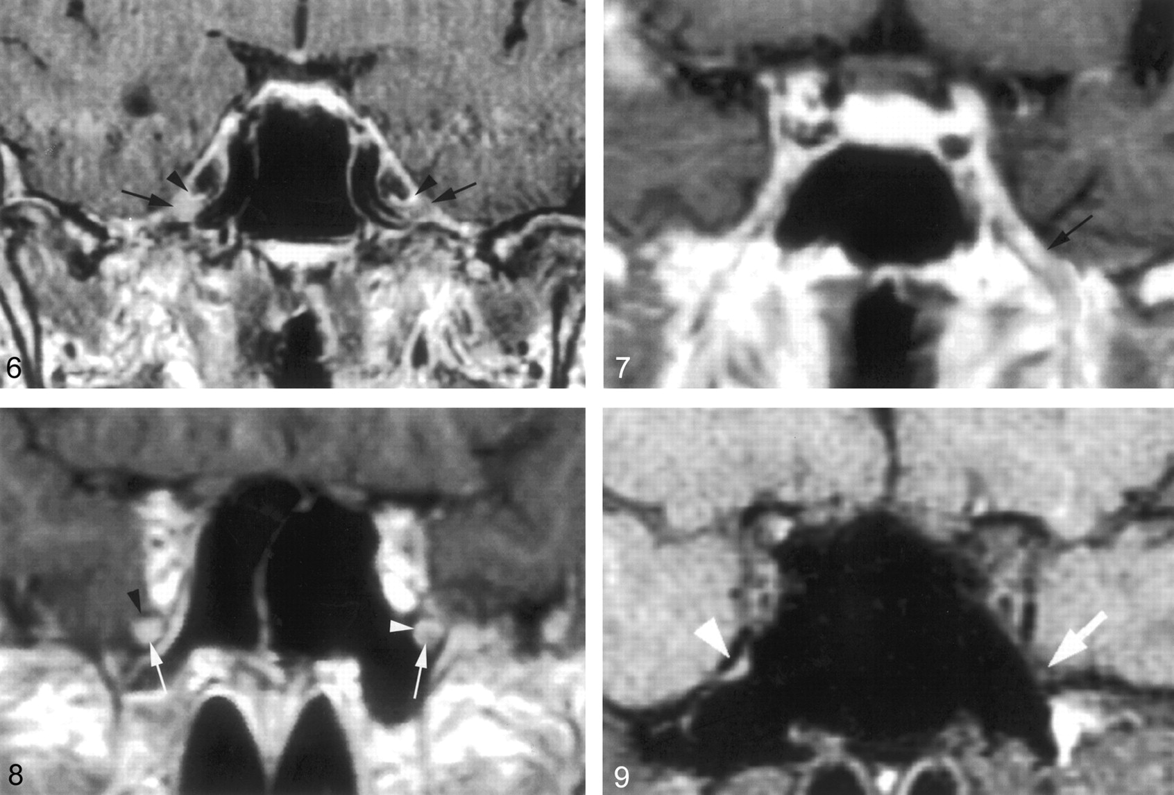
  • Fig 6.
    • Download figure
    • Open in new tab
    • Download powerpoint
    Fig 6.

    Coronal gadolinium-enhanced T1-weighted image (TR/TE/NEX, 400/15/2) suggests a significantly enhancing right trigeminal ganglion (right arrow). The perineural venous plexus (right arrowhead) and the trigeminal ganglion cannot be identified as separate structures. This appearance is thought to be due to obscuration of the ganglion by a more extensive perineural venous plexus than that typically seen. The subject did not have any symptoms related to trigeminal nerve or a history of malignancy. Therefore, this is considered a normal variation in imaging appearance. The contralateral side demonstrates the typical appearance of the trigeminal ganglion (left arrow) and of adjacent venous plexus (left arrowhead).

  • Fig 7.
    • Download figure
    • Open in new tab
    • Download powerpoint
    Fig 7.

    Coronal gadolinium-enhanced T1-weighted image (TR/TE/NEX, 500/15/2) at the level of the foramen ovale shows a significantly enhancing V3 branch (arrow) on the left as a normal anatomic variation. The patient did not have any symptoms of V3 dysfunction. The perineural venous plexus and V3 cannot be seen as individual structures.

  • Fig 8.
    • Download figure
    • Open in new tab
    • Download powerpoint
    Fig 8.

    Coronal gadolinium-enhanced T1-weighted image (TR/TE/NEX, 500/15/2) at the level of the foramen rotundum demonstrates an enhancing V2 branch on the right when compared with the left (arrows). This is considered to represent a normal anatomic variation, because the subject did not have any symptoms related to V2 dysfunction or history of malignancy. The perineural venous plexus and V2 cannot be separated from each other. This appearance might be caused by a more prominent venous plexus. On the left, the perineural venous plexus (white arrowhead) is incomplete and absent in the inferior lateral aspect. The black arrowhead indicates the bony boundary of the foramen rotundum.

  • Fig 9.
    • Download figure
    • Open in new tab
    • Download powerpoint
    Fig 9.

    Coronal T1-weighted image (TR/TE/NEX, 753/14/2) through the foramen rotundum demonstrating normal appearing V2 on the left (arrow). No nerve can be visualized within the foramen rotundum on the right (arrowhead).

Tables

  • Figures
    • View popup
    TABLE 1:

    Qualitative findings and measurements

    FindingGanglionV2V3
    Visualized98% (95–100)93% (88–100) (Fig 9)93% (88–100)
    Only nerve seenNA9% (2–17)3% (0–7)
    Nerve and plexus seen as distinct structuresNA91% (83–98)97% (93–100)
    Enhancing ganglion or nerve seen with no distinct vascular plexus4% (0–10)18% (9–28)3% (0–7)
    Plexus completely surrounding V2 or seen on both sides of V3NA41% (29–53)75% (64–87)
    Combined diameter of nerve and plexus when seen as distinct structures (mm)NA3.0 ± 0.53 (2.9–3.1)3.9 ± 0.84 (3.7–4.1)
    Diameter of the nerve alone (mm)NA1.6 ± 0.48 (1.5–1.7)1.9 ± 0.67 (1.8–2.0)
    • Note.—Qualitative results are reported as percentages. Sizes are reported as mean ±SD. CI (95% lower-upper) are provided for all results in parenthese.

    • View popup
    TABLE 2:

    Interobserver reliability of qualitative findings and measurements

    FindingGanglionV2V3
    Visualization*0.66 (98%)0.64 (95%)0.41 (92%)
    Morphology and enhancement*0.79 (98%)0.56 (81%)0.55 (95%)
    Plexus completely surrounding V2 or seen on both sides of V3*NA0.74 (86%)0.65 (87%)
    Diameter of nerve and plexus (mm)†NA0.830.79
    Diameter of nerve alone (mm)†NA0.720.85
    • ↵* κ statistic and percent agreement (in parentheses) for qualitative findings.

    • ↵† Intraclass correlation coefficient for measured diameters.

    • View popup
    TABLE 3:

    Left-right symmetry of qualitative findings and measurements

    FindingGanglionV2V3
    Visualized*P = 1.0, agree = 0.92P = .51, agree = 0.86P = .51, agree = 0.86
    Morphology and enhancement*P = .37, agree = 0.92P = .16, agree = 0.75P = .39, agree = 0.91
    Plexus completely surrounding V2 or seen on both sides of V3NAP = 1.0, agree = 0.49P = 1.0, agree = 0.62
    Diameter of nerve and plexus (mm)NAP = .36, diff = 0.07P = .23, diff = 0.16
    Diameter of nerve and alone (mm)NAP = .12, diff = 0.13P = .57, diff = 0.05
    • Note.—Reported P values for significant different between left and right from McNemar’s test for qualitative findings (indicated by asterisks) and paired t tests for measurements. Agree indicates proportion concordant (sides in agreement/total); diff, mean difference in millimeters (right-left).

PreviousNext
Back to top

In this issue

American Journal of Neuroradiology: 24 (7)
American Journal of Neuroradiology
Vol. 24, Issue 7
1 Aug 2003
  • Table of Contents
  • Index by author
Advertisement
Print
Download PDF
Email Article

Thank you for your interest in spreading the word on American Journal of Neuroradiology.

NOTE: We only request your email address so that the person you are recommending the page to knows that you wanted them to see it, and that it is not junk mail. We do not capture any email address.

Enter multiple addresses on separate lines or separate them with commas.
MR Imaging of the Trigeminal Ganglion, Nerve, and the Perineural Vascular Plexus: Normal Appearance and Variants with Correlation to Cadaver Specimens
(Your Name) has sent you a message from American Journal of Neuroradiology
(Your Name) thought you would like to see the American Journal of Neuroradiology web site.
CAPTCHA
This question is for testing whether or not you are a human visitor and to prevent automated spam submissions.
Cite this article
Lorna Sohn Williams, Ilona M. Schmalfuss, Christopher L. Sistrom, Takuya Inoue, Ryusui Tanaka, Eduardo R. Seoane, Anthony A. Mancuso
MR Imaging of the Trigeminal Ganglion, Nerve, and the Perineural Vascular Plexus: Normal Appearance and Variants with Correlation to Cadaver Specimens
American Journal of Neuroradiology Aug 2003, 24 (7) 1317-1323;

Citation Manager Formats

  • BibTeX
  • Bookends
  • EasyBib
  • EndNote (tagged)
  • EndNote 8 (xml)
  • Medlars
  • Mendeley
  • Papers
  • RefWorks Tagged
  • Ref Manager
  • RIS
  • Zotero
0 Responses
Respond to this article
Share
Bookmark this article
MR Imaging of the Trigeminal Ganglion, Nerve, and the Perineural Vascular Plexus: Normal Appearance and Variants with Correlation to Cadaver Specimens
Lorna Sohn Williams, Ilona M. Schmalfuss, Christopher L. Sistrom, Takuya Inoue, Ryusui Tanaka, Eduardo R. Seoane, Anthony A. Mancuso
American Journal of Neuroradiology Aug 2003, 24 (7) 1317-1323;
del.icio.us logo Twitter logo Facebook logo Mendeley logo
  • Tweet Widget
  • Facebook Like
  • Google Plus One

Jump to section

  • Article
    • Abstract
    • Methods
    • Results
    • Discussion
    • Conclusion
    • References
  • Figures & Data
  • Info & Metrics
  • Responses
  • References
  • PDF

Related Articles

  • No related articles found.
  • PubMed
  • Google Scholar

Cited By...

  • Malignant lymphomatous invasion of Meckels cave: pathoanatomical considerations of the petrous apex
  • Visualization of the Peripheral Branches of the Mandibular Division of the Trigeminal Nerve on 3D Double-Echo Steady-State with Water Excitation Sequence
  • Crossref
  • Google Scholar

This article has not yet been cited by articles in journals that are participating in Crossref Cited-by Linking.

More in this TOC Section

  • Correlation of Apparent Diffusion Coefficient at 3T with Prognostic Parameters of Retinoblastoma
  • Parathyroid Lesions: Characterization with Dual-Phase Arterial and Venous Enhanced CT of the Neck
  • Comparison of Contrast Effect on the Cochlear Perilymph after Intratympanic and Intravenous Gadolinium Injection
Show more Head and Neck

Similar Articles

Advertisement

Indexed Content

  • Current Issue
  • Accepted Manuscripts
  • Article Preview
  • Past Issues
  • Editorials
  • Editors Choice
  • Fellow Journal Club
  • Letters to the Editor

Cases

  • Case Collection
  • Archive - Case of the Week
  • Archive - Case of the Month
  • Archive - Classic Case

Special Collections

  • Special Collections

Resources

  • News and Updates
  • Turn around Times
  • Submit a Manuscript
  • Author Policies
  • Manuscript Submission Guidelines
  • Evidence-Based Medicine Level Guide
  • Publishing Checklists
  • Graphical Abstract Preparation
  • Imaging Protocol Submission
  • Submit a Case
  • Become a Reviewer/Academy of Reviewers
  • Get Peer Review Credit from Publons

Multimedia

  • AJNR Podcast
  • AJNR SCANtastic
  • Video Articles

About Us

  • About AJNR
  • Editorial Board
  • Not an AJNR Subscriber? Join Now
  • Alerts
  • Feedback
  • Advertise with us
  • Librarian Resources
  • Permissions
  • Terms and Conditions

American Society of Neuroradiology

  • Not an ASNR Member? Join Now

© 2025 by the American Society of Neuroradiology All rights, including for text and data mining, AI training, and similar technologies, are reserved.
Print ISSN: 0195-6108 Online ISSN: 1936-959X

Powered by HighWire