Advertisement

Main menu

  • Home
  • Content
    • Current Issue
    • Accepted Manuscripts
    • Article Preview
    • Past Issue Archive
    • AJNR Case Collection
    • Case of the Week Archive
    • Classic Case Archive
    • Case of the Month Archive
  • Special Collections
    • Spinal CSF Leak Articles (Jan 2020-June 2024)
    • 2024 AJNR Journal Awards
    • Most Impactful AJNR Articles
  • Multimedia
    • AJNR Podcast
    • AJNR Scantastics
    • Video Articles
  • For Authors
    • Submit a Manuscript
    • Author Policies
    • Fast publishing of Accepted Manuscripts
    • Graphical Abstract Preparation
    • Manuscript Submission Guidelines
    • Imaging Protocol Submission
    • Submit a Case for the Case Collection
  • About Us
    • About AJNR
    • Editorial Board
  • More
    • Become a Reviewer/Academy of Reviewers
    • Subscribers
    • Permissions
    • Alerts
    • Feedback
    • Advertisers
    • ASNR Home
  • Other Publications
    • ajnr

User menu

  • Alerts
  • Log in

Search

  • Advanced search
American Journal of Neuroradiology

American Journal of Neuroradiology

ASHNR American Society of Functional Neuroradiology ASHNR American Society of Pediatric Neuroradiology ASSR
  • Alerts
  • Log in

Advanced Search

  • Home
  • Content
    • Current Issue
    • Accepted Manuscripts
    • Article Preview
    • Past Issue Archive
    • AJNR Case Collection
    • Case of the Week Archive
    • Classic Case Archive
    • Case of the Month Archive
  • Special Collections
    • Spinal CSF Leak Articles (Jan 2020-June 2024)
    • 2024 AJNR Journal Awards
    • Most Impactful AJNR Articles
  • Multimedia
    • AJNR Podcast
    • AJNR Scantastics
    • Video Articles
  • For Authors
    • Submit a Manuscript
    • Author Policies
    • Fast publishing of Accepted Manuscripts
    • Graphical Abstract Preparation
    • Manuscript Submission Guidelines
    • Imaging Protocol Submission
    • Submit a Case for the Case Collection
  • About Us
    • About AJNR
    • Editorial Board
  • More
    • Become a Reviewer/Academy of Reviewers
    • Subscribers
    • Permissions
    • Alerts
    • Feedback
    • Advertisers
    • ASNR Home
  • Follow AJNR on Twitter
  • Visit AJNR on Facebook
  • Follow AJNR on Instagram
  • Join AJNR on LinkedIn
  • RSS Feeds

Welcome to the new AJNR, Updated Hall of Fame, and more. Read the full announcements.


AJNR is seeking candidates for the position of Associate Section Editor, AJNR Case Collection. Read the full announcement.


OtherHEAD AND NECK

Pre- and Posttreatment MR Imaging in AIDS-Related Kaposi Sarcoma of the Conjunctiva and Lacrimal Gland

A. Scheschonka, M. Mösch, S. Krieglsteiner, B. Turowski and F. E. Zanella
American Journal of Neuroradiology August 2003, 24 (7) 1327-1329;
A. Scheschonka
aInstitut für Neuroradiologie, Klinikum der J. W. Goethe—Universität Frankfurt am Main, Frankfurt am Main, Germany
  • Find this author on Google Scholar
  • Find this author on PubMed
  • Search for this author on this site
M. Mösch
bMedizinische Klinik III, Klinikum der J. W. Goethe—Universität Frankfurt am Main, Frankfurt am Main, Germany
  • Find this author on Google Scholar
  • Find this author on PubMed
  • Search for this author on this site
S. Krieglsteiner
cKlinik für Augenheilkunde, Klinikum der J. W. Goethe—Universität Frankfurt am Main, Frankfurt am Main, Germany
  • Find this author on Google Scholar
  • Find this author on PubMed
  • Search for this author on this site
B. Turowski
aInstitut für Neuroradiologie, Klinikum der J. W. Goethe—Universität Frankfurt am Main, Frankfurt am Main, Germany
  • Find this author on Google Scholar
  • Find this author on PubMed
  • Search for this author on this site
F. E. Zanella
aInstitut für Neuroradiologie, Klinikum der J. W. Goethe—Universität Frankfurt am Main, Frankfurt am Main, Germany
  • Find this author on Google Scholar
  • Find this author on PubMed
  • Search for this author on this site
  • Article
  • Figures & Data
  • Info & Metrics
  • Responses
  • References
  • PDF
Loading

Abstract

Summary: AIDS-related Kaposi sarcoma can affect the eye: the (bulbar) conjunctiva and lacrimal gland being rare sites of occurrence. We present a case of AIDS-related Kaposi sarcoma of the conjunctiva in which MR imaging was effective in suggesting the diagnosis and aiding therapeutic strategy. We also discuss advances in imaging of ocular and orbital malignancies presented in the recent literature.

The ocular manifestations of AIDS have received relatively little attention in the literature. The most common lesions seen are retinal. They can be either infectious (mostly as cytomegalovirus retinitis) or noninfectious retinopathy (microvasculopathy on progression of AIDS). Cerebral toxoplasmosis is only rarely associated with ocular involvement (1). A less common involvement of the eye in AIDS-related disease is Kaposi sarcoma (KS) of the conjunctiva (2), and one should be aware that this can be an initial manifestation of HIV infection, although it is rarely the case (3). Isolated KS of the conjunctiva was first reported in 1967 (4).

KS, now known as angioproliferative disease, occurs in several clinico-epidemiologic forms, all of which are associated with infection by human herpesvirus eight (HHV-8) (5). The current concept of KS pathophysiology is complex. In a context of immune dysregulation, a latent HHV8 infection of endothelial cells can occur. Progression to KS is most likely caused by two factors: deregulated expression of oncogenes and oncosuppressor genes by HHV-8 and, for AIDS-related KS, the proliferative and angiogenic effects of the HIV-tat protein (5).

Relatively few reports provide information on the imaging characteristics of KS in bone, pharynx, lung, or liver. In 2000, Collaco et al (6) presented a case of disseminated KS with orbital involvement and reported sonographic and CT findings. To our knowledge, no information on MR findings in KS of the eye has yet been published. We present an MR imaging study of a patient with histologically confirmed KS of the eye and also include results of follow-up MR imaging after therapy.

Case Report

We report the case of a 38-year-old male patient with a 3-week history of progressive reddening and swelling of his left eye (Fig 1) followed by blurred vision. Microbiological tests did not reveal an infection. Eighteen years earlier, he was diagnosed with HIV infection and has received antiretroviral therapy for 9 years. On presentation, his CD4 count was 4/ μL after a 7-month period of noncompliance under highly active antiretroviral therapy (HAART, consisting of trizivir [abacavir sulfate, lamivudine, and zidovudine] and viread [tenofovir disoproxil fumarate]). An orbital MR imaging study (Fig 2 A–C) revealed abnormal enhancing tissue with low signal intensity on T1-weighted images and high signal intensity on turbo inversion recovery magnitude (TIRM) images involving mostly the conjunctiva and lacrimal gland of the left eye. As a differential diagnosis, a lymphoproliferative disorder was considered, including a conjunctival manifestation of KS with involvement of the lacrimal gland. Histologic analysis confirmed the diagnosis of KS. Mucocutaneous examinations revealed additional oral lesions, whereas no gastrointestinal disorder could be found. After repeated therapy with liposomal doxorubicin, serial MR imaging (Fig 2D–F) did not show a contrast-enhancing mass in the previously affected tissues of the left eye.

Fig 1.
  • Download figure
  • Open in new tab
  • Download powerpoint
Fig 1.

Clinical aspect of left eye before therapy, conjunctival injection and infiltration.

Fig 2.
  • Download figure
  • Open in new tab
  • Download powerpoint
Fig 2.

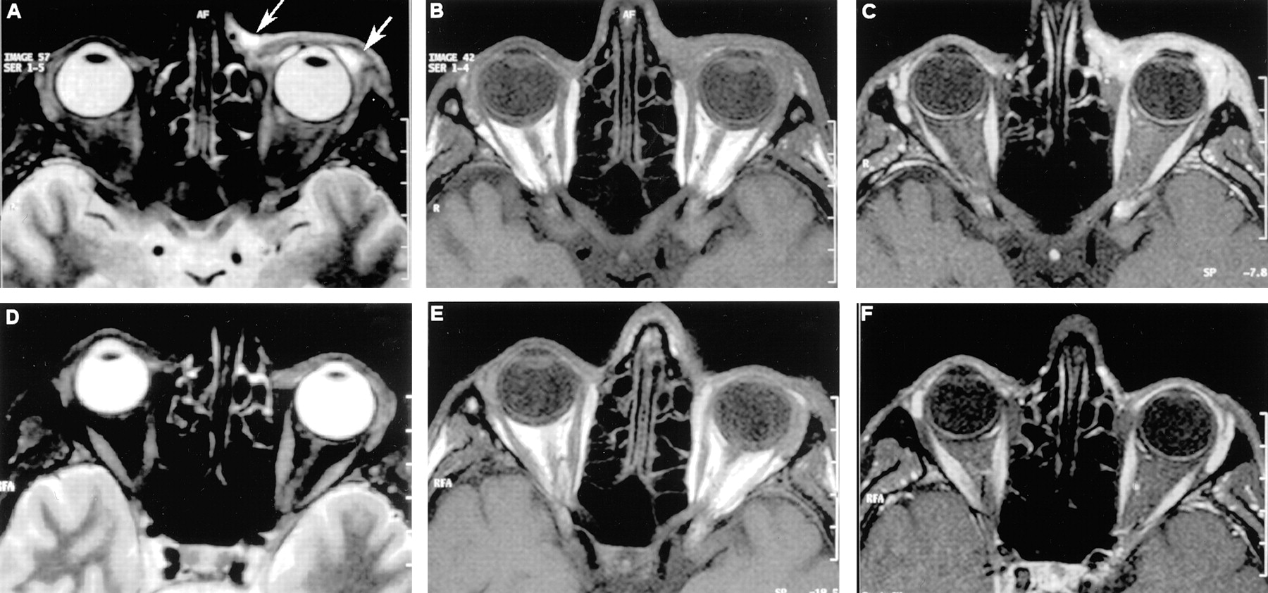
MR images obtained a 38-year-old man who developed AIDS-related Kaposi sarcoma of the conjunctiva and lacrimal gland. All axial sections were tilted to the level of the optic nerve; coronal sections were perpendicular to axial sections. Posttreatment images were obtained 3 months after pretreatment images.

A, Axial turbo inversion recovery magnitude (4000/30 [TR/TE]) MR image of the orbita shows an abnormal mass (arrows) with high signal intensity that involves the conjunctiva and lacrimal gland.

B, Axial T1-weighted (735/12) MR image of the orbita shows an abnormal mass that is isointense relative to muscular tissue (arrows) and involves the conjunctiva and lacrimal gland.

C, Corresponding postcontrast T1-weighted (735/12) MR image shows a marked enhancement of the abnormal tissue of the lower eyelid.

D, Corresponding posttreatment axial turbo inversion recovery magnitude (4000/30).

E, Corresponding posttreatment axial T1-weighted (735/12).

F, Corresponding posttreatment postcontrast T1-weighted (735/12)

Discussion

KS is the most common tumor arising in HIV-infected patients and is an AIDS-defining illness according to Centers for Disease Control guidelines. The clinical course of AIDS-related KS is highly variable, and skin lesions often coincide with pulmonal or gastrointestinal KS. When KS is suspected, mucocutaneous and gastrointestinal investigation should be initiated. In the case of our patient, KS was present in one eye, the pharynx, and lung. KS of the eye is an entity that has not been widely described in the literature.

Although clinical examination and biopsy are still required to confirm the diagnosis of KS, knowledge about the radiologic presentation could aid diagnosis in the orbita or other intracranial locations with limited access for evaluation. To our knowledge, this is the first publication of information on MR imaging results of KS of the eye. In the case of our patient, an abnormal mass was seen with high signal intensity on T2-weighted (in this case, turbo inversion recovery magnitude) and low signal intensity on T1-weighted images with a marked enhancement on postcontrast images due to rich vascularization. The findings were interpreted as an infiltrative process of the bulbar conjunctiva and diffuse involvement of the left lacrimal gland. Although those findings are nonspecific and raise the possibility of a lymphoproliferative disorder, pseudotumor, or infection, the diagnosis of KS is prompted by the context of HIV infection. Bulbar conjunctival KS can be confused with caroticocavernous fistula, hemangioma, or lymphangioma. Caroticocavernous fistula would be accompanied by edemateous swelling or chronic subconjunctival hemorrhage. A fistula would show a pronounced vascular congestion of the superior ophthalmic vein, in contrast to KS. Cavernous hemangiomas occurring in adults are almost always intraconal in location. They are usually hypointense on T1-weighted images and markedly hyperintense on T2-weighted images. Capillary hemangiomas can be located in the preseptal region and show an infiltrative pattern, but they are usually pediatric tumors affecting infants up to 2 years of age. Lymphangioma occurs in children between 1 and 15 years old and involves muscle tissue. In addition to MR imaging, orbital tumors can be further differentiated by clinical history, examination, CT, and angiography. In vascular tumors, Doppler sonography may be helpful.

We recommend use of the following sequences: axial T2-weighted images (3 mm), axial T1-weighted images without fat saturation (3 mm), postcontrast axial and coronal T1-weighted images with fat saturation (3 mm), and postcontrast T1-weighted parasagittal images if spatial information is required. KS may not present with distinct imaging criteria but may have characteristic tissue distribution (glands, mucosal tissues).

If precise spatial information on the dimensions of ocular KS is required, advanced MR imaging techniques may be employed. Ultra thin-section 3D fast spin-echo (3D-FSE) T2-weighted imaging, a new MR imaging technique (7), can be used as a supplementary method and has been proposed recently for the study of ocular and orbital malignancies. The 3D-FSE T2-weighted sequence offers superior resolution of intraocular and orbital structures compared with that of conventional MR imaging. The main advantage of the 3D-FSE T2-weighted sequence is the option to perform multiplanar reconstructions. This method allows a subtle analysis of anatomic structures and thus can be helpful in localizing pre- or postseptal tumors and checking for bulbar infiltration. Although this new technique is particularly useful in the evaluation of intraocular tumors and the nerve-sheath complex, it may also improve diagnosis and therapeutic management in patients with ocular and orbital malignancies as KS.

Conclusion

In our patient, a regression of ocular KS was seen after a therapy with liposomal doxorubicin. Thus, we observe that a marked reduction of KS can been seen at follow-up MR imaging, but this observation is not specific to any particular therapeutic or pathogenetic mechanism. Nevertheless, knowledge of the MR imaging features of KS are useful not only in diagnosis but also in noninvasive assessment of the impact of emerging therapeutic strategies (8) for KS.

References

  1. ↵
    Maclean H, Hall AJ, McCombe MF, Sandland AM. AIDS and the eye: a 10-year experience. Aust N Z J Ophthalmol 1996;24:61–67
  2. ↵
    Murray N, McCluskey P, Wakefield D, Beaumont P. Isolated bulbar conjunctival Kaposi’s sarcoma. Aust N Z J Ophthalmol 1994;22:81–82
    CrossRefPubMed
  3. ↵
    Kurumety UR, Lustbader JM. Kaposi’s sarcoma of the bulbar conjunctiva as an initial clinical manifestation of acquired immunodeficiency syndrome. Arch Ophthalmol 1995;113:978
    CrossRefPubMed
  4. ↵
    Mortada A. Conjunctival regressing Kaposi sarcoma. Br J Ophthalmol 1967;51:275–280
    FREE Full Text
  5. ↵
    Ensoli B, Sgadari C, Barillari G, et al. Biology of Kaposi’s sarcoma. Eur J Cancer 2001;37:1251–1269
    CrossRefPubMed
  6. ↵
    Collaco L, Goncalves M, Gomes L, Miranda R. Orbital Kaposi’s sarcoma in acquired immunodeficiency syndrome. Eur J Ophthalmol 2000;10:88–90
    PubMed
  7. ↵
    McCaffery S, Simon EM, Fischbein NJ, et al. Three-dimensional high-resolution magnetic resonance imaging of ocular and orbital malignancies. Arch Ophthalmol 2002;120:747–754
    CrossRefPubMed
  8. ↵
    Dezube BJ. New therapies for the treatment of AIDS-related Kaposi sarcoma. Curr Opin Oncol 2000;12:445–449
    CrossRefPubMed
  • Received September 3, 2002.
  • Accepted after revision November 19, 2002.
  • Copyright © American Society of Neuroradiology
View Abstract
PreviousNext
Back to top

In this issue

American Journal of Neuroradiology: 24 (7)
American Journal of Neuroradiology
Vol. 24, Issue 7
1 Aug 2003
  • Table of Contents
  • Index by author
Advertisement
Print
Download PDF
Email Article

Thank you for your interest in spreading the word on American Journal of Neuroradiology.

NOTE: We only request your email address so that the person you are recommending the page to knows that you wanted them to see it, and that it is not junk mail. We do not capture any email address.

Enter multiple addresses on separate lines or separate them with commas.
Pre- and Posttreatment MR Imaging in AIDS-Related Kaposi Sarcoma of the Conjunctiva and Lacrimal Gland
(Your Name) has sent you a message from American Journal of Neuroradiology
(Your Name) thought you would like to see the American Journal of Neuroradiology web site.
CAPTCHA
This question is for testing whether or not you are a human visitor and to prevent automated spam submissions.
Cite this article
A. Scheschonka, M. Mösch, S. Krieglsteiner, B. Turowski, F. E. Zanella
Pre- and Posttreatment MR Imaging in AIDS-Related Kaposi Sarcoma of the Conjunctiva and Lacrimal Gland
American Journal of Neuroradiology Aug 2003, 24 (7) 1327-1329;

Citation Manager Formats

  • BibTeX
  • Bookends
  • EasyBib
  • EndNote (tagged)
  • EndNote 8 (xml)
  • Medlars
  • Mendeley
  • Papers
  • RefWorks Tagged
  • Ref Manager
  • RIS
  • Zotero
0 Responses
Respond to this article
Share
Bookmark this article
Pre- and Posttreatment MR Imaging in AIDS-Related Kaposi Sarcoma of the Conjunctiva and Lacrimal Gland
A. Scheschonka, M. Mösch, S. Krieglsteiner, B. Turowski, F. E. Zanella
American Journal of Neuroradiology Aug 2003, 24 (7) 1327-1329;
del.icio.us logo Twitter logo Facebook logo Mendeley logo
  • Tweet Widget
  • Facebook Like
  • Google Plus One

Jump to section

  • Article
    • Abstract
    • Case Report
    • Discussion
    • Conclusion
    • References
  • Figures & Data
  • Info & Metrics
  • Responses
  • References
  • PDF

Related Articles

  • No related articles found.
  • PubMed
  • Google Scholar

Cited By...

  • No citing articles found.
  • Crossref
  • Google Scholar

This article has not yet been cited by articles in journals that are participating in Crossref Cited-by Linking.

More in this TOC Section

  • Correlation of Apparent Diffusion Coefficient at 3T with Prognostic Parameters of Retinoblastoma
  • Parathyroid Lesions: Characterization with Dual-Phase Arterial and Venous Enhanced CT of the Neck
  • Efficacy of Diffusion-Weighted Imaging for the Differentiation between Lymphomas and Carcinomas of the Nasopharynx and Oropharynx: Correlations of Apparent Diffusion Coefficients and Histologic Features
Show more Head and Neck

Similar Articles

Advertisement

Indexed Content

  • Current Issue
  • Accepted Manuscripts
  • Article Preview
  • Past Issues
  • Editorials
  • Editors Choice
  • Fellow Journal Club
  • Letters to the Editor

Cases

  • Case Collection
  • Archive - Case of the Week
  • Archive - Case of the Month
  • Archive - Classic Case

Special Collections

  • Special Collections

Resources

  • News and Updates
  • Turn around Times
  • Submit a Manuscript
  • Author Policies
  • Manuscript Submission Guidelines
  • Evidence-Based Medicine Level Guide
  • Publishing Checklists
  • Graphical Abstract Preparation
  • Imaging Protocol Submission
  • Submit a Case
  • Become a Reviewer/Academy of Reviewers
  • Get Peer Review Credit from Publons

Multimedia

  • AJNR Podcast
  • AJNR SCANtastic
  • Video Articles

About Us

  • About AJNR
  • Editorial Board
  • Not an AJNR Subscriber? Join Now
  • Alerts
  • Feedback
  • Advertise with us
  • Librarian Resources
  • Permissions
  • Terms and Conditions

American Society of Neuroradiology

  • Not an ASNR Member? Join Now

© 2025 by the American Society of Neuroradiology All rights, including for text and data mining, AI training, and similar technologies, are reserved.
Print ISSN: 0195-6108 Online ISSN: 1936-959X

Powered by HighWire