
Abstract
Summary: A dural arteriovenous fistula was discovered incidentally in a 58-year-old man with activated protein C resistance who underwent routine outpatient cerebral angiography for workup of multiple intracranial aneurysms.
The development of a dural arteriovenous fistula (DAVF) is a well-recognized sequela of dural sinus thrombosis. Among the most common preexisting conditions for development of sinus thrombosis and subsequent fistula formation are trauma, infection with thrombophlebitis (often related to middle ear or mastoid sepsis), severe dehydration, and thrombophilic states (1).
Among the thrombophilic conditions, activated protein C resistance (APCR) has received relatively little attention in the literature in relation to DAVF, despite its being the most common genetic cause of thrombophilia and, therefore, a significant predisposing factor for sinus thrombosis. Although several reports have documented an increased prevalence of APCR in patients with DAVF (2–4), here we present what is to the best of our knowledge the first case of DAVF discovered incidentally in a patient with APCR.
Case Report
A 58-year-old man presented as an outpatient for cerebral angiography. He had initially been referred to another institution for cerebral MR imaging and MR angiography for investigation of vague symptoms of intermittent unsteadiness, occipital paraesthesia, and vertigo over the preceding 6–9 months. These symptoms were thought to be suspicious for vertebrobasilar insufficiency. MRA disclosed two aneurysms in the cavernous segment of the right internal carotid artery and an additional aneurysm in the region of the anterior communicating artery. No significant atheromatous disease was evident. On the strength of these findings, he was referred to our institution for further investigation of his aneurysms by cerebral angiography.
Consultation with the patient before angiography was arranged to obtain informed consent. During the interview, the patient volunteered a history of APCR (subsequently confirmed on serologic testing for factor V Leiden [FVL]), from which he claimed he had suffered no ill effects; however, his sister, who also has APCR, had a history of deep venous thrombosis and pulmonary thromboembolism.
Knowing that this patient had APCR, we consulted the hematology service for advice regarding prophylactic anticoagulant therapy during conventional angiography. The hematologists advised that no additional measures were necessary over and above our standard practice of administering 5000 IU of heparin intravenously following cannulation of the common femoral artery. After consent was obtained, angiography was performed as usual under systemic heparinization.
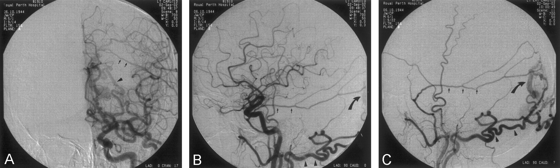
Initial selective injection of the right internal carotid artery confirmed the presence of the three aneurysms shown during MR angiography. Because of a small amount of atheroma at the left carotid bulb, it was decided initially to perform a common carotid injection to minimize the risk of embolic complications. This injection revealed a DAVF fed by the left occipital and middle meningeal arteries, confirmed on subsequent selective injection of the left external carotid artery (Fig 1). On the basis of this finding, injection of the right external carotid artery was also performed. This demonstrated further supply to the fistula by the right occipital and middle meningeal arteries. Left vertebral artery injection also disclosed supply to the fistula from its posterior meningeal branches. No supply to the fistula from either internal carotid artery was present. In retrospect, review of the MR imaging and MR angiography findings shows evidence of the fistula, with large external carotid artery branches seen bilaterally and a large venous channel seen running parallel to the distal segment of the superior sagittal sinus (not shown).

Anteroposterior (A) and lateral (B) angiograms, obtained via left common carotid artery injection, and lateral angiogram (C), obtained via left external carotid artery injection, show enlarged external carotid artery branches and an early draining vein that was confirmed during selective injection of the left external carotid artery. Middle meningeal artery, posterior branch (small arrows); occipital artery (arrowheads); early draining vein (curved arrows).
Discussion
Protein C is the key element in a vital natural anticoagulant pathway. Protein C is activated by thrombin bound to thrombomodulin on the surface of endothelial cells. When activated, protein C inhibits coagulation by cleaving, and thereby inactivating, coagulation factors Va and VIIIa (5). In hereditary APCR, protein C is activated in the normal fashion, but cleavage of activated clotting factors is either reduced or absent because of mutation(s) in the genes encoding the clotting factors themselves. More than 95% of hereditary cases are due to FVL, in which a single point mutation at position 506 in the gene encoding factor V results in substitution of glutamine for arginine (FVR506Q) (6). This substitution removes a cleavage site for protein C in the factor V molecule, rendering FVL506Q resistant to protein C and, therefore, its anticoagulant effects; however, the procoagulant effect of this mutation appears to confer increased risk of venous, but not arterial, thrombosis (5). It is estimated that around 3% of the world’s population is heterozygous for this mutation (7), which appears to be unique to whites (5). Inheritance of APCR follows an autosomal dominant pattern. Heterozygotes carry a sevenfold increased lifetime risk of venous thrombosis, whereas homozygotes have a 20-fold increased risk. It appears that concomitant procoagulant factors, such as oral contraception, pregnancy, and coexistent low-penetrance defects such as protein S or C deficiency are additive in terms of the additional risk conferred on the affected individual (7). In the case of transient procoagulant factors, such as the presence of an intravascular foreign body during conventional angiography, our advice from the consulting hematology team was that no special precautions should be taken over and above our standard practice of prophylactic heparinization. On the basis of our discussions with the hematologists, however, we would certainly advise systemic heparinization for patients with APCR who are undergoing conventional angiography in institutions where prophylactic heparinization is not routine.
It is now widely accepted that cerebral venous sinus thrombosis is one of the main underlying etiologic factors in the development of DAVF (1). Sinus thrombosis has many potential causes, including trauma, infection, dehydration, and the procoagulant state associated with pregnancy, the oral contraceptive pill (and other drugs), and congenital or acquired defects in the coagulation pathways. Among the last factors mentioned, it has now been well established that APCR is a significant risk factor for development of dural sinus thrombosis (8, 9). Moreover, a significant increase in the prevalence of APCR in patients with DAVF has been documented in the literature (2–4). Thus, it seems logical to conclude that patients with APCR have an initial predisposition to dural sinus thrombosis, and, therefore, to the pathophysiologic sequence which culminates in DAVF.
In our case, the discovery of the patient’s DAVF was incidental. Although we had knowledge before angiography that he had APCR, his symptomatology was sufficiently vague and his mode of referral to our institution so specific that we had not considered the potential relevance of performing common carotid or selective external carotid angiography. Indeed, it could be argued that the on-table discovery of his DAVF was somewhat fortuitous: had the presence of atheroma at his left carotid bulb not prompted us to perform a common, rather than internal, carotid injection, we may not have recognized the smaller feeders from his vertebral artery until we reported the study.
Dural sinus thrombosis and DAVF may have a multitude of presenting symptoms and signs, making their diagnosis on clinical grounds difficult. In many cases, these conditions are minimally symptomatic or even asymptomatic. In some patients, as in our case, the symptoms may be vague or mimic other conditions such as transient ischemic attack, cervical spondylosis, or middle or inner ear disease (10, 11). In our case, the patient’s symptoms were not explained by the aneurysms detected on the basis of MR and MR angiography findings. This should perhaps have prompted a more thorough search for cerebral venous abnormality on the part of the clinician or the radiologist concerned, although no mention of the patient’s APCR was made in the MR imaging referral. It could be argued that at the time of angiography, armed with the knowledge of his APCR and his unusual symptoms, perhaps we should have been looking more actively for a problem such as dural sinus thrombosis or DAVF. APCR is relatively common and has confirmed associations with both dural sinus thrombosis and dural arteriovenous fistulas.
Conclusion
We believe that this case is of significant interest from two separate standpoints. First, to the best of our knowledge, it is the only reported case of incidental discovery of DAVF in a patient with APCR and thus seems to lend further weight to the hypothesis that DAVF is a sequela of dural sinus thrombosis. Second, it serves as a salutary warning that, when dealing with patients with APCR, clinicians and radiologists alike should be aware of the association with dural sinus thrombosis and therefore DAVF. This awareness should prompt active investigation for these potentially dangerous conditions when other causes for the patients’ symptoms have not been found.
References
- Received November 13, 2002.
- Accepted after revision December 4, 2002.
- Copyright © American Society of Neuroradiology
In this issue


Jump to section
Related Articles
Cited By...
- No citing articles found.