
Article Figures & Data
Figures
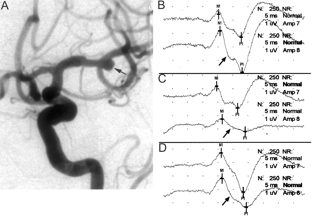
- Fig 1.
Images from the case of a 78-year-old woman who presented with symptoms of a subarachnoid hemorrhage.
A, Left internal carotid artery injection in the anterolateral projection shows a 5-mm left middle cerebral artery bifurcation aneurysm (arrow). Intra-aneurysmal coiling was attempted while the patient was systemically heparinized.
B, Baseline cerebral SSEPs after bilateral median nerve stimulation. Top tracing, left median nerve stimulation (ie, right brain); bottom tracing, right median nerve stimulation (ie, left brain) (arrow).
C, One minute after coil placement into the aneurysm, a >50% decrease in amplitude of the right median nerve SSEP was noted (arrow). This is consistent with significant left cerebral ischemia. Fluoroscopic evaluation suggested the coil was partially prolapsed into the parent artery, and considering the change in potentials, it was decided to quickly remove this coil. Formal angiographic assessment may well have shown significant compromise in the parent vessel; however, because the changes were rapid and profound, the coil was removed. Top tracing, left median nerve stimulation (ie, right brain); bottom tracing, right median nerve stimulation (ie, left brain) (arrow).
D, Left cerebral evoked potential (arrow) returned to baseline levels after removal of the coil. Because coil embolization could not be performed safely, the patient subsequently underwent surgical clipping of the aneurysm. Top tracing, left median nerve stimulation (ie, right brain); bottom tracing, right median nerve stimulation (ie, left brain) (arrow).
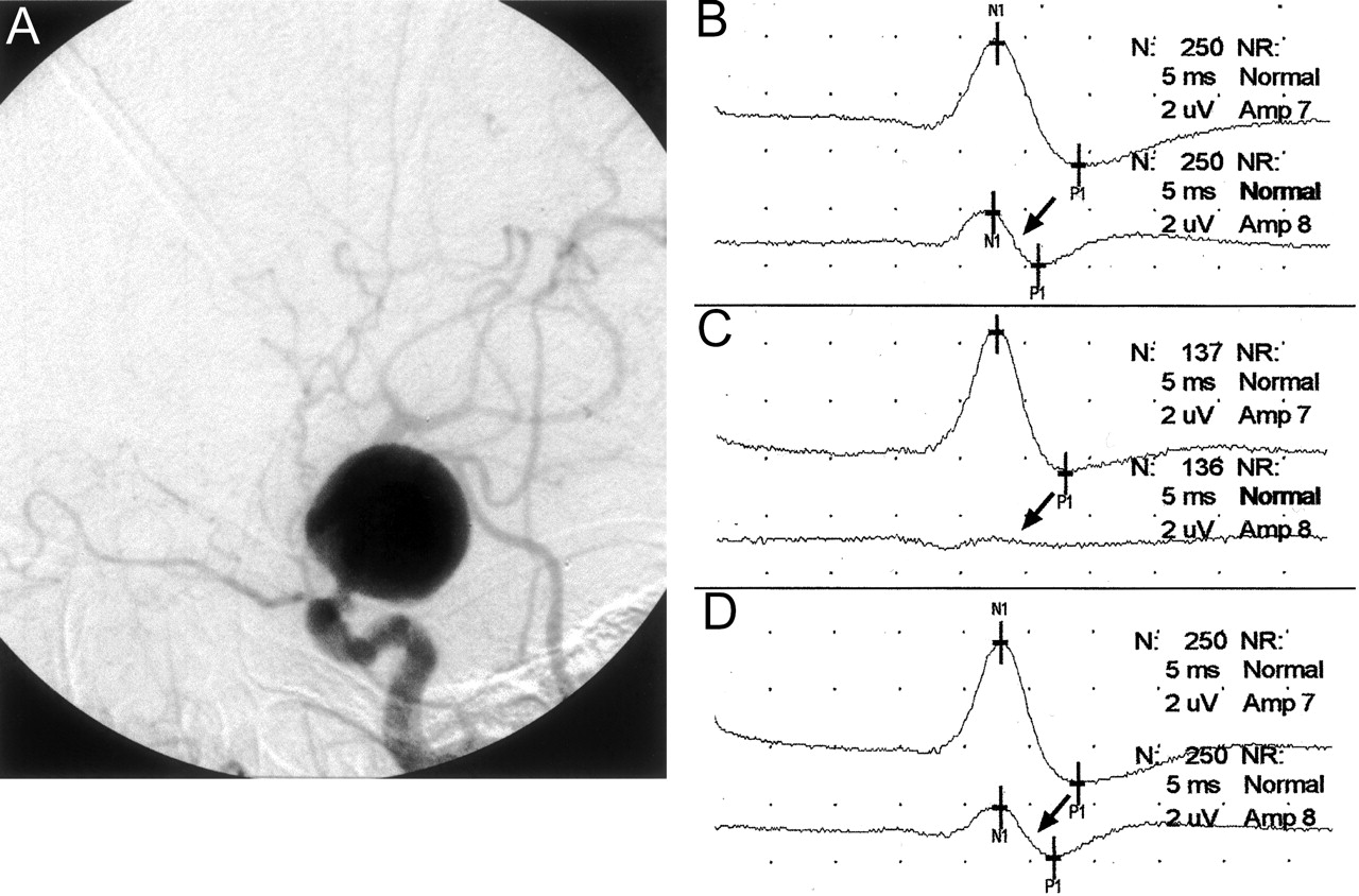
- Fig 2.
Images from the case of a 75-year-old woman who presented with a ruptured giant left internal carotid artery aneurysm.
A, Lateral projection angiogram of the left internal carotid artery shows the aneurysm arising in the supraclinoid segment shortly after the takeoff of the ophthalmic artery. The aneurysm was thought to be unfavorable for GDC embolization, and balloon test occlusion was thus performed in anticipation of permanent vessel occlusion. Because of the patient’s physical condition, the procedure could be performed only with the patient under general anesthesia.
B, Baseline cerebral SSEPs after bilateral median nerve stimulation. Note the baseline asymmetry, with the right median nerve SSEP (arrow) being smaller in amplitude than the left. Top tracing, left median nerve stimulation (ie, right brain); bottom tracing, right median nerve stimulation (ie, left brain) (arrow).
C, Left internal carotid artery balloon test occlusion was performed, resulting in gradual amplitude reduction of the left cerebral (right median nerve stimulation) SSEP over 5 min and precipitous decrease in the 6th min. The SSEP obtained 6 min after balloon occlusion shows a nearly complete loss of the left cerebral SSEP (arrow). Top tracing, left median nerve stimulation (ie, right brain); bottom tracing, right median nerve stimulation (ie, left brain) (arrow).
D, Balloon was deflated immediately after the SSEP tracing shown in 2C. The cerebral SSEP returned to baseline amplitude levels after 1 min (arrow). Top tracing, left median nerve stimulation (ie, right brain); bottom tracing, right median nerve stimulation (ie, left brain) (arrow).


Tables
Patient No. Age (yr) Sex Aneurysm Location Ruptured (Y/N) Procedure NPM Type 1 80 F LICA N Aneurysm coiling SSEP 2 33 F R cavemous ICA N Aneurysm coiling SSEP 3 41 F Basilar trunk N Aneurysm coiling SSEP, BAEP 4 54 M LICA N Aneurysm coiling SSEP, EEG 5 40 F RICA N BTO s/p intracranial bypass SSEP, EEG, neuro exam BTO and RICA occulusion SSEP, EEG, neuro exam 6 52 F RICA ophthalmic Y BTO and aneurysm coiling SSEP, EEG 7 59 M Distal L PICA Y Sodium amytal testing and glue embolization of L PICA SSEP, BAEP 8 48 F MCA Y BTO and RICA occlusion* SSEP, BAEP, neuro exam 9 52 F Basilar tip Y Attempted aneurysm coiling SSEP, BAEP 10 81 F RICA N BTO SSEP, EEG, neuro exam 11 80 F Basilar N BTO and RVA occlusion SSEP, BAEP 12 74 F RICA N BTO and aneurysm coiling SSEP, EEG, neuro exam 13 72 M R AICA Y Aneurysm coiling SSEP, EEG 14 76 F R supraclinoid ICA N BTO and aneurysm coiling SSEP, BAEP, neuro exam 15 76 M Mid-basilar N BTO and LVA occlusion† SSEP, BAEP, neuro exam 16 54 F R Pcomm N Pre-op BTO EEG, neuro exam 17 67 F L para-ophthalmic ICA N BTO and aneurysm coiling SSEP, EEG 18 52 F LICA N BTO and aneurysm coiling SSEP, EEG, neuro exam 19 41 F L paraclinoid N Aneurysm coiling SSEP, EEG 20 52 M LICA N Aneurysm coiling SSEP, EEG 21 49 F L cavernous ICA N Pre-op BTO SSEP, neuro exam LICA occlusion SSEP, EEG, neuro exam 22 72 F R cavernous ICA N BTO and RICA occlusion SSEP, EEG 23 76 F R MCA bifurcation N Aneurysm coiling SSEP, EEG 24 43 M R distal vertebral N BTO and RVA occlusion SSEP, BAEP, neuro exam 25 60 M L para-ophthalmic ICA N BTO and aneurysm coiling SSEP, EEG, neuro exam 26 78 F L MCA bifurcation Y Attempted aneurysm coiling SSEP, EEG 27 75 F L distal ICA Y BTO and attempted aneurysm coiling SSEP, EEG 28 54 M Basilar tip Y Aneurysm coiling SSEP, BAEP 29 73 F Basilar tip, Acomm, R Pcomm N Aneurysm coiling SSEP, BAEP 30 40 F R MCA bifurcation Y Aneurysm coiling SSEP 31 68 F Basilar tip N Aneurysm coiling SSEP, BAEP 32 62 F L paraclinoid ICA Y Aneurysm coiling SSEP, EEG 33 44 M R P2 segment N Sodium amytal testing SSEP, BAEP, neuro exam Aneurysm coiling SSEP, BAEP, EEG 34 69 F LICA Y BTO and aneurysm coiling SSEP, EEG 35 51 M L distal VA N BTO and LVA occlusion SSEP, BAEP, neuro exam Note.—Y indicates yes; N, no; NPM, neurophysiological monitoring; F, female; M, male; L, left; R, right; ICA, internal carotid artery; PICA, posterior inferior cerebellar artery; MCA, middle cerebral artery; AICA, anterior inferior cerebellar artery; Pcomm, posterior communicating artery; Acomm, anterior communicating artery; VA, vertebral artery; BTO, balloon test occlusion; s/p, status-post; pre-op, preoperative; SSEP, somatosensory evoked potential; BAEP, brain stem auditory evoked potential; neuro exam, neurologic examination.
* Surgical ligation of the left internal carotid artery.
† Basilar artery test occlusion failed for this patient; permanent left vertebral artery occlusion was performed.
- TABLE 2:
Patients with changes in results of neurophysiological monitoring or NPM or clinical examination during endovascular procedures
Patient No. General Anesthesia (Y/N)* NPM Changes (Y/N) PE Changes (Y/N) Altered Management (Y/N) Type of Alteration/Reason for No Alteration 4 Y Y N N Coil loop protruding from aneurysm; coil not withdrawn 5 N Y N Y Delayed PVO to allow EC-IC graft maturation 7 Y Y N Y Increased mean arterial pressure to improve perfusion 9 Y Y N N Aneurysm perforation; proceeded with treatment 11 Y Y N Y PVO of right vertebral instead of basilar artery 26 Y Y N Y No coil detachment 27 Y Y N Y No coiling or PVO 29 Y Y N N Aneurysm perforation; proceeded with treatment 30 Y Y N N NPM changes at end of procedure 10 N N Y Y No PVO 33 N N Y Y No PVO Note.—Y indicates yes; N, no; NPM, neurophysiological monitoring; PE, physical examination; PVO, permanent vessel occlusion; EC-IC, extracranial-intracranial.
* Balloon test occlusion was performed with the patient under conscious sedation when possible; permanent vessel occlusion or aneurysm coiling was then performed with the patient under general anesthesia.
In this issue


Jump to section
Related Articles
Cited By...
- Intraoperative vascular complications during 2278 cerebral endovascular procedures with multimodality IONM: relationship between signal change, complication, intervention and postoperative outcome
- Role of Anesthesia for Endovascular Treatment of Ischemic Stroke: Do We Need Neurophysiological Monitoring?
- A checklist for cerebral aneurysm embolization complications
- Feasibility and Efficacy of Transcranial Motor-Evoked Potential Monitoring in Neuroendovascular Surgery