
Article Figures & Data
Figures
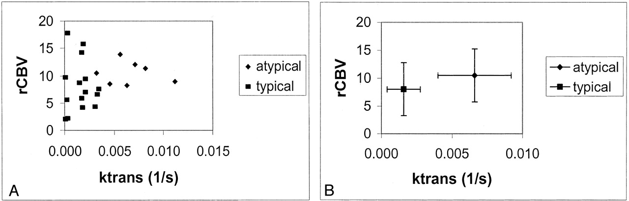
- Fig 1.
Scatterplots of rCBV versus Ktrans.
A, Grade II (diamonds) versus grade I (squares) meningiomas. The groups are well separated in the permeability measure but not in rCBV.
B, rCBV versus Ktrans for the two grades. Data points are the mean values. Error bars indicate the SD.
- Fig 2.
Photomicrographs (hematoxylin-eosin).
A, Medium-power image demonstrates tumor cells, which form a lobulated pattern with whorls (arrows) in a typical meningioma (original magnification ×100).
B, High-power images demonstrates at least three mitotic figures (arrows) in this atypical meningioma (original magnification ×200).
- Fig 3.
Pathologically confirmed meningioma (WHO grade I/III).
A, Axial contrast-enhanced T1-weighted (600/14) MR image.
B, Axial gradient-echo (1000/54) pMRI image with an rCBV color overlay demonstrates high rCBV throughout the lesion.
C, Axial gradient-echo (1000/54) axial pMRI image with SD25 depicts a decrease in signal intensity after 25 seconds.
D, Normalized signal intensity plotted against time for the white matter (squares) and the lesion (triangles). The lines represent the fitted curves derived by using the first-pass PM algorithm. The return to baseline is more rapid in typical meningiomas than in atypical meningiomas.
- Fig 4.
Pathologically confirmed atypical meningioma (WHO grade II/III).
A, Axial contrast-enhanced T1-weighted (600/14) MR image.
B, Axial gradient-echo (1000/54) pMRI with an rCBV color overlay demonstrates high rCBV throughout the lesion.
C, Axial gradient-echo (1000/54) pMRI with an SD25 overlay. Red indicates regions of greatest decrease in signal intensity, which are correlated to areas of increased permeability. Qualitatively, this is the best way to distinguish this lesion from that shown in Figure 2.
D, Normalized signal intensity plotted against time for the white matter (squares) and the lesion (triangles). The lines represent the fitted curve derived by using the first-pass PM algorithm. Increased permeability results in a slower return to baseline.


Tables
rCBV and Ktrans data in 22 patients with typical or atypical meningiomas
Patient/Age, y/Sex rCBV* Ktrans, seconds−1† vp, %‡ ve, %§ kep‖ Location Typical meningiomas 1/27/F 9.3 2.1 × 10⁻³ 0.87 3.06 6.9 × 10⁻⁴ Right temporal 2/33/M 17.8 2.8 × 10⁻⁴ 10.94 0.20 1.4 × 10⁻³ Left lateral ventricle 3/38/F 14.2 1.8 × 10⁻³ 9.9 0.19 9.1 × 10⁻³ Left frontal 4/41/F 8.7 1.5 × 10⁻³ 2.79 8.19 1.8 × 10⁻⁴ Left parietal 5/42/F 4.1 1.8 × 10⁻³ 1.23 1.61 1.2 × 10⁻³ Right cerebellopontine 6/42/F 15.8 1.9 × 10⁻³ 12.3 0.80 2.3 × 10⁻³ Right frontal 7/47/F 9.7 0.9 × 10⁻⁵ 3.88 1.9 × 10⁻⁴ 0.47 Right parasagittal 8/47/F 5.83 1.7 × 10⁻³ 1.63 0.38 4.5 × 10⁻³ Right occipital 9/50/M 4.19 3.0 × 10⁻³ 5.92 5.36 5.8 × 10⁻⁴ Right frontal 10/51/F 6.54 3.3 × 10⁻³ 2.63 9.63 3.4 × 10⁻⁴ Right lateral ventricle 11/53/F 1.99 9.3 × 10⁻⁵ 8.30 2.3 × 10⁻² 4.0 × 10⁻³ Left parietal 12/55/M 2.15 2.7 × 10⁻⁴ 2.11 5.7 × 10⁻² 4.7 × 10⁻³ Left parafalcine 13/56/F 7.5 3.5 × 10⁻³ 2.56 7.56 4.6 × 10⁻⁴ Left sphenoid wing 14/57/F 6.94 2.1 × 10⁻³ 2.47 8.09 2.6 × 10⁻⁴ Left frontal 15/59/M 5.59 2.0 × 10⁻⁴ 6.78 3.5 × 10⁻² 5.7 × 10⁻³ Right parafalcine Mean 8.02 1.6 × 10⁻³ 4.95 3.01 3.4 × 10⁻² Not applicable SD 4.74 1.2 × 10⁻³ 3.80 3.66 1.2 × 10⁻¹ Not applicable Atypical meningiomas 16/46/F 8.97 1.1 × 10⁻² 1.57 13.7 8.2 × 10⁻⁴ Right parafalcine 17/49/F 8.2 6.3 × 10⁻³ 3.09 5.17 1.2 × 10⁻³ Left parasagittal 18/52/F 11.4 8.2 × 10⁻³ 9.80 36.5 2.2 × 10⁻⁴ Left frontal 19/58/F 12.07 7.1 × 10⁻³ 12.02 32.2 2.2 × 10⁻⁴ Right parietal 20/58/F 10.45 3.2 × 10⁻³ 12.3 1.44 2.2 × 10⁻³ Left frontal 21/69/F 8.52 4.6 × 10⁻³ 2.78 1.68 2.7 × 10⁻³ Right temporal 22/79/F 13.9 5.6 × 10⁻³ 3.58 11.49 4.9 × 10⁻⁴ Right frontal Mean 10.50 6.6 × 10⁻³ 6.45 14.6 1.1 × 10⁻³ Not applicable SD 2.1 2.59 × 10⁻³ 4.71 14.3 9.9 × 10⁻⁴ Not applicable * P = .10.
† P = .002.
‡ P = .48.
§ P = .08.
‖ P = .31.
In this issue


Jump to section
Related Articles
Cited By...
- Preoperative MR Imaging to Differentiate Chordoid Meningiomas from Other Meningioma Histologic Subtypes
- Differentiation between Treatment-Induced Necrosis and Recurrent Tumors in Patients with Metastatic Brain Tumors: Comparison among 11C-Methionine-PET, FDG-PET, MR Permeability Imaging, and MRI-ADC--Preliminary Results
- Effects of Microvascular Permeability Changes on Contrast-Enhanced T1 and Pharmacokinetic MR Imagings After Ischemia
- Imaging biomarkers of angiogenesis and the microvascular environment in cerebral tumours
- Increased Blood-Brain Barrier Permeability on Perfusion CT Might Predict Malignant Middle Cerebral Artery Infarction