
Article Figures & Data
Figures
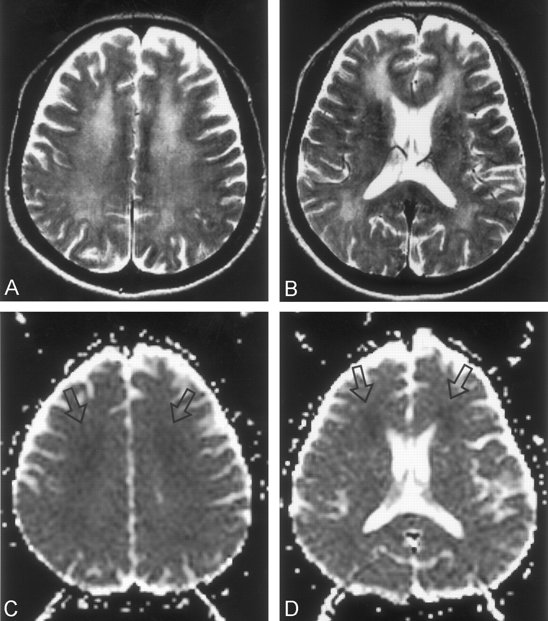
- Fig 1.
Case 1. MR images obtained in a 54-year-old woman on day 25 after CO exposure.
A and B, T2-weighted images (TR/TE/NEX, 4000/98/1) show bilateral, symmetric, confluent areas of high signal intensity in both the centrum semiovale (A) and periventricular white matter (B). The high intensity appears more prominent in the frontal lobes than elsewhere.
C and D, ADC maps (calculated from DWIs [6500/98/1, b = 0 and 1000 s/mm2]) obtained at the same levels as in A and B, respectively, show focal areas of low signal intensity (decreased diffusion) in the bilateral frontal lobes. Most of the remaining centrum semiovale and periventricular white matter appears isointense. DWIs at the same levels showed confluent high signal intensity in the centrum semiovale and periventricular white matter. The appearance was similar to that on the T2-weighted images (not shown) and mainly resulted from a T2 shine-through effect.
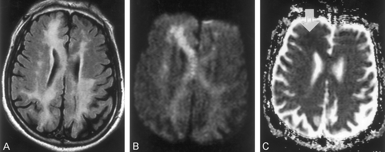
- Fig 2.
Case 2. MR images obtained in a 71- year-old man on day 41 after CO exposure.
A, FLAIR image (TR/TE/TI/NEX, 9000/110/2200/1) shows asymmetric distribution of the high-intensity lesions in the periventricular white matter and corpus callosum.
B, DWI (TR/TE/NEX, 6500/98/1; b = 1000 s/mm2) shows similar high signal intensity in the periventricular white matter and corpus callosum. The high intensity is more prominent in the right frontal lobe and on the right side of the genu of the corpus callosum than elsewhere.
C, ADC map shows a focal area of subtle low signal intensity in the right frontal lobe (arrow). Other white matter lesions appear isointense.
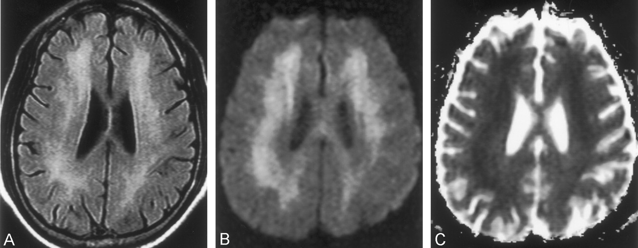
- Fig 3.
Case 4. MR images obtained in a 65-year-old woman on day 55 after CO exposure.
A, FLAIR image (TR/TE/TI/NEX, 9000/110/2200/1) shows diffuse high-intensity lesions in the periventricular white matter.
B, DWI (TR/TE/NEX, 6500/98/1; b = 1000 s/mm2) shows confluent high signal intensity in the same periventricular white matter.
C, ADC map shows diffuse low signal intensity in the periventricular white matter.


Tables
- TABLE 1:
Summary of clinical features in five patients with delayed encephalopathy of CO intoxication
Patient/Age, y/Sex* Initial Manifestations of Acute CO Intoxication Lucid Interval, wk Major Clinical Findings of Delayed Encephalopathy 1/54/F Coma with multiple burn injury 3 Short-term memory loss, confabulation; much improved at 7-mo follow-up 2/71/M Sudden decrease of verbal output, free voiding 1 Aphasia, gait disturbance; much improved at 5-mo follow-up 3/63/M Abnormal repetitive behavior, progressive abulia 3 Abulia, akinetic mutism; much improved at 8-mo follow-up 4/65/F Coma, decrease of verbal output and cognitive dysfunction 4 Urinary/fecal incontinence, short-stepped gait; persistent clinical symptoms at 6-mo follow-up 5/63/F Coma, free voiding, and defecation 4 Abulia, bradykinesia, free voiding; much improved at 5-mo follow-up * In all patients except patient 4, the cause of their exposure to CO gas was a gas leakage from an under-the-floor home heating system. Patient 4 was exposed to exhaust gas from a car.
- TABLE 2:
Summary of findings on conventional MR images, DWIs, and ADC maps, with ADC values in five patients with delayed encephalopathy of CO intoxication
Patient Interval to Imaging, d* Findings on T2-Weighted and FLAIR Images Findings on DWIs Findings on ADC Maps Mean ADC Values, ×10−3 mn 1 25 Diffuse high intensity in the PVWM and centrum semiovale, more prominent in both frontal lobes; high intensity in both globi pallidus Diffuse high intensity in the PVWM and centrum semiovale, more prominent in both frontal lobes Focal areas of low intensity in both frontal lobes 0.61 (0.53–0.70)/0.89 2 41 Asymmetric high intensity in the PVWM, centrum semiovale, and corpus callosum; more prominent on R side Asymmetric high intensity in the PVWM, centrum semiovale, and corpus callosum; more prominent on the R side Focal areas of low intensity in R frontal lobe 0.65 (0.53–0.75)/0.91 3 56 Diffuse high intensity in the PVWM and centrum semiovale Diffuse high intensity in the PVWM and centrum semiovale Focal areas of low intensity in both frontal lobes 0.64 (0.55–0.73)/0.93 4 55 Diffuse high intensity in the PVWM and centrum semiovale Diffuse high intensity in the PVWM and centrum semiovale Diffuse low intensity in the PVWM 0.56 (0.46–0.71)/0.86 4‡ 82 No change No change No change 0.53 (0.41–0.71)/0.91 5 95 Diffuse high intensity in the PVWM and centrum semiovale, more prominent in both frontal lobes; high intensity in both globi pallidus Diffuse high intensity in the PVWM and centrum semiovale Focal areas of low intensity in both frontal lobes 0.75 (0.68–0.85)/0.94 Note.—PVWM indicates periventricular white matter.
* Interval between CO Exposure and MR Imaging.
†Data are the value in the WM lesions (range)/value in normal-looking WM on T2-weighted images.
‡ Follow-up MR examination.
In this issue


Jump to section
Related Articles
Cited By...
- Dynamic MR Imaging Patterns of Cerebral Fat Embolism: A Systematic Review with Illustrative Cases
- The Role of MR Imaging in Assessment of Brain Damage from Carbon Monoxide Poisoning: A Review of the Literature
- Delayed toxic-hypoxic encephalopathy
- Neuropsychiatric aspects of carbon monoxide poisoning: diagnosis and management
- 1H-magnetic resonance spectroscopy indicates damage to cerebral white matter in the subacute phase after CO poisoning
- Longitudinal study of carbon monoxide intoxication by diffusion tensor imaging with neurospsychiatric correlation
- White Matter Damage in Carbon Monoxide Intoxication Assessed in Vivo Using Diffusion Tensor MR Imaging
- The Anatomy of Object Recognition--Visual Form Agnosia Caused by Medial Occipitotemporal Stroke