Advertisement

Main menu

  • Home
  • Content
    • Current Issue
    • Accepted Manuscripts
    • Article Preview
    • Past Issue Archive
    • AJNR Case Collection
    • Case of the Week Archive
    • Classic Case Archive
    • Case of the Month Archive
  • Special Collections
    • Spinal CSF Leak Articles (Jan 2020-June 2024)
    • 2024 AJNR Journal Awards
    • Most Impactful AJNR Articles
  • Multimedia
    • AJNR Podcast
    • AJNR Scantastics
    • Video Articles
  • For Authors
    • Submit a Manuscript
    • Author Policies
    • Fast publishing of Accepted Manuscripts
    • Graphical Abstract Preparation
    • Manuscript Submission Guidelines
    • Imaging Protocol Submission
    • Submit a Case for the Case Collection
  • About Us
    • About AJNR
    • Editorial Board
  • More
    • Become a Reviewer/Academy of Reviewers
    • Subscribers
    • Permissions
    • Alerts
    • Feedback
    • Advertisers
    • ASNR Home
  • Other Publications
    • ajnr

User menu

  • Alerts
  • Log in

Search

  • Advanced search
American Journal of Neuroradiology

American Journal of Neuroradiology

ASHNR American Society of Functional Neuroradiology ASHNR American Society of Pediatric Neuroradiology ASSR
  • Alerts
  • Log in

Advanced Search

  • Home
  • Content
    • Current Issue
    • Accepted Manuscripts
    • Article Preview
    • Past Issue Archive
    • AJNR Case Collection
    • Case of the Week Archive
    • Classic Case Archive
    • Case of the Month Archive
  • Special Collections
    • Spinal CSF Leak Articles (Jan 2020-June 2024)
    • 2024 AJNR Journal Awards
    • Most Impactful AJNR Articles
  • Multimedia
    • AJNR Podcast
    • AJNR Scantastics
    • Video Articles
  • For Authors
    • Submit a Manuscript
    • Author Policies
    • Fast publishing of Accepted Manuscripts
    • Graphical Abstract Preparation
    • Manuscript Submission Guidelines
    • Imaging Protocol Submission
    • Submit a Case for the Case Collection
  • About Us
    • About AJNR
    • Editorial Board
  • More
    • Become a Reviewer/Academy of Reviewers
    • Subscribers
    • Permissions
    • Alerts
    • Feedback
    • Advertisers
    • ASNR Home
  • Follow AJNR on Twitter
  • Visit AJNR on Facebook
  • Follow AJNR on Instagram
  • Join AJNR on LinkedIn
  • RSS Feeds

Welcome to the new AJNR, Updated Hall of Fame, and more. Read the full announcements.


AJNR is seeking candidates for the position of Associate Section Editor, AJNR Case Collection. Read the full announcement.


Getting new auth cookie, if you see this message a lot, tell someone!
Research ArticleBRAIN

Anatomy and Frequency of Large Pontomesencephalic Veins on 3D CT Angiograms of the Circle of Willis

Mehmet Teksam, Sean Casey, Alexander McKinney, Eduard Michel and Charles L. Truwit
American Journal of Neuroradiology September 2003, 24 (8) 1598-1601;
Mehmet Teksam
  • Find this author on Google Scholar
  • Find this author on PubMed
  • Search for this author on this site
Sean Casey
  • Find this author on Google Scholar
  • Find this author on PubMed
  • Search for this author on this site
Alexander McKinney
  • Find this author on Google Scholar
  • Find this author on PubMed
  • Search for this author on this site
Eduard Michel
  • Find this author on Google Scholar
  • Find this author on PubMed
  • Search for this author on this site
Charles L. Truwit
  • Find this author on Google Scholar
  • Find this author on PubMed
  • Search for this author on this site
  • Article
  • Figures & Data
  • Info & Metrics
  • Responses
  • References
  • PDF
Loading

Article Figures & Data

Figures

  • Fig 1.
    • Download figure
    • Open in new tab
    • Download powerpoint
    Fig 1.

    34-year-old man who presented to the emergency department with headache.

    A, Nonenhanced CT scan of the head demonstrates a focal hyperattenuation (arrow) suggestive of subarachnoid hemorrhage in the interpeduncular cistern.

    B, CT angiographic source image demonstrates a large anterior PMV (arrow) in the interpeduncular fossa posterior to basilar artery (arrowhead).

    C–E, Sagittal slab MIP image (C) and 3D volume-rendered craniocaudal (D) and posteroanterior (E) views demonstrate the large anterior PMV (white arrowhead) posterior to the basilar artery (black arrowhead) and the interpeduncular veins (black arrows). A indicates anterior; P, posterior; S, superior; I, inferior.

  • Fig 2.
    • Download figure
    • Open in new tab
    • Download powerpoint
    Fig 2.

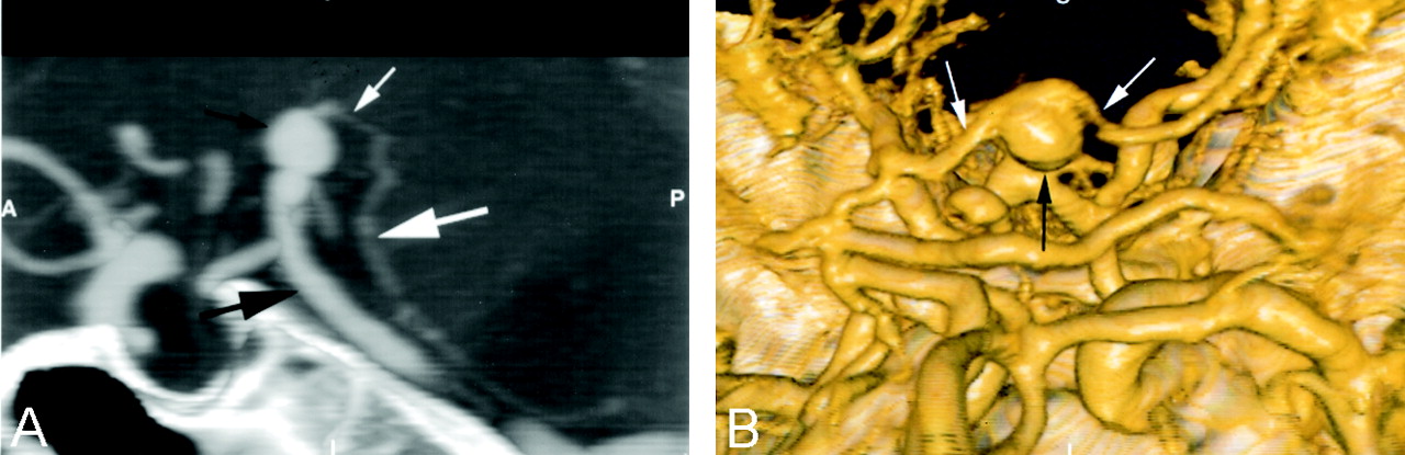
    68-year-old woman with basilar tip aneurysm.

    A and B, Sagittal slab MIP (A) and 3D volume-rendered (Towne’s view) (B) images demonstrate an anterior PMV (large white arrow in A) posterior to the basilar artery (large black arrow in A). Note that the interpeduncular veins (small white arrows), which connect the anterior PMV with the basal vein of Rosenthal, are draping over the basilar tip aneurysm (small black arrows). A indicates anterior; P, posterior; S, superior; I, inferior.

  • Fig 3.
    • Download figure
    • Open in new tab
    • Download powerpoint
    Fig 3.

    56-year-old woman evaluated with CT angiography for aneurysm of the supraclinoid segment of the internal carotid artery.

    A, Three-dimensional volume-rendered image (posteroanterior view) demonstrates a transverse pontine vein (large black arrows) that appears to be arising from the basilar artery (thin black arrow). This vein could potentially be confused with the anterior inferior cerebellar artery. The actual anterior inferior cerebellar artery is located more inferiorly (black arrowhead). Note that the interpeduncular vein (white arrow), which drains into the basal vein of Rosenthal (white arrowhead), appears to be coming out of the basilar artery.

    B and C, Three-dimensional volume-rendered images in anteroposterior (B) and posteroanterior (C) views better demonstrate the anterior PMV (white arrowhead) behind the basilar artery (black arrowhead). Note that the hypoplastic P1 segment of the right posterior cerebral artery (green arrow in C) and right interpeduncular vein (large white arrow) draining into the basal vein of Rosenthal (small white arrows in B) are now clearly separated. R indicates right; L, left; S, Superior; I, inferior.

  • Fig 4.
    • Download figure
    • Open in new tab
    • Download powerpoint
    Fig 4.

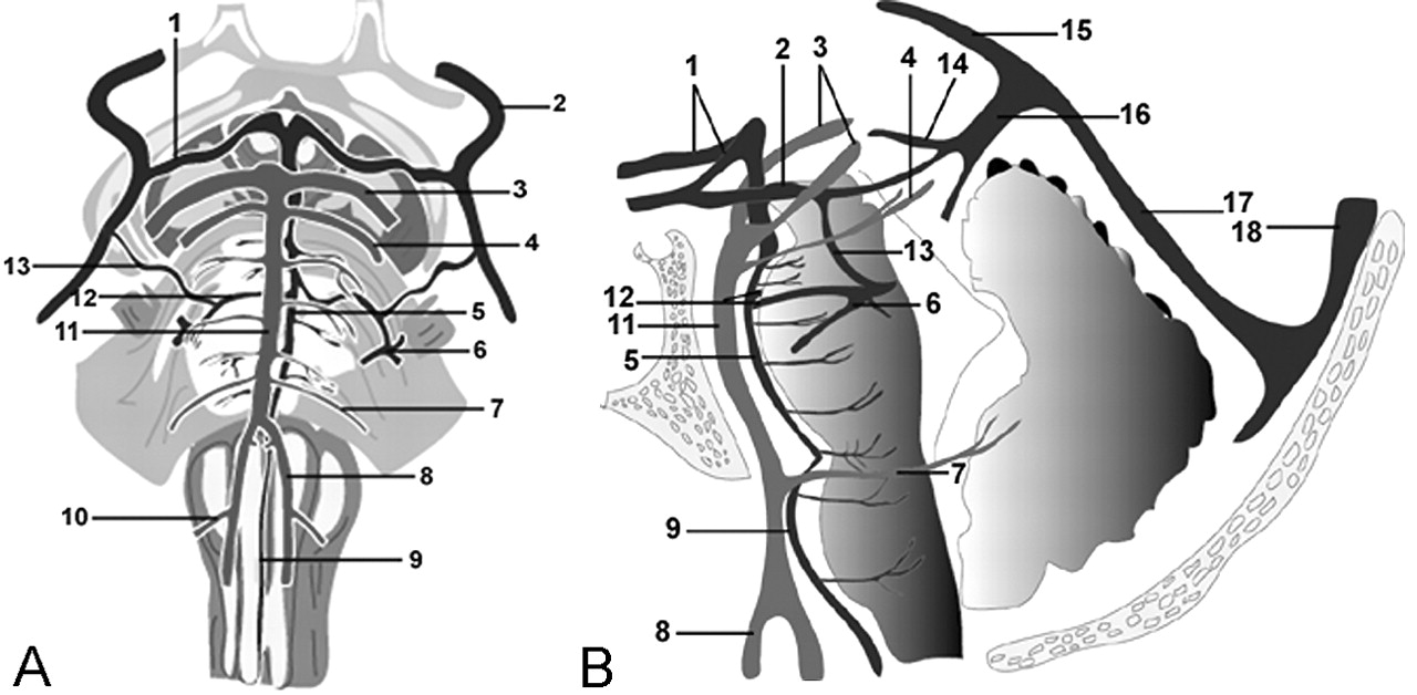
    A and B, Anteroposterior (A) and lateral (B) anatomic diagrams demonstrate the anatomy of the PMVs in relation to the vertebrobasilar system. 1 indicates interpeduncular vein; 2, basal vein of Rosenthal; 3, posterior cerebral artery; 4, superior cerebellar artery; 5, anterior PMV; 6, petrosal vein; 7, anterior inferior cerebellar artery; 8, vertebral artery; 9, anterior medullary vein; 10, posterior inferior cerebellar artery; 11, basilar artery; 12, transverse pontine vein; 13, lateral mesencephalic vein; 14, internal cerebral vein; 15, inferior sagittal sinus; 16, vein of Galen; 17, sraight sinus; 18, superior sagittal sinus.

Back to top

In this issue

American Journal of Neuroradiology: 24 (8)
American Journal of Neuroradiology
Vol. 24, Issue 8
1 Sep 2003
  • Table of Contents
  • Index by author
Advertisement
Print
Download PDF
Email Article

Thank you for your interest in spreading the word on American Journal of Neuroradiology.

NOTE: We only request your email address so that the person you are recommending the page to knows that you wanted them to see it, and that it is not junk mail. We do not capture any email address.

Enter multiple addresses on separate lines or separate them with commas.
Anatomy and Frequency of Large Pontomesencephalic Veins on 3D CT Angiograms of the Circle of Willis
(Your Name) has sent you a message from American Journal of Neuroradiology
(Your Name) thought you would like to see the American Journal of Neuroradiology web site.
CAPTCHA
This question is for testing whether or not you are a human visitor and to prevent automated spam submissions.
Cite this article
Mehmet Teksam, Sean Casey, Alexander McKinney, Eduard Michel, Charles L. Truwit
Anatomy and Frequency of Large Pontomesencephalic Veins on 3D CT Angiograms of the Circle of Willis
American Journal of Neuroradiology Sep 2003, 24 (8) 1598-1601;

Citation Manager Formats

  • BibTeX
  • Bookends
  • EasyBib
  • EndNote (tagged)
  • EndNote 8 (xml)
  • Medlars
  • Mendeley
  • Papers
  • RefWorks Tagged
  • Ref Manager
  • RIS
  • Zotero
0 Responses
Respond to this article
Share
Bookmark this article
Anatomy and Frequency of Large Pontomesencephalic Veins on 3D CT Angiograms of the Circle of Willis
Mehmet Teksam, Sean Casey, Alexander McKinney, Eduard Michel, Charles L. Truwit
American Journal of Neuroradiology Sep 2003, 24 (8) 1598-1601;
del.icio.us logo Twitter logo Facebook logo Mendeley logo
  • Tweet Widget
  • Facebook Like
  • Google Plus One

Jump to section

  • Article
    • Abstract
    • Methods
    • Results
    • Discussion
    • Conclusion
    • Footnotes
    • References
  • Figures & Data
  • Info & Metrics
  • Responses
  • References
  • PDF

Related Articles

  • No related articles found.
  • PubMed
  • Google Scholar

Cited By...

  • No citing articles found.
  • Crossref
  • Google Scholar

This article has not yet been cited by articles in journals that are participating in Crossref Cited-by Linking.

More in this TOC Section

  • Fast Contrast-Enhanced 4D MRA and 4D Flow MRI Using Constrained Reconstruction (HYPRFlow): Potential Applications for Brain Arteriovenous Malformations
  • Quiet PROPELLER MRI Techniques Match the Quality of Conventional PROPELLER Brain Imaging Techniques
  • Predictors of Reperfusion in Patients with Acute Ischemic Stroke
Show more Brain

Similar Articles

Advertisement

Indexed Content

  • Current Issue
  • Accepted Manuscripts
  • Article Preview
  • Past Issues
  • Editorials
  • Editors Choice
  • Fellow Journal Club
  • Letters to the Editor

Cases

  • Case Collection
  • Archive - Case of the Week
  • Archive - Case of the Month
  • Archive - Classic Case

Special Collections

  • Special Collections

Resources

  • News and Updates
  • Turn around Times
  • Submit a Manuscript
  • Author Policies
  • Manuscript Submission Guidelines
  • Evidence-Based Medicine Level Guide
  • Publishing Checklists
  • Graphical Abstract Preparation
  • Imaging Protocol Submission
  • Submit a Case
  • Become a Reviewer/Academy of Reviewers
  • Get Peer Review Credit from Publons

Multimedia

  • AJNR Podcast
  • AJNR SCANtastic
  • Video Articles

About Us

  • About AJNR
  • Editorial Board
  • Not an AJNR Subscriber? Join Now
  • Alerts
  • Feedback
  • Advertise with us
  • Librarian Resources
  • Permissions
  • Terms and Conditions

American Society of Neuroradiology

  • Not an ASNR Member? Join Now

© 2025 by the American Society of Neuroradiology All rights, including for text and data mining, AI training, and similar technologies, are reserved.
Print ISSN: 0195-6108 Online ISSN: 1936-959X

Powered by HighWire