
Article Figures & Data
Figures
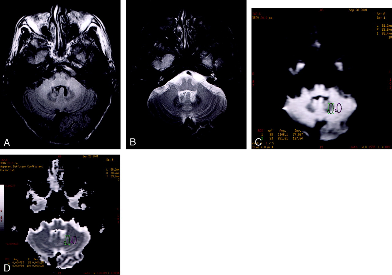
- Fig 1.
Axial FLAIR image (A) at the level of the midcerebellum demonstrates homogeneously increased signal intensity within the dentate nuclei, presumably representing acute changes related to metronidazole toxicity. Postgadolinium T1-weighted image (B) demonstrates mildly decreased T1 signal intensity within the dentate nuclei, without evidence of enhancement. There is associated high DW signal intensity within the dentate nuclei (C). The ADC map (D) confirms that the high signal intensity on the DW image (C) is due to edema, with quantitative ADC measurements demonstrating an elevated ADC value of 989 × 10−6 mm2/s within the left dentate nucleus. There was no evidence of restricted diffusion on the ADC maps. Axial T2-weighted spin-echo image (E) at the level of the lateral ventricles demonstrates focal and confluent areas of nonenhancing signal intensity abnormality in the periventricular regions believed to be related to chronic small vessel ischemic changes. These changes were stable at follow-up imaging (not shown) and presumably not related to metronidazole toxicity.
- Fig 2.
Follow-up MR imaging performed on a 3.0-T system approximately 8 weeks after discontinuation of metronidazole. FLAIR image (A) and corresponding T2-weighted spin-echo image (B) demonstrate near-complete resolution of previously noted areas of signal intensity abnormality (Fig 1). The observed low signal intensity within the dentate nucleus is normal and related to increased susceptibility effects at 3.0 T, which increases proportional to the square of the main magnetic field strength. The DW image (C) and ADC map (D) have also normalized in the interim. Quantitative ADC measurements (D) now demonstrate an ADC value of 739 × 10−6 mm2/s, which is within normal limits.


