
Article Figures & Data
Figures
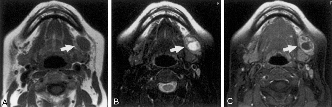
- Fig 1.
66-year-old woman with well-differentiated squamous cell carcinoma of lower gingiva.
A, Axial T1-weighted MR image of metastatic submandibular node (arrow) shows homogeneous intermediate signal intensity.
B, Axial T2-weighted, fat-suppressed MR image of the same metastatic node shows focal hyperintensity in the node (arrow).
C, Axial fat-suppressed, gadolinium-enhanced T1-weighted MR image shows a nonenhanced area in the node (arrow).
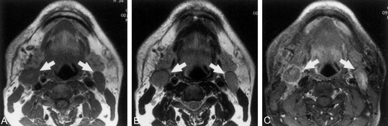
- Fig 2.
34-year-old man with benign lymphadenopathy (lymphoid hyperplasia) of the bilateral jugulodigastric nodes.
A, Axial T1-weighted MR image shows homogeneous (except for hilum signal intensity) nodes (arrows).
B, Axial T2-weighted MR image shows homogeneous nodes (arrows).
C, Axial fat-suppressed, gadolinium-enhanced T1-weighted MR image shows homogeneously enhanced (except for hilum signal intensity) nodes (arrows).
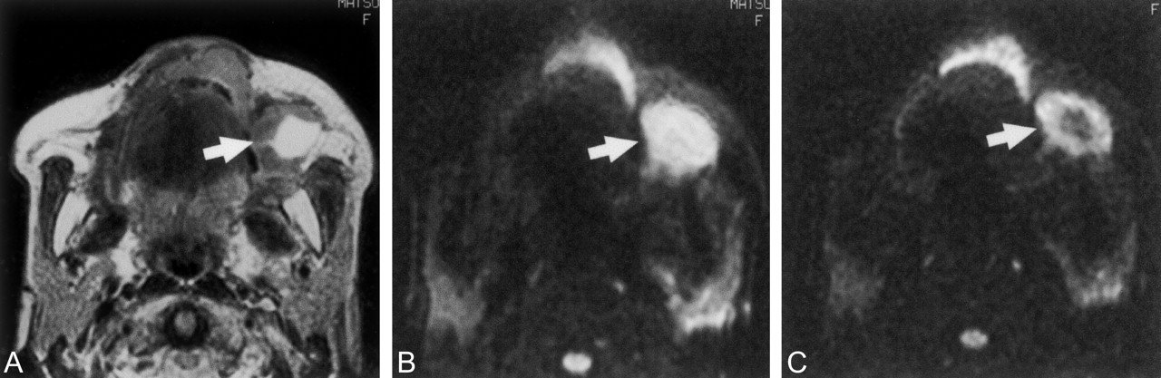
- Fig 3.
71-year-old woman with well-differentiated squamous cell carcinoma of maxilla.
A, Axial T2-weighted MR image of metastatic buccinator node (arrow) shows a large, hyperintense necrotic area.
B, Axial diffusion-weighted echo-planar MR image, at b factor of 500 s/mm2, of metastatic node (arrow) shows hyperintensity of the node.
C, Axial diffusion-weighted echo-planar MR image, at b factor of 1000 s/mm2, of metastatic node (arrow) shows nodal signal intensity with a central hypointense area. A large reduction in signal intensity on the image at b 1000 s/mm2 compared with the image at b 500 s/mm2 results in an elevated ADC (0.410 × 10⁻³ mm2/s).
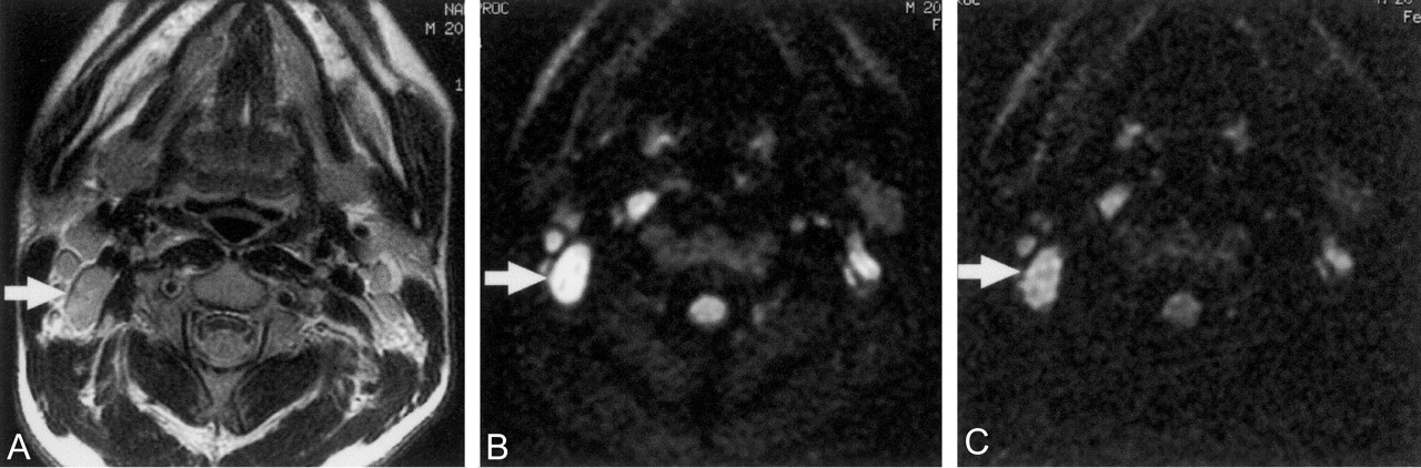
- Fig 4.
20-year-old man with benign lymphadenopathy in neck.
A, Axial T2-weighted MR image shows an inflammatory jugulodigastric node (arrow) with homogeneous signal intensity.
B, Axial diffusion-weighted MR image at b factor of 500 s/mm2 of benign lymphadenopathy (arrow) shows homogeneous hyperintensity.
C, Axial diffusion-weighted echo-planar MR image at b factor of 1000 s/mm2 of benign lymphadenopathy (arrow) shows a defined area of hyperintensity. An intermediate reduction in signal intensity on the image at b 1000 s/mm2 compared with the image at b 500 s/mm2 results in a moderate ADC (0.335 × 10⁻³ mm2/s).
- Fig 5.
56-year-old woman with diffuse large B cell lymphoma.
A, Axial T2-weighted MR image shows lymphoma in jugulodigastric node (arrow) with homogeneous signal intensity.
B, Axial diffusion-weighted echo-planar MR image at b factor of 500 s/mm2 of nodal lymphoma (arrow) shows hyperintensity.
C, Axial diffusion-weighted echo-planar MR image at b factor of 1000 s/mm2 of nodal lymphoma (arrow) shows a defined area of hyperintensity. A small reduction in signal intensity on the image at b 1000 s/mm2 compared with the image at b 500 s/mm2 results in a low ADC (0.258 × 10⁻³ mm2/s).
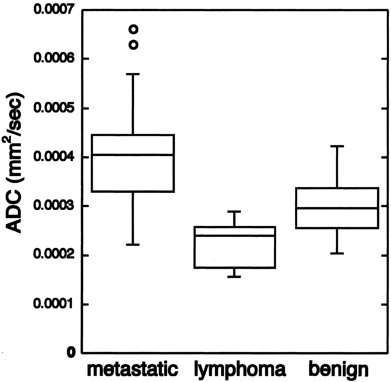
- Fig 6.
Graph (box plots) shows ADCs of metastatic nodes, nodal lymphomas, and nodes with benign lymphadenopathy. The horizontal line is a median (50th percentile) of the measured values, the top and bottom of the box represent 25th and 75th percentiles, respectively, and whiskers indicate the range from the largest to smallest observed data points within 1.5 interquartile range presented by the box. Note that ADCs of metastatic nodes are significantly higher than those of nodes with benign lymphadenopathy (P < .01, Mann-Whitney U test).
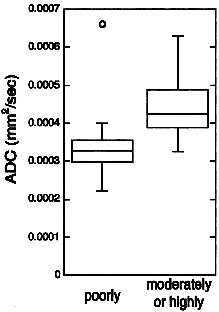
- Fig 7.
Graph (box plots) shows ADCs of poorly differentiated and of moderately or highly differentiated squamous cell carcinomas metastasized to cervical lymph nodes. Note that ADCs of metastatic nodes from poorly differentiated cancers are significantly lower than those from moderately or highly differentiated cancers (P < .01, Mann-Whitney U test).
- Fig 8.
Photomicrographs show metastatic lymph nodes from poorly (A) and highly (B) differentiated squamous cell carcinomas.
A, Poorly differentiated cancer cells with high nuclear-to-cytoplasmic ratios are densely packed in metastatic node.
B, Highly differentiated cancer cells are loosely packed, and amorphous materials (*), probably produced by cancer cells, and fibrous tissues (**) are frequently observed. (Hematoxylin and eosin, original magnification ×200)
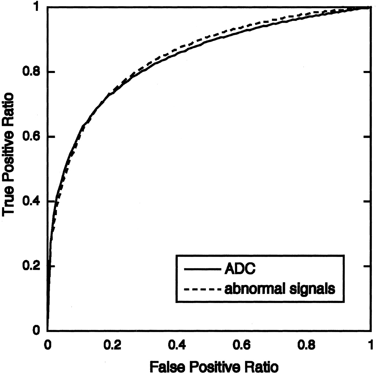
- Fig 9.
Graph shows averaged ROC curves for MR diagnostic criteria by using ADC or internal architectural abnormality (abnormal signal intensity) of nodes. Az values calculated from ROC curves indicate that diagnostic ability using either of these criteria are at similar levels: Az value (ADC) = 0.8440 ± 0.0538, and Az value (abnormal signal intensity) = 0.8437 ± 0.0230.


Tables
Univariate and multivariate logistic regression analysis of MR imaging features of metastatic nodes, benign lymphadenopathy, and nodal lymphomas in the neck
Analysis ADC Abnormal Signal Intensity Short Axis Diameter Long Axis Diameter Univariate Coefficient 1.919 1,467 1.312 1.234 SE 0.545 0.365 0.485 0.436 P value .0004 <.0001 .0068 .0047 Multivariate Coefficient 2.170 1.406 SE 0.656 0.408 P value .0009 .0006 NS NS Note.—NS indicates not significant; SE, standard error.
In this issue


Jump to section
Related Articles
Cited By...
- Quantitative Diffusion-Weighted MRI Parameters and Human Papillomavirus Status in Oropharyngeal Squamous Cell Carcinoma
- Diffusion-Weighted Imaging with Dual-Echo Echo-Planar Imaging for Better Sensitivity to Acute Stroke
- Differentiation of Recurrent Tumor and Posttreatment Changes in Head and Neck Squamous Cell Carcinoma: Application of High b-Value Diffusion-Weighted Imaging
- Prediction of Nodal Metastasis in Head and Neck Cancer Using a 3T MRI ADC Map
- Efficacy of Diffusion-Weighted Imaging for the Differentiation between Lymphomas and Carcinomas of the Nasopharynx and Oropharynx: Correlations of Apparent Diffusion Coefficients and Histologic Features
- Multiparametric MR Imaging of Sinonasal Diseases: Time-Signal Intensity Curve- and Apparent Diffusion Coefficient-Based Differentiation between Benign and Malignant Lesions
- Correlation of 18F-FDG Uptake with Apparent Diffusion Coefficient Ratio Measured on Standard and High b Value Diffusion MRI in Head and Neck Cancer
- Apparent Diffusion Coefficient Mapping for Sinonasal Diseases: Differentiation of Benign and Malignant Lesions
- Complementary Roles of Whole-Body Diffusion-Weighted MRI and 18F-FDG PET: The State of the Art and Potential Applications
- Can diffusion-weighted imaging distinguish between normal and squamous cell carcinoma of the palatine tonsil?
- Non-Gaussian Analysis of Diffusion-Weighted MR Imaging in Head and Neck Squamous Cell Carcinoma: A Feasibility Study
- Diffusion-Weighted Magnetic Resonance Imaging for Predicting and Detecting Early Response to Chemoradiation Therapy of Squamous Cell Carcinomas of the Head and Neck