
Article Figures & Data
Figures
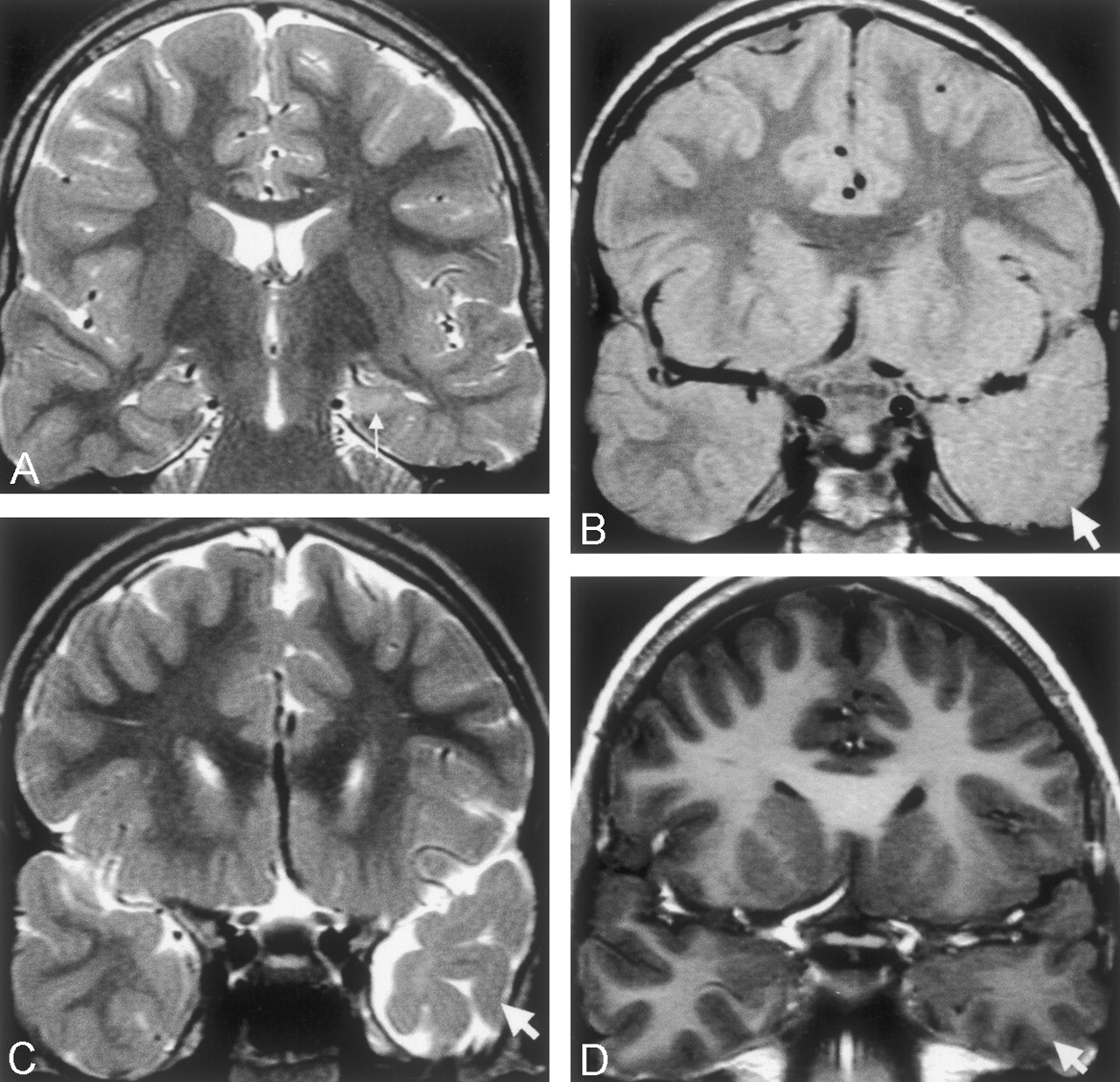
- Fig 1.
Images of a 10-year-old male patient who had meningitis at age 6 months and then onset of temporal lobe epilepsy at age 3 years.
A, Coronal view fast spin-echo image (2975/98) shows typical left hippocampal sclerosis, with loss of volume and increased signal intensity in the left hippocampus (arrow).
B and C, Coronal view proton attenuation-weighted images (2975/17) show lack of anterior temporal gray-white matter definition (arrows) and atrophy compared with the normal right side.
D, Coronal view fast spoiled gradient-recalled volume acquisition T1-weighted image shows subtle decreased signal intensity within the anterior temporal white matter compared with the left side, with no cortical thickening.
- Fig 2.
Coronal view fast spin-echo T2-weighted images (2975/98) of an 11-year-old female patient who had experienced complex partial seizures since the age of 4 years after prolonged febrile convulsions during infancy. At age 12 years, the patient underwent a temporal lobectomy. Histologic examination showed hippocampal sclerosis, with no dysplasia in the temporal specimen.
A and B, Subtle asymmetry of signal intensity in the white matter of the left temporal pole compared with the right (arrows) is shown, with apparent shrinkage of the white matter due to the slightly increased white matter signal intensity. The appearances on coronal view T1-weighted fast spoiled gradient-recalled volume acquisition images were normal.
C, More posteriorly, right hippocampal sclerosis is shown (arrow), with normal signal intensity within posterior right temporal white matter.
- Fig 3.
Coronal view T2-weighted fast spin-echo images (2975/98) of a male patient who experienced onset of epilepsy at age 4 months, soon after drainage of a large left frontal subdural empyema. Clusters of complex partial seizures occurred every week during the first year of epilepsy.
A and B, Images obtained when the patient was 18 months old. An immature appearance persists in both temporal poles, but the ipsilateral left temporal pole is small (arrows), with abnormal increased white matter signal intensity.
C, Image obtained when the patient was 18 months old. Left hippocampal sclerosis with volume loss and increased signal intensity is shown (arrow). Note also the abnormal increased signal intensity in the temporal lobe white matter, compared with the delayed myelination seen elsewhere.
D and E, Images obtained at similar section positions when the patient was 5 years old. The myelination pattern in the frontal lobes and right temporal lobes is now mature. The left temporal pole is atrophic, and the ipsilateral left temporal white matter is now of similar signal intensity to gray matter, although still slightly hyperintense (arrows).
F, Image obtained when the patient was 5 years old. Typical left hippocampal sclerosis is shown (arrow), with a slight increase in white matter signal intensity on the left compared with the right on this more posterior section. Atrophy of the parahippocampal and fusiform gyri can be seen.
- Fig 4.
Images of a 22-month-old male patient with severe left temporal lobe epilepsy that was recognized at age 9 months after bacterial meningitis at age 6 months.
A and B, Coronal view T2-weighted fast spin-echo images (2975/98) show an immature myelin pattern in the right temporal pole and frontal poles. The left anterior temporal lobe white matter shows abnormal increased signal intensity (arrows) compared with the normal right side with subtle ipsilateral atrophy.
C, Coronal view heavily T1-weighted inversion recovery image (8000/68; inversion time, 400 ms) shows left hippocampal sclerosis (arrowhead) and abnormal decreased signal intensity within temporal white matter (asterisk). Abnormal decreased signal intensity within the temporal, insular, and inferior frontal cortex also can be seen, compared with the right side (small arrows), with no cortical thickening.


Tables
- TABLE 1:
Clinical findings for 54 children with hippocampal sclerosis, with and without anterior temporal changes
Clinical Finding n=54 AT Signal Changes n = 31 No AT Signal Changes n=23 Mean age at onset of epilepsy (years ±SD) 4.3 (±2.8) 6.7 (±3.6) P < .01 Age at previous cerebral insult (years ±SD) 0.9 (±0.7) 3.2 (±3.6) P < .03 Duration of epilepsy (years ±SD)* 6.9 (±4.1) 4.4 (±4.5) P < .04 Mean number of seizures (±SD)* 1730 (±3730) 325 (±480) P < .05 Number treated surgically (%)* 19 (61%) 5 (22%) P < .004 Mean age at time of MR imaging (years ±SD) 11.2 (±4.2) 11.4 (±4.6) ns Gender (male:female) 15:16 8:15 ns Number with previous cerebral insult 27 17 ns Time from insult to epilepsy onset (years ±SD) 3.1 (±2.8) 3.9 (±3.5) ns Overall seizure frequency (sz/wk ±SD) 7 (±15.8) 3 (±6.2) ns Seizure frequency before MR imaging (sz/wk ±SD) 4.6 (±7.6) 4.4 (±8.7) ns Number with previous febrile convulsions (%) 15 (48%) 9 (39%) ns Number with family history of epilepsy (%) 10 (32%) 7 (30%) ns Note.—AT indicates anterior temporal; sz, seizures; wk, week; ns, not significant.
* Dependent on the age at onset of epilepsy.
Type of Antecedent Insult n=44 AT Signal Changes n = 27 No AT Signal Changes n=17 Febrile convulsions 13 (13)* 5 (5) Meningitis 6 (6) 1 (1) Encephalitis 1 (0) 2 (2) Early ischemic/hypoxic/hypoglycemic event(s) 6 (2) 3 (1) Head injury 3 (2) Other 1 (1) 3 (1) Subdural empyema 1 (1) Cavernoma complicated by hemorrhage 1 (1) Hypertensive encephalopathy 1 (0) Leukemia and encephalopathy 1 (0) Note.—AT indicates anterior temporal.
* Numbers in parentheses indicate the number of patients with seizures during the illness.


