Advertisement

Main menu

  • Home
  • Content
    • Current Issue
    • Accepted Manuscripts
    • Article Preview
    • Past Issue Archive
    • AJNR Case Collection
    • Case of the Week Archive
    • Classic Case Archive
    • Case of the Month Archive
  • Special Collections
    • Spinal CSF Leak Articles (Jan 2020-June 2024)
    • 2024 AJNR Journal Awards
    • Most Impactful AJNR Articles
  • Multimedia
    • AJNR Podcast
    • AJNR Scantastics
    • Video Articles
  • For Authors
    • Submit a Manuscript
    • Author Policies
    • Fast publishing of Accepted Manuscripts
    • Graphical Abstract Preparation
    • Manuscript Submission Guidelines
    • Imaging Protocol Submission
    • Submit a Case for the Case Collection
  • About Us
    • About AJNR
    • Editorial Board
  • More
    • Become a Reviewer/Academy of Reviewers
    • Subscribers
    • Permissions
    • Alerts
    • Feedback
    • Advertisers
    • ASNR Home
  • Other Publications
    • ajnr

User menu

  • Alerts
  • Log in

Search

  • Advanced search
American Journal of Neuroradiology

American Journal of Neuroradiology

ASHNR American Society of Functional Neuroradiology ASHNR American Society of Pediatric Neuroradiology ASSR
  • Alerts
  • Log in

Advanced Search

  • Home
  • Content
    • Current Issue
    • Accepted Manuscripts
    • Article Preview
    • Past Issue Archive
    • AJNR Case Collection
    • Case of the Week Archive
    • Classic Case Archive
    • Case of the Month Archive
  • Special Collections
    • Spinal CSF Leak Articles (Jan 2020-June 2024)
    • 2024 AJNR Journal Awards
    • Most Impactful AJNR Articles
  • Multimedia
    • AJNR Podcast
    • AJNR Scantastics
    • Video Articles
  • For Authors
    • Submit a Manuscript
    • Author Policies
    • Fast publishing of Accepted Manuscripts
    • Graphical Abstract Preparation
    • Manuscript Submission Guidelines
    • Imaging Protocol Submission
    • Submit a Case for the Case Collection
  • About Us
    • About AJNR
    • Editorial Board
  • More
    • Become a Reviewer/Academy of Reviewers
    • Subscribers
    • Permissions
    • Alerts
    • Feedback
    • Advertisers
    • ASNR Home
  • Follow AJNR on Twitter
  • Visit AJNR on Facebook
  • Follow AJNR on Instagram
  • Join AJNR on LinkedIn
  • RSS Feeds

Welcome to the new AJNR, Updated Hall of Fame, and more. Read the full announcements.


AJNR is seeking candidates for the position of Associate Section Editor, AJNR Case Collection. Read the full announcement.


Research ArticlePEDIATRICS

Orbit Deformities in Craniofacial Neurofibromatosis Type 1

Claude Jacquemin, Thomas M. Bosley and Helena Svedberg
American Journal of Neuroradiology September 2003, 24 (8) 1678-1682;
Claude Jacquemin
  • Find this author on Google Scholar
  • Find this author on PubMed
  • Search for this author on this site
Thomas M. Bosley
  • Find this author on Google Scholar
  • Find this author on PubMed
  • Search for this author on this site
Helena Svedberg
  • Find this author on Google Scholar
  • Find this author on PubMed
  • Search for this author on this site
  • Article
  • Figures & Data
  • Info & Metrics
  • Responses
  • References
  • PDF
Loading

Article Figures & Data

Figures

  • Tables
  • Fig 1.
    • Download figure
    • Open in new tab
    • Download powerpoint
    Fig 1.

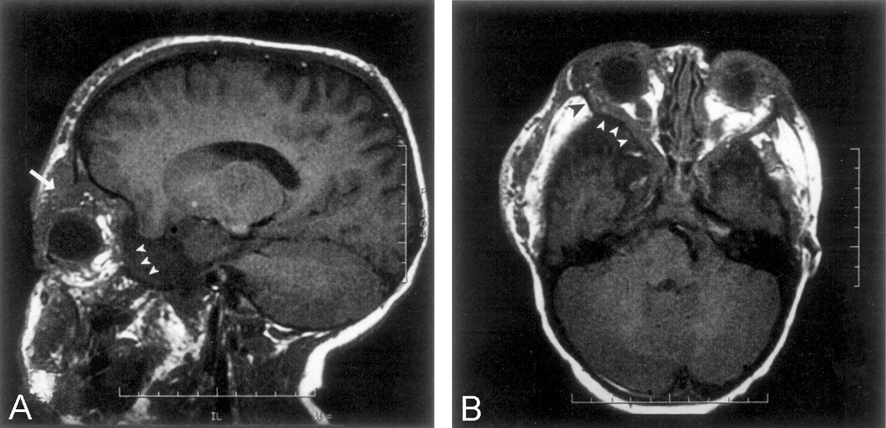
    T1-weighted MR images.

    A, Parasagittal image shows a temporal arachnoid cyst on the left that pushes the orbital contents, including the globe, forward (arrowheads), while a tumor mass in the superior quadrant of the orbit pushes the globe downward (arrow).

    B, Axial image of the same patient with a temporal arachnoid cyst on the left (white arrowheads) shows shortening of the lateral wall of the orbit (black arrowhead) and flattening of the orbital angle. Also note the abnormal concave posterior aspect of the lateral orbital wall and the large wing of the sphenoid.

  • Fig 2.
    • Download figure
    • Open in new tab
    • Download powerpoint
    Fig 2.

    Coronal CT scans.

    A, Image of the classic egg-shaped, orbital enlargement and deformity. Note the phthisical eye (asterisk) and tumoral infiltration of superior rectus muscle (arrow) and orbital septum (arrowheads). The left paranasal sinuses are underdeveloped. The nasal septum and crista galli are deviated.

    B, Image of the posterior orbit shows that the tumors in the pterygoid fossa (asterisk) infiltrates the left orbital apex, enlarging the inferior orbital fissure and distorting the posterior orbit.

  • Fig 3.
    • Download figure
    • Open in new tab
    • Download powerpoint
    Fig 3.

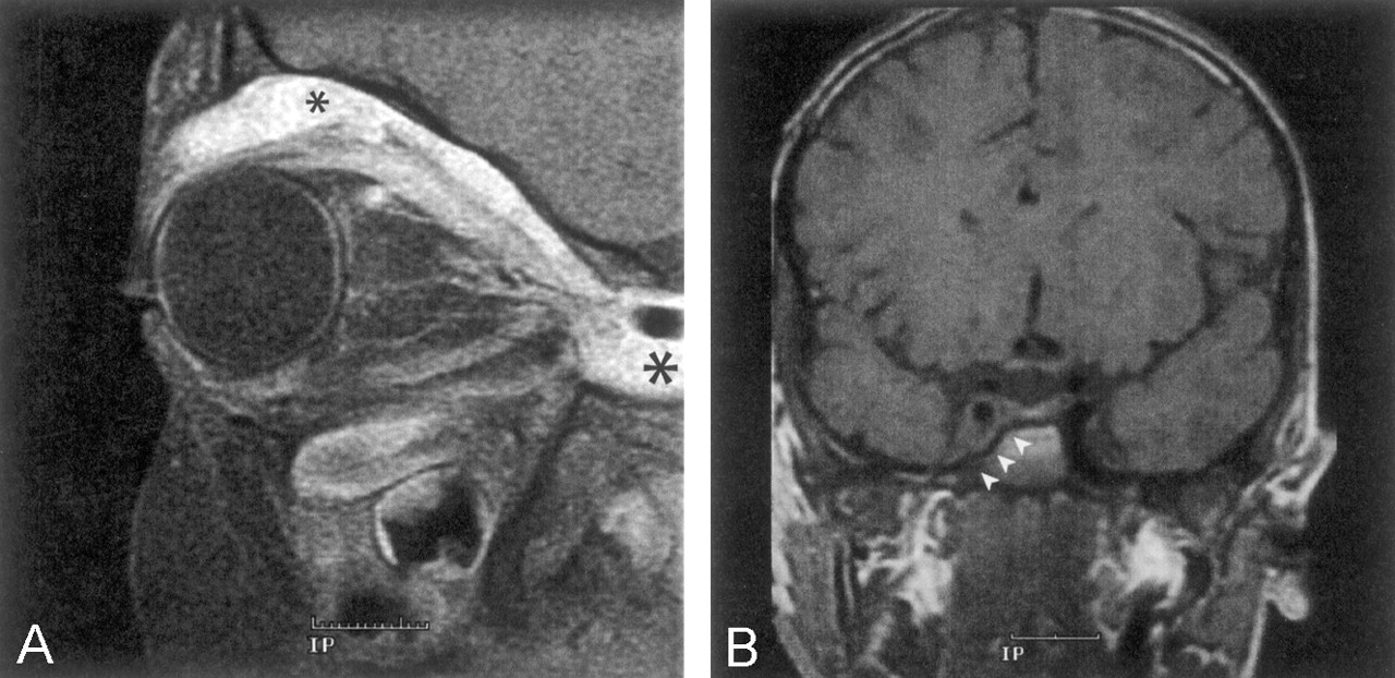
    A, Sagittal T1-weighted, contrast-enhanced, fat-suppressed MR image shows infiltration of the superior extraconal space (small asterisk), superior rectus muscle, and levator palpebrae muscle by PNFs. Posterior extension of the tumor into the cavernous sinus is marked by the large asterisk. Intraconal sensory nerves are contrast enhanced and thickened because of tumor infiltration. Ipsilateral maxillary sinus is small compared with that on the contralateral side.

    B, Coronal T1-weighted MR image of the same patient shows extension of the tumor into the cavernous sinus, with discrete remodeling of the sphenoid body (arrowheads).
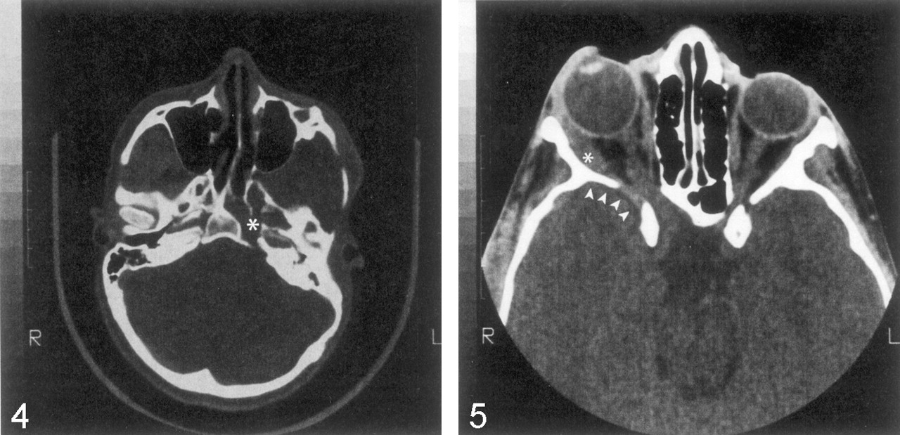
  • Fig 4.
    • Download figure
    • Open in new tab
    • Download powerpoint
    Fig 4.

    Axial CT image of the skull base shows expanded cranial foramina (foramen ovale, rotundum, and lacerum) on the left (asterisk) due to tumor enlargement of branches of the trigeminal nerve.

  • Fig 5.
    • Download figure
    • Open in new tab
    • Download powerpoint
    Fig 5.

    Axial CT image through the orbits shows unilateral enlargement of the right optic canal due to tumor infiltration of the optic nerve sheath without an optic nerve glioma. The ipsilateral globe is buphthalmic, and the lateral rectus (asterisk) is enlarged as a result of tumoral infiltration. The greater wing of the sphenoid is partially decalcified and bowed forward (arrowheads), without an obvious arachnoid cyst.

Tables

  • Figures
  • Patients with NF1

    LocationNo. of Patients (n=31)
    Soft-tissue tumor without orbital involvement7 (22.6)
    Radiologic orbital changes24 (77.4)
    Distortion due to an enlarged middle cranial fossa13 (41.9)
    Orbital-rim enlargement18 (58.1)
     Associated with buphthalmos13
     Associated with a small maxillary sinus13
     Associated with tumor in the anterior orbit2
    Imaging evidence of tumor within the orbit19 (61.3)
     Intraconal tumor13
     Globe (buphthalmos with or without enucleation)13
     Lacrimal gland11
     Extraocular muscle13
     Posterior orbit16
     Orbital bone18
    Expansion of the orbital foramen16 (51.6)
     Expansion of the superior orbital fissure15
     Expansion of the inferior orbital fissure12
     Expansion of the optic canal8
      Optic nerve glioma only6
      PNF infiltration of the optic nerve sheath2
    • Note.—Data in parentheses are percentages.

Back to top

In this issue

American Journal of Neuroradiology: 24 (8)
American Journal of Neuroradiology
Vol. 24, Issue 8
1 Sep 2003
  • Table of Contents
  • Index by author
Advertisement
Print
Download PDF
Email Article

Thank you for your interest in spreading the word on American Journal of Neuroradiology.

NOTE: We only request your email address so that the person you are recommending the page to knows that you wanted them to see it, and that it is not junk mail. We do not capture any email address.

Enter multiple addresses on separate lines or separate them with commas.
Orbit Deformities in Craniofacial Neurofibromatosis Type 1
(Your Name) has sent you a message from American Journal of Neuroradiology
(Your Name) thought you would like to see the American Journal of Neuroradiology web site.
CAPTCHA
This question is for testing whether or not you are a human visitor and to prevent automated spam submissions.
Cite this article
Claude Jacquemin, Thomas M. Bosley, Helena Svedberg
Orbit Deformities in Craniofacial Neurofibromatosis Type 1
American Journal of Neuroradiology Sep 2003, 24 (8) 1678-1682;

Citation Manager Formats

  • BibTeX
  • Bookends
  • EasyBib
  • EndNote (tagged)
  • EndNote 8 (xml)
  • Medlars
  • Mendeley
  • Papers
  • RefWorks Tagged
  • Ref Manager
  • RIS
  • Zotero
0 Responses
Respond to this article
Share
Bookmark this article
Orbit Deformities in Craniofacial Neurofibromatosis Type 1
Claude Jacquemin, Thomas M. Bosley, Helena Svedberg
American Journal of Neuroradiology Sep 2003, 24 (8) 1678-1682;
del.icio.us logo Twitter logo Facebook logo Mendeley logo
  • Tweet Widget
  • Facebook Like
  • Google Plus One

Jump to section

  • Article
    • Abstract
    • Methods
    • Results
    • Discussion
    • Conclusion
    • References
  • Figures & Data
  • Info & Metrics
  • Responses
  • References
  • PDF

Related Articles

  • No related articles found.
  • PubMed
  • Google Scholar

Cited By...

  • Posterior-Anterior Cephalometric Study of Neurofibromatosis Type 1 Patients With Facial Plexiform Neurofibroma: Analysis of Skeletal Symmetry Concerning Midfacial and Skull Base Reference Points (Zygomatic Arch, Mastoid, and Juga)
  • Sphenoid Bone Pneumatisation on Lateral Cephalograms of Patients With Neurofibromatosis Type 1
  • Periapical Cemento-osseous Dysplasia Is Rarely Diagnosed on Orthopantomograms of Patients with Neurofibromatosis Type 1 and Is Not a Gender-specific Feature of the Disease
  • Optic Pathway Glioma and Cerebral Focal Abnormal Signal Intensity in Patients with Neurofibromatosis Type 1: Characteristics, Treatment Choices and Follow-up in 134 Affected Individuals and a Brief Review of the Literature
  • Analysis of Orbital Plain Radiographs for Orbital Deformities in Neurofibromatosis Type 1 Patients, with Special Reference to Alterations of the Orbital Rim as Indicators of Adjacent Plexiform Neurofibroma
  • Reconstruction of the Sphenoid Wing in a Case of Neurofibromatosis Type 1 and Complex Unilateral Orbital Dysplasia with Pulsating Exophthalmos
  • Dysplasia of the Orbit and Adjacent Bone Associated with Plexiform Neurofibroma and Ocular Disease in 42 NF-1 Patients
  • Crossref
  • Google Scholar

This article has not yet been cited by articles in journals that are participating in Crossref Cited-by Linking.

More in this TOC Section

  • Comparison of Image Quality and Radiation Dose in Pediatric Temporal Bone CT Using Photon-Counting Detector CT and Energy-Integrating Detector CT
  • SyMRI & MR Fingerprinting in Brainstem Myelination
  • Dual-Layer Detector CT for PEDS Image Quality
Show more Pediatrics

Similar Articles

Advertisement

Indexed Content

  • Current Issue
  • Accepted Manuscripts
  • Article Preview
  • Past Issues
  • Editorials
  • Editors Choice
  • Fellow Journal Club
  • Letters to the Editor

Cases

  • Case Collection
  • Archive - Case of the Week
  • Archive - Case of the Month
  • Archive - Classic Case

Special Collections

  • Special Collections

Resources

  • News and Updates
  • Turn around Times
  • Submit a Manuscript
  • Author Policies
  • Manuscript Submission Guidelines
  • Evidence-Based Medicine Level Guide
  • Publishing Checklists
  • Graphical Abstract Preparation
  • Imaging Protocol Submission
  • Submit a Case
  • Become a Reviewer/Academy of Reviewers
  • Get Peer Review Credit from Publons

Multimedia

  • AJNR Podcast
  • AJNR SCANtastic
  • Video Articles

About Us

  • About AJNR
  • Editorial Board
  • Not an AJNR Subscriber? Join Now
  • Alerts
  • Feedback
  • Advertise with us
  • Librarian Resources
  • Permissions
  • Terms and Conditions

American Society of Neuroradiology

  • Not an ASNR Member? Join Now

© 2025 by the American Society of Neuroradiology All rights, including for text and data mining, AI training, and similar technologies, are reserved.
Print ISSN: 0195-6108 Online ISSN: 1936-959X

Powered by HighWire